This is an open access journal, and articles are distributed under the terms of the Creative Commons Attribution-Non Commercial-ShareAlike 4.0 License, which allows others to remix, tweak, and build upon the work non-commercially, as long as appropriate credit is given and the new creations are licensed under the identical terms.
© 2018 Journal of Advanced Pharmacy Education & Research | Published by SPER Publication 121
Serum concentrations of CD4+ and CD8+ in patients infected
with scabies caused by
Sarcoptes scabiei
Ali A. Mohy, Ahmed Abduljabbar Jaloob Aljanaby, Saleem Khteer Al-Hadraawy
Department of Biology, Faculty of science, University of Kufa, Iraq.
Correspondence:Ahmed Abduljabbar Jaloob Aljanaby, Department of Biology, Faculty of science, University of Kufa, Iraq. E-mail: ahmedaj.aljanabi @ uokufa.edu .iq
ABSTRACT
Background: Scabies is one of the most important epidemic diseases in developing countries infecting males and females especially in rural areas. Therefore, this study aimed to evaluate the cellular immune response in patients infected with scabies caused by Sarcoptes scabiei depended on the measurement serum concentration of T-helper cell (CD4+) and T-cytotoxic cell (CD8+). Materials and Methods: Sixty patients (30 male and 30 female) infected with chronic scabies were included in this study and 30 healthy individuals were considered as control group. CD4+ and CD8+ levels in serum of patients and healthy individuals were measurement by using ELISA device. Results: The results proved that there was a significant increase (P < 0.05) in serum concentrations of CD4+ and CD8+ as compared with control group. On the other hand, the results demonstrated that there was a positive correlation between CD4+ and CD8+ (R2 = 0.0301). Serum concentration of CD8+ was higher than CD4+ with significant increase (P value=0.0001). Conclusion: Human infected with S. scabiei lead to induce higher amounts of Th and Tc cells due to the high levels production of CD8+ and CD4+ in serum of patients.
Keywords:Scabies, Sarcoptes scabiei, T-helper, T-cytotoxic, CD4+, CD8+.
Introduction
Sarcoptes scabiei is one of the most important causative agents of scabies in worldwide. Scabies is a one of the most prevalent infection in all countries especially in developing countries [1, 2]. Patients infected with chronic scabies suffering from very strong itching due to allergic and inflammatory reactions mounted by the host against the parasite and its product such as eggs [3]. The clinical features in patients infected with scabies were characterized between mild to severe destructive [4]. The
immune and inflammatory response features in patients are remaining not fully understood [5]. T-lymphocyte (T-cell) CD4+ T-helper (Th) and CD8+ T-cytotoxic (Tc) have an important role in immune response in scabies due to cytokines secreted such as interferon-γ (INF-γ), tumor necrotic factor-α (TNF-α) and interlukine-2 (IL-2) [6, 7]. T-helper cell secrete IL-13, IL-5
and IL-4 and mediated humoral immunity by inducing antibody production to fight extracellular parasite [8]. T-helper 2 cells are also dominant effector cells in the pathogenesis of IgE-mediated hypersensitivity in scabies and other allergic inflammatory infections [9]. Peripheral blood mononuclear cells isolated from patients infected with scabies secreted increased levels of Th2 cytokines include IL-13, IL-5 and IL-4 but decreased secretion of the Th1 cytokines INF-γ [10]. Cellular immune response in patients infected with scabies is still unclear. In Iraq, there are no studies focusing on the rule of T-cells (Th and Tc) in patients infected with scabies. Therefore, in this study we aimed to evaluate the cellular immune response in patient infected with scabies caused by S. scabiei due to the measurement serum concentration of CD4+ and CD8+ in these patients.
Materials and Methods
Patients:
Sixty patients (30 male and 30 Female, ages range18 to 40 years old) infected with chronic scabies cause by Sarcoptes scabiei (duration of infection 6 weeks) were included in this study, admitted to Al-Najaf general hospital in Al-Najaf City, Iraq, during the period from March to December 2017. Thirty healthy persons (15 Males and 15 females, ages range 18 to 40 years old), included in the current study were considered as the control group.
Access this article online
Website: www.japer.in E-ISSN: 2249-3379
How to cite this article:Ali A. Mohy, Ahmed Abduljabbar Jaloob Aljanaby, Saleem Khteer Al-Hadraawy. Serum concentrations of CD4+ and CD8+ in patients infected with scabies caused by Sarcoptes scabiei. J Adv Pharm Edu Res 2018;8(1):121-126.
Ali A. Mohy, et al.: Serum concentrations of CD4+ and CD8+ in patients
122 Journal of Advanced Pharmacy Education & Research | Jan-Mar 2018 | Vol 8 | Issue 1
Detection of patients infected with chronic
scabies cause by Sarcoptes scabiei:
Sarcoptesscabiei was diagnosed in patients according to physical exam and inspecting the affected area of skin or removing a S. scabiei from the skin with a needle, taking a scrape off a small section of skin to obtain a tissue sample and examining this sample under a microscope to confirm the presence of scabies mites or their eggs
[11].
Serum collection:
Two ml of serum were collectedfrom patients and healthy individuals as follow: 5 ml of blood were collected from two groups and centrifugation (Memmert Company, Germany) was done at 3000rpm for 5min. Serum was collected in sterile tube and stored at -15C until use [2].
Measurement of human CD4+ and CD8+:
Human CD4 and CD8 kits were provided from Elabscience Company, Bulgaria. CD4+ and CD8+ levels in serum of patients and healthy individuals were measurement by using ELISA device (Human Reader Company, Germany) according to instructions of the manufacturer.
Statistical Analysis:
T-test was used in the currentstudy for comparison between the samples by using SPSS V.10 computer software. P value less than 0.05 was considered statistically significant [12, 13].
Results
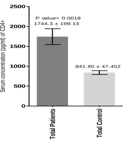
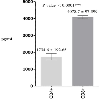
The results of the current study proved that there was a significant increase (P value= 0.0018) in serum concentration of CD4 in total patients infected with chronic scabies (1744.3 ± 199.13 pg/ml) as compared with control (841.80 ± 47.452 pg/ml) (Figure 1). Also, the results demonstrated that there was a significant increase in serum concentration of CD4 in male (1926.8 ± 328.99 pg/ml) and female (1561.9 ± 225.45 pg/ml) infected with chronic scabies as compared with control with P value; 0.0308 and 0.0197, respectively (Figure 2). Serum concentration of CD8 in total patients infected with chronic scabies (4046.2 ± 97.963 pg/ml) was higher than in total control (3464.9 ± 131.25 pg/ml) with high significant increase (P=0.0007) (Figure 3). On the other hand, serum concentration of CD8 in male patients was in high level (4082.6 ± 135.34 pg/ml) as compared with male control (3483.8 ± 191.21 pg/ml) with significant increase (P value= 0.0138). The same serum concentration of CD8 was increased in female patients (4009.7 ± 143.74 pg/ml) as compared with female control (3446.1 ± 186.43 pg/ml) with significant increase (P value= 0.0241) (Figure 4). On the other hand, there was a highly significant increase in serum concentration of CD8+ (4078.7 ± 97.399 pg/ml) in total patients (P value=< 0.0001) as compared with serum concentration of CD4+ (1734.6 ± 192.65 pg/ml) (Figure 5). But there was a positive correlation between CD4+ and CD8+ (R2=0.0301) (Figure 6).
To
ta
l P
at
ie
nt
s
To
ta
l C
on
tro
l
0 500 1000 1500 2000 2500
1744.3 ± 199.13
841.80 ± 47.452 P value= 0.0018
Se
ru
m
c
on
ce
nt
ra
tio
n
(p
g/
m
l)
of
CD4
+
Journal of Advanced Pharmacy Education & Research | Jan-Mar 2018 | Vol 8 | Issue 1 123 Male Co nt ro l
Female Cont
ro l 0 500 1000 1500 2000 2500
1926.8 ± 328.99
889.93 ± 84.046
1561.9 ± 225.45
793.67 ± 43.973 P value=0.0308* P value=0.0197*** S er um c on ce nt ra tio n (p g/ m l) of CD4 +
Figure 2:Serum concentration (pg/ml) of CD4+ in male and female infected with scabies
To ta l p at ie nt s To ta l c on tr ol 0 1000 2000 3000 4000 5000
4046.2 ± 97.963
3464.9 ± 131.25 P value= 0.0007
Se ru m c on ce nt ra tio n (p g/ m l) of CD8 +
Figure 3:Serum concentration (pg/ml) of CD8+ in total patients infected with scabies
Ma le C o n tr o l F e ma le C o n tr o l 0 1000 2000 3000 4000 5000
4082.6 ± 135.34
3483.8 ± 191.21 P value= 0.0138*
4009.7 ± 143.74
3446.1 ± 186.43
P value= 0.0241*
S e ru m c o n c e n tr a ti o n ( p g /m l) o f CD8 +
Ali A. Mohy, et al.: Serum concentrations of CD4+ and CD8+ in patients
124 Journal of Advanced Pharmacy Education & Research | Jan-Mar 2018 | Vol 8 | Issue 1
C
D
4
+
C
D
8
+
0 1000 2000 3000 4000 5000
1734.6 ± 192.65
4078.7 ± 97.399 P value=< 0.0001***
pg/ml
Figure 5:Serum concentration (pg/ml) of CD4+ and CD8+ in total patients infected with scabies
Figure 6:Correlation between serum concentration of CD4+ and CD8+ in patients infected with scabies
Discussion
Scabies is a one of the most important skin infections mainly caused by Sarcoptes Scabiei. There are over one hundred million infections all over the world [14]. Cellular immune response in
human infected with chronic scabies is still not fully understood. In this study, we aimed to compare between serum concentration of CD4+ and CD8+ in patients infected with scabies caused by S. Scabiei. The results proved that there was a high significant increase in CD4+ and CD8+ as compared with control and there was a positive coloration between CD4+ and CD8+. On the other hand, serum concentration of CD8+ was higher than CD4+. T-cells play an important role in cell mediated immune response. CD4+ T-cell has been demonstrated as the most prevalent T-lymphocytes in skin lesions in human infected with scabies [15, 16]. These results are not
in agreement with Akdis et al. (1999) [17] that they suggested that the corresponds to inflammatory cells in the skin lesions from patients with scabies have a significantly greater number of infiltrating CD4+ lymphocytes compared with CD8+. While
the results of the current study are in agreement with Walton et al. (2008) [18] and Liu et al. (2014) [19] when they demonstrated that there was an increased number of infiltrating CD8+ T-cell as compared with CD4+cells according to immune-histology and flow cytometry study. Other studies by Roberts et al (2005) [20] and Walton et al (2008) [18] indicated that the number of
T-cell and B-T-cell in the blood of patients infected with scabies have been within normal ranges. Bovenschen et al (2005) [21] and Walton et al (2010) [10] suggested that the greater number of CD8+ T-cell is due to a selective movement of CD8+ T-cell leading to epidermal hyper proliferation. Therefore, the cytotoxic T-cell is responsible for the imbalance of inflammatory response and may contribute to the failure of the skin immune system to induce an effective response resulting in uncontrolled growth of this mite [22, 23]. Patients infected with Sarcoptes scabies hurt from powerful inflammatory reactions itching and mediated through allergic mounted by the host against the mite and its products, a comprehensive range of clinical symptoms
y = 0.0877x + 3926.6 R² = 0.0301
0 1000 2000 3000 4000 5000 6000
0 2000 4000 6000 8000 10000
Seru
m
co
n
cen
trat
io
n
(p
g
/m
l)o
f
CD
4
+
Journal of Advanced Pharmacy Education & Research | Jan-Mar 2018 | Vol 8 | Issue 1 125
from acute destructive to minor occurs in scabies but despite the significant worldwide impact of the disease, the inflammatory and immune responses related with the diverse symptoms of this disease remain poorly understood [14]. This increase in serum
concentration of CD4 and CD8 may be due to inflammation of skin and produce the gamma interferon (INF-γ) and interleukin-10 (IL-interleukin-10) in the patients of scabies or may increase the eosinophil in skin of scabies’ infection [24, 25]. In skin infection and
inflammatory site high numbers of eosinophils are produced and for this reason, the concentration levels of IL-4 and IL-13 increased [26-28].
Conclusions and Future
Recommendations
Infection by chronic scabies for 6 weeks caused by Sarcoptes scabiei in human leading to inducement of higher amounts of T-cytotoxic cell and T-helper cell due to the high levels production of CD8+ and CD4+ in serum of patients. It is recommended to evaluate other immune cells and other CD markers such as T-lymphocyte cell (CD3+), Granulocyte cell (CD15+) and Monocyte cell (CD14+) to provide complete and clear immune vision against Sarcoptes scabiei.
Significant Statements
This study discovered that T-cytotoxic and T-helper cells have an important role in immune response in patients infected with Sarcoptes scabiei which was not proved previously. This study will help researchers to discover more details about new mechanisms of immune response in individuals infected with scabies.
References
1. Hay RJ, Johns NE, Williams HC, Bolliger IW, Dellavalle RP, Margolis DJ, Marks R, Naldi L, Weinstock MA, Wulf SK, Michaud C. The global burden of skin disease in 2010: an analysis of the prevalence and impact of skin conditions. J Invest Dermatol. 2014 Jun 1; 134(6):1527-34.
2. Al-Hadraawy SK, Hessen HB. Hematological and Epidemiological Study for Patients Infected with Scabies. J Pharm Sci & Res. 2017 Jun 1;9(6):897.
3. Marotta M, Toni F, Dallolio L, Toni G, Leoni E. Management of a family outbreak of scabies with high risk of spread to other community and hospital facilities. Am J Infect Control. 17. 2018 Feb 1.
4. Cohen PR. Scabies masquerading as bullous pemphigoid: scabies surrepticius. Clin Cosmet Investig Dermatol. 2017, 10.
5. Morgan MS, Rider Jr SD, Arlian LG. Identification of antigenic Sarcoptes scabiei proteins for use in a diagnostic test and of non-antigenic proteins that may be immunomodulatory. PLoS Negl Trop Dis. 2017 Jun 12;11(6): e0005669.
6. Romagnani S. T-cell subsets (Th1 versus Th2). Annals of allergy, asthma & immunol. 2000 Jul 1; 85(1):9-18.
7. Kidd P. Th1/Th2 balance: the hypothesis, its limitations, and implications for health and disease. Altern Med Rev. 2003 Aug 1; 8(3):223-46.
8. Rampton M, Walton SF, Holt DC, Pasay C, Kelly A, Currie BJ, McCarthy JS, Mounsey KE. Antibody responses to Sarcoptes scabiei apolipo protein in a porcine model: relevance to immunodiagnosis of recent infection. PloS one. 2013 Jun 6;8(6): e65354.
9. Walton SF, Slender A, Pizutto S, Mounsey KE, Opresecu F, Thomas WR, Hales BJ, Currie BJ. Analysis of IgE binding patterns to house dust mite allergens in scabies‐
endemic communities: insights for both diseases. Clin Exp Allergy. 2015 Dec 1;45(12):1868-72.
10. Walton SF, Pizzutto S, Slender A, Viberg L, Holt D, Hales BJ, Kemp DJ, Currie BJ, Rolland JM, O'Hehir R. Increased allergic immune response to Sarcoptes scabiei antigens in crusted versus ordinary scabies. Clin Vaccine Immunol. .2010 Sep 1;17(9):1428-38.
11. Chosidow O. Clinical practices. Scabies. N Engl J Med. 2006; 354(16).
12. Aljanaby AA, Alhasnawi HM. Research Article Phenotypic and Molecular Characterization of Multidrug Resistant Klebsiella pneumoniae Isolated from Different Clinical Sources in Al-Najaf Province-Iraq. Pak J Biol Sci. 2017; 20 (5).
13. Aljanaby AA, Medhat AR. Research Article Prevalence of Some Antimicrobials Resistance Associated-genes in Salmonella typhi Isolated from Patients Infected with Typhoid Fever. J. Biol. Sci. 2017; 17 (4).
14. Bhat SA, Mounsey KE, Liu X, Walton SF. Host immune responses to the itch mite, Sarcoptes scabiei, in humans. Parasites & vectors. 2017 Dec; 10(1):385.
15. Falk ES, Eide TJ. Histologic and clinical findings in human scabies. Int J Dermatol. 1981 Nov 1; 20(9):600-5. 16. Falk ES, Matre R. In situ characterization of cell infiltrates
in the dermis of human scabies. Am J Dermatopathol. 1982 Feb; 4(1):9-15.
17. Akdis CA, Akdis M, Simon D, Dibbert B, Weber M, Gratzl S, Kreyden O, Disch R, Wüthrich B, Blaser K, Simon HU. T cells and T cell-derived cytokines as pathogenic factors in the nonallergic form of atopic dermatitis. J Invest Dermatol. 1999 Oct 1; 113(4):628-34. 18. Walton SF, Beroukas D, Roberts‐Thomson P, Currie BJ. New insights into disease pathogenesis in crusted (Norwegian) scabies: the skin immune response in crusted scabies. Br J Dermatol. 2008 Jun 1; 158(6):1247-55. 19. Liu X, Walton SF, Murray HC, King M, Kelly A, Holt DC,
Currie BJ, McCarthy JS, Mounsey KE. Crusted scabies is associated with increased IL‐17 secretion by skin T cells. Parasite immunolo. 2014 Nov 1; 36(11):594-604. 20. Roberts LJ, Huffam SE, Walton SF, Currie BJ. Crusted
Ali A. Mohy, et al.: Serum concentrations of CD4+ and CD8+ in patients
126 Journal of Advanced Pharmacy Education & Research | Jan-Mar 2018 | Vol 8 | Issue 1
21. Bovenschen HJ, Seyger MM, Van De Kerkhof PC. Plaque psoriasis vs. atopic dermatitis and lichen planus: A comparison for lesional T‐cell subsets, epidermal proliferation and differentiation. Br J Dermatol. 2005 Jul 1; 153(1):72-8.
22. Orkin M. Scabies in AIDS. Semin Dermatol. 1993; 12(1). 23. Fuchs BS, Sapadin AN, Phelps RG, Rudikoff D. Diagnostic dilemma: crusted scabies superimposed on psoriatic erythroderma in a patient with acquired immunodeficiency syndrome. SKIN med: Dermatology for the Clinician. 2007 Mar 1; 6(3):142-4.
24. Shi M, Pan W, Tabel H. Experimental African trypanosomiasis: IFN-gamma mediates early mortality. Eur J Immuno. 2003; 33(1).
25. Guilliams M, Oldenhove G, Noel W, Hérin M, Brys L, Loi P, Flamand V, Moser M, De Baetselier P, Beschin A.
African trypanosomiasis: naturally occurring regulatory T cells favor trypanotolerance by limiting pathology associated with sustained type 1 inflammation. J Immunol. 2007 Sep 1;179(5):2748-57.
26. Prussin C, Metcalfe D D. IgE mast cells, basophils, and eosinophils. J Allergy Clin Immunol. 2006. 117 (Suppl 2 Mini-Primer): S450–6.
27. Aljanaby AAJ. Antibacterial activity of an aqueous extracts of Alkanna tinctoria roots against drug resistant aerobic pathogenic bacteria isolated from patients with burns infections. romj.2018 (7):1; 1-6.
28. Aljanaby AAJ and aljanaby IAJ. Profile of Antimicrobial Resistance of Aerobic Pathogenic Bacteria isolated from Different Clinical Infections in Al-Kufa Central Hospital–