Funda Ünsal
B, C, E, F, Mehmet Fatih Sönmez
A, D–FThe Effects of Ovariectomy
on Ghrelin Expression in the Rat Uterus*
Department of Histology and Embryology, Faculty of Medicine, Erciyes University, Kayseri, Turkey
A – research concept and design; B – collection and/or assembly of data; C – data analysis and interpretation; D – writing the article; E – critical revision of the article; F – final approval of article; G – other
Abstract
Background. Ghrelin is a hormone which has effects on the secretion of growth hormone, the gastrointestinal system, the cardiovascular system, cell proliferation and the reproductive system.
Objectives. The aim of this study is to investigate structural changes in the uterine tissue and to assess ghrelin immunoreactivity in the endometrium as a result of bilateral ovariectomization of rats.
Material and Methods. In this study, 28 adult female albino Wistar rats were used. The rats were randomly divided into four groups. Group I was the control group; Group II was the placebo group; Group III was ovariectomized; and Group IV was ovariectomized with 2mg/kg estrogen administered per day. Age-matched diestrous intact rats were used as controls. At the end of the experiment, the rats were decapitated 1, 3, 5, 7, and 15 days after ovariec-tomy under ketamine anesthesia and their uterine tissue was removed.
Results. In the ovariectomized rats, reductions in the sizes of both the uterine epithelium and the endometrial glands were observed, as well as a loss of connective tissue. Ghrelin-positive cells in the endometrial surface and the gland epithelium were visualized by immunohistochemistry. After ovariectomization, ghrelin expression was found to be decreased in a time dependent manner.
Conclusions. Bilateral ovariectomization leads to histological changes in the uterine tissue. Ovariectomization was observed to decrease endometrium ghrelin immunoreactivity (Adv Clin Exp Med 2014, 23, 3, 363–370).
Key words: endometrium, ghrelin, immunoreactivity, ovariectomy, rats.
Adv Clin Exp Med 2014, 23, 3, 363–370 ISSN 1899–5276
ORIGINAL PAPERS
© Copyright by Wroclaw Medical University
Ghrelin is known to be an endogenous ligand for the GH-secretagogue receptor (GHS-R) [1]. Both ghrelin and the GHS-R are also expressed in quite a lot of tissues other than the hypothal-amus and pituitary, including the brain [2], kid-ney [3], stomach [4] and uterus [5]. This molecule was originally noticed for its ability to elicit GH se-cretion in vivo and in vitro in various species, such as humans and rodents [6–8]. This unique expres-sion strongly indicates that ghrelin, which can be produced locally, is also involved in hormone spe-cific actions such as paracrine and autocrine ef-fects, as well as in the actions of the gut-derived peptide [9]. Previous studies have demonstrated that ghrelin is involved in hormone-specific roles, including endocrine and non-endocrine reactions.
These reactions involve corticotropic, lactotropic, and gonadotropic axes controls, the cardiovascu-lar and gastrointestinal systems, as well as carbo-hydratrate metabolism via pancreatic insulin se-cretion, cell growth and proliferation in tissues and tumors [10, 11].
A mounting body of evidence indicates that ghrelin has a role in the control of reproduc-tive physiology by two distinct, probably overlap-ping actions: (a) by means of systemic release of the stomach-derived peptide, which has effects on the reproductive system; and (b) through biologi-cal actions on reproductive organs via lobiologi-cally ex-pressed ghrelin [12, 13]. More recently, circulat-ing ghrelin has been reported to act at different levels on the rat hypothalamic-pituitary gonadal
axis (HPGA), affecting gonadotropin releasing hormone (GnRH) pulsatility, as well as follicle stimulating hormone (FSH) and luteinizing hor-mone (LH) production and secretion [14, 15]. Ghrelin administration stops LH secretion central-ly in ovariectomized (OVX) female rats [16], mon-keys [17] and sheep [18].
The mammalian ovary is known to be a com-plex endocrine organ. It is responsible for oo-cyte release during ovulation and hormonogene-sis. Ovarian steroids are one of the most important factors influencing uterine morphology and motil-ity. Thus, the aim of this study was to investigate alterations in the ghrelin producing cells of the uterus induced by ovariectomy.
Material and Methods
Animals
The study was conducted at the Erciyes Uni-versity Hakan Çetinsaya Experimental and Clin-ic Research Center. EthClin-ical approval was obtained from the Erciyes University Animal Research Local Ethics Committee and all procedures conformed to the “Guide for the Care and Use of Laboratory Animals”. Twenty-eight adult female Wistar rats, 200–250 g in weight at the beginning of the ex-periments, were used. They were housed in a qui-et and temperature-and humidity-controlled room (21 ± 3°C and 60 ± 5%, respectively) in which a 12-h-light/dark cycle was maintained (light from 7:00 am to 7:00 pm). The rats were randomly di-vided into four groups. Group I: Control (n = 4); Group II: Sham (n = 4); Group III: OVX (n = 10); and Group IV: 2 mg/kg/day estrogen given after OVX (n = 10).
Experimental Procedure
The OVX procedure was performed when the animals were 12 weeks old. The rats were anesthe-tized with a combination of intra-peritoneal ket-amine (21.2 mg/kg) and xylazine (4.2 mg/kg). The OVX was preceded by a midline dorsal skin incision approximately 3 cm long. The ovarian vessels were then clamped and the ovaries removed. Afterwards, the uterine tubes were ligated and the muscles and skin were sutured [19]. For the replacement study, estrogen was administered to OVX rats intraperi-toneally. Synthetic estrogen 17 b-estradiol (Sigma Chemical Co., Steinheim, Germany) was dissolved in a mix of ethyl alcohol and sesame oil (Sigma Chemical Co., Steinheim, Germany) and 2 mg/ /kg/0.1 mL injection doses were prepared. After the surgery, the Group IV rats were given subcutaneous
2 mg/kg/day/0.1 ml 17 b-estradiol. The placebo
group was given 0.1 mL sesame oil subcutaneous-ly. The animals were sacrificed by decapitation 1, 3, 5, 7 and 15 days after OVX. Age-matched diestrous intact rats were used as controls.
Immunohistochemistry
The immunohistochemistry procedure was performed as previously described [20]. Brief-ly; five to six micrometer thick sections were cut by microtome, dewaxed and rehydrated following routine protocol, rinsed in deionized water and treated with 3% H2O2 in methanol for 10 min to inhibit endogenous peroxidase activity. The sec-tions were pre-incubated with normal rabbit se-rum (NRS) (S20-100, Chemicon International, Hampshire, UK), diluted 1 : 5 in phosphate-buff-ered saline (PBS) for 20 min at room temperature in a humidified chamber. The primary polyclonal antibody rose against ghrelin (Ghrelin goat poly-clonal IgG, Santa Cruz Biotechnology, California, USA) was diluted 1 : 100 in 20% NRS. The sections were incubated with primary antiserum overnight at 41°C. The sections were incubated with second-ary antibody (rabbit anti-goat IgG, Santa Cruz Bio-technology, California, USA) for 30 min at room temperature in a humidified chamber. The sections were exposed to streptavidin horseradish peroxi-dase (HRP) conjugate (43-4323, Zymed Laborato-ries Inc., South San Francisco, CA, USA), diluted 1:200, for 30 min. The substrate reaction was vi-sualized with 0.5 mg/mL 3,30-diaminobenzidine (DAB) (Liquid DAB Plus Substrate Kit; 00-2020, Zymed Laboratories Inc., South San Francisco, CA, USA) plus 0.1% hydrogen peroxide for 3 min. All dilutions and thorough washes between steps were performed using PBS, pH 7.6, unless other-wise specified. The sections were counterstained with hematoxylin, dehydrated through a graded ethanol series and mounted with EntellanR (Mer-ck, Darmstadt, Germany). For the negative con-trols, incubation with the primary antibody was omitted. Stomach tissue was used as the positive control.
Semi-Quantitative Evaluation
of Staining
26–50% = 0.4, 51–75% = 0.6, 76–100% = 0.9). They were all analyzed individually and the histoscores were obtained as area xintensity [21, 22].
Statistical Analysis
All analyses were performed using a statisti-cal software package (SPSS for Windows®, v. 11.5).
The data were expressed as mean ± SEM. The data were analyzed statistically using one-way analysis of variance (ANOVA). Post hoc analyses were car-ried out with the Tukey test. Statistical significance was set at P < 0.05
Results
Light microscopic examinations showed nor-mal uteruses in the control group (Fig. 1a). In both the placebo group and the first-day group (Fig. 1b) no histological changes were observed after OVX. The endometrium, which was com-posed of simple columnar epithelium and myome-trium, appeared normal. Decreases in the height of surface and glandular epithelium, connective tissue and the number of glandula in the lamina
propria were observed in the third-day group after
OVX. In addition, thinner uterine walls were not-ed. Moreover, the endometrium was composed of low columnar epithelium, and thinning of the
lam-ina propria was observed on the 5th (Fig. 1c) and
7th day after OVX. The thickness of the uterine
walls was also decreased. On the fifteenth day after OVX, thinning of the uterine walls was more evi-dent (Fig. 1d). The endometrium was composed of simple cuboidal epithelium and a thin
lami-na propria. A reduction in collagen staining and
a decrease in the number of glandula in the
lam-ina propria was observed. No histological
disor-der was observed in the experimental group that was administered estrogen after OVX (Fig. 2). The measured thicknesses of uterine walls are present-ed in Fig. 5. Uterine wall thickness was shown to decrease after OVX and this decreases was signifi-cantly different than in the group that was admin-istered estrogen (p < 0.05) (Fig. 5B).
In the control group (Fig. 3a) ghrelin expres-sion was seen in the surface epithelium (++), glan-dular epithelium (++), stroma (+) and muscle layer (+). No difference in ghrelin expression was ob-served between the placebo group and the first-day group after OVX (Fig. 3b). In these groups, ghrelin
Fig. 1. The effects of ovariectomy on uterus morphology: (a) Group I (control), (b) Group III (1st day after
ovariecto-my), (c) Group III (5th day after ovariectomy), (d) Group III (15th day after ovariectomy). The endometrium (e),
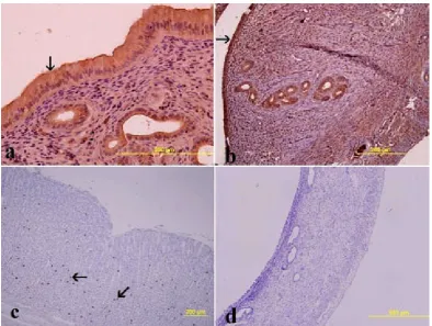
Fig. 3. The effects of ovariectomy on ghrelin expression in the uterus: (a) Group I (control), (b) Group III (1st day after
ovariectomy), (c) Group III (5th day after ovariectomy), (d) Group III (15th day after ovariectomy). Ghrelin
immuno-reactivity (arrow) is strongly expressed in the luminal (L) and glandular (G) epithelium in (a) and (b). A decrease of ghrelin expression over time after OVX is visible in (c) and (d). Sections stained with immunoperoxidase
Fig. 2. The effects of estrogen supplementation on the uteruses of ovariectomized rats: (a) Group IV (3rd day after
ovariectomy), (b) Group IV (5th day after ovariectomy), (c) Group IV (7th day after ovariectomy), (d) Group IV
(15th day after ovariectomy). Endometrium (e), myometrium (m) and perimetrium (arrow head) are normal in the
expression was noted in the surface and glandular epithelium, stroma and muscular layer. A decrease in ghrelin expression was observed over time af-ter OVX in Group III (Fig. 3c and 3d). However, in the group that was administered estrogen after OVX, the ghrelin expression level was same as in the control group (Fig. 4). The expression of ghre-lin was assessed according to the intensity and am-plitude of expression. Figure 5 shows the expres-sion levels of ghrelin in the luminal and glandular epithelium. Ghrelin expression in the OVX group was significantly different than that of the group administered estrogen (p < 0.05, Fig. 5a).
Discussion
Ghrelin is an acylated polypeptide hormone secreted predominantly by the endocrine cells of the stomach [1, 23]. Quite a lot of evidence clear-ly shows that ghrelin is responsible for regulating many things, such as energy balance, GH release, food intake, G protein-coupled receptors and the GH secretagogue receptor (GHSR) type 1a [16]. Interestingly, ghrelin is also involved in reproduc-tion, by influencing the synthesis and release of re-productive factors by the brain and the pituitary, and by influencing gonadal physiology [12, 13].
Fig. 4. The effects of ovariectomy on ghrelin expression in the uterus: (a) Group II (placebo), (b) Group IV (5th day
after ovariectomy) (c) positive controls (ghrelin immunopositive cells in stomach tissue); (d) negative controls (Group III, 15th day after ovariectomy). Ghrelin expression (arrow) is at similar levels in both the control group and the group
administered estrogen (Group IV). Sections stained with immunoperoxidase
As noted above, ghrelin administration stops LH secretion centrally in ovariectomized rats [16]. Furthermore, Fernandez et al. [14] demonstrat-ed that ghrelin is capable of inhibiting LH secre-tion in vivo in prepubertal males and in gonadec-tomized animals, both male and female, but FSH release was not affected.
A number of growth factors and cytokines secreted by the reproductive tract and the pre-implantation embryo exert a paracrine and/or autocrine influence on the rate of embryo devel-opment [24]. Both ghrelin and GHS-R mRNAs have been detected in the morula and in further stages of embryo development; in addition, ghre-lin protein is produced by the reproductive tract and released into the uterine fluid [25]. Ghrelin levels in uterine fluid were strikingly augmented over a fasting period in mice, and ghrelin stopped the development of preimplantation murine em-bryos in vitro [26]. Aghajanova et al. demonstrat-ed that ghrelin and GHSR expression is less inten-se in the mid-inten-secretory endometrium of infertile women than in fertile controls [27]. Furthermore, it was demonstrated that in rats, chronic ghrelin treatment throughout the first half of pregnancy decreases litter size [28].
Ghrelin also negatively modulates cell viabili-ty and proliferation [11]. While GHS-R1a mRNA is detected in the endometrium during the normal menstrual cycle, ghrelin expression is observed in the secretory endometrium, particularly in luminal and glandular epithelial cells. Since specific prod-ucts secreted by these cells play an important role in embryo implantation, these data indicate a role for ghrelin as a probable paracrine/autocrine regulator of human endometrial receptivity. In this study, ghrelin expression was seen in the surface epitheli-um, glandular epitheliepitheli-um, stroma and muscle layer in the rat uterus. These findings agree with the dis-tribution patterns and the possible paracrine/auto-crine regulator role of ghrelin in the rat uterus that have been reported previously [27].
Analyses using in situ hybridization and im-munohistochemical methods have shown the lo-calization of the ghrelin in rat and human repro-ductive tissues. Immunohistochemical and mRNA profiling studies have suggested that rat gonadal ghrelin expression is a function of the estrous cy-cle, and that its levels increase in the cytoplasm of luteal phase steroidogenic luteal cells [29]. A sim-ilar expression pattern has been found in the hu-man ovary, where hilus interstitial cells and young and mature corpus lutei were found to be ghrelin-immunoreactive [30]. Ghrelin inhibits estradiol and progesterone secretion from granulose cells of the human ovary, but this effect was blocked in the presence of a ghrelin receptor antagonist [31].
endometrium was noted. This diversity suggests that the mechanism by which ghrelin expression is regulated is based on several factors including es-trogen, age and tissue. The direct effect of the ova-ries on the uterus might be responsible of the de-crease of ghrelin expression in the endometrium.
In conclusion, the present study shows that (a) morphological changes were observed in the rat uterus after OVX; (b) ghrelin expression was
distributed throughout the endometrium; and (c) ghrelin expression was reduced in the endometri-um following OVX, probably due to the decrease in the estrogen level. Overall, these data reinforce the concept that ghrelin may have a paracrine/au-tocrine regulatory role in the endometrium. How-ever, further study is warranted to determine the factors involved in the regulation of the ghrelin expression.
Acknowledgements. The authors wish to thank Dr. Eser Kılıç, Dr.Cagrı Sakalar and Dr. Metin Aytekin for technical support.
References
[1] Kojima M, Hosoda H, Date Y, Nakazato M, Matsuo H, Kangawa K: Ghrelin is a growth-hormone-releasing acyl-ated peptide from stomach. Nature 1999, 402, 656–660.
[2] Nakazato M, Murakami N, Date Y, Kojima M, Matsuo H, Kangawa K, Matsukura S: A role for ghrelin in the central regulation of feeding. Nature 2001, 409, 194–198.
[3] Mori K, Yoshimoto A, Takaya K, Hosoda K, Ariyasu H, Yahata K, Mukoyama M, Sugawara A, Hosoda H, Kojima M, Kangawa K, Nakao K: Kidney produces a novel acylated peptide, ghrelin. FEBS Lett 2000, 486, 213–216. [4] Sonmez MF, Ozan E: Determination of ghrelin immunoreactivity in the rat stomach after fasting and refeeding.
Acta Histochem 2007, 109, 193–199.
[5] Tanaka K, Minoura H, Isobe T, Yonaha H, Kawato H, Wang DF, Yoshida T, Kojima M, Kangawa K, Toyoda N: Ghrelin is involved in the decidualization of human endometrial stromal cells. J Clin Endocrinol Metab 2003, 88, 2335–2340.
[6] Arvat E, Maccario M, Di Vito L, Broglio F, Benso A, Gottero C, Papotti M, Muccioli G, Dieguez C, Casanueva FF, Deghenghi R, Camanni F, Ghigo E: Endocrine activities of ghrelin, a natural growth hormone secretagogue (ghs), in humans: Comparison and interactions with hexarelin, a nonnatural peptidyl ghs, and gh-releasing hormone. J Clin Endocrinol Metab 2001, 86, 1169–1174.
[7] Tschop M, Smiley DL, Heiman ML: Ghrelin induces adiposity in rodents. Nature 2000, 407, 908–913.
[8] Wren AM, Small CJ, Ward HL, Murphy KG, Dakin CL, Taheri S, Kennedy AR, Roberts GH, Morgan DG, Ghatei MA, Bloom SR: The novel hypothalamic peptide ghrelin stimulates food intake and growth hormone secretion. Endocrinology 2000, 141, 4325–4328.
[9] Korbonits M, Goldstone AP, Gueorguiev M, Grossman AB: Ghrelin – a hormone with multiple functions. Front Neuroendocrinol 2004, 25, 27–68.
[10] Broglio F, Arvat E, Benso A, Papotti M, Muccioli G, Deghenghi R, Ghigo E: Ghrelin: Endocrine and non-endocrine actions. J Pediatr Endocrinol Metab 2002, 15, 1219–1227.
[11] Jeffery PL, Herington AC, Chopin LK: The potential autocrine/paracrine roles of ghrelin and its receptor in hormone-dependent cancer. Cytokine Growth Factor Rev 2003, 14, 113–122.
[12] Barreiro ML, Tena-Sempere M: Ghrelin and reproduction: A novel signal linking energy status and fertility? Mol Cell Endocrinol 2004, 226, 1–9.
[13] Tena-Sempere M: Ghrelin: Novel regulator of gonadal function. J Endocrinol Invest 2005, 28, 26–29.
[14] Fernandez-Fernandez R, Tena-Sempere M, Aguilar E, Pinilla L: Ghrelin effects on gonadotropin secretion in male and female rats. Neurosci Lett 2004, 362, 103–107.
[15] Lorenzi T, Meli R, Marzioni D, Morroni M, Baragli A, Castellucci M, Gualillo O, Muccioli G: Ghrelin: A meta-bolic signal affecting the reproductive system. Cytokine Growth Factor Rev 2009, 20, 137–152.
[16] Furuta M, Funabashi T, Kimura F: Intracerebroventricular administration of ghrelin rapidly suppresses pulsatile luteinizing hormone secretion in ovariectomized rats. Biochem Biophys Res Commun 2001, 288, 780–785. [17] Vulliemoz NR, Xiao E, Xia-Zhang L, Germond M, Rivier J, Ferin M: Decrease in luteinizing hormone pulse
frequency during a five-hour peripheral ghrelin infusion in the ovariectomized rhesus monkey. J Clin Endocrinol Metab 2004, 89, 5718–5723.
[18] Iqbal J, Kurose Y, Canny B, Clarke IJ: Effects of central infusion of ghrelin on food intake and plasma levels of growth hormone, luteinizing hormone, prolactin, and cortisol secretion in sheep. Endocrinology 2006, 147, 510–519.
[19] Lasota A, Danowska-Klonowska D: Experimental osteoporosis – different methods of ovariectomy in female white rats. Rocz Akad Med Bialymst 2004, 49, 129–131.
[20] Sonmez MF, Narin F, Balcioglu E: Melatonin and Vitamin C Attenuates Alcohol-Induced Oxidative Stress in Aorta. Basic Clin Pharmacol Toxicol 2009, 105, 410–415.
[21] Hawkins NJ, Ward RL: Sporadic colorectal cancers with microsatellite instability and their possible origin in hyperplastic polyps and serrated adenomas. J Natl Cancer Inst 2001, 93, 1307–1313.
[23] Hayashida T, Murakami K, Mogi K, Nishihara M, Nakazato M, Mondal MS, Horii Y, Kojima M, Kangawa K, Murakami N: Ghrelin in domestic animals: Distribution in stomach and its possible role. Domest Anim Endocrinol 2001, 21, 17–24.
[24] Hardy K, Spanos S: Growth factor expression and function in the human and mouse preimplantation embryo. J Endocrinol 2002, 172, 221–236.
[25] Gualillo O, Caminos JE, Nogueiras R, Seoane LM, Arvat E, Ghigo E, Casanueva FF, Dieguez C: Effect of food restriction on ghrelin in normal-cycling female rats and in pregnancy. Obes Res 2002, 10, 682–687.
[26] Kawamura K, Sato N, Fukuda J, Kodama H, Kumagai J, Tanikawa H, Nakamura A, Honda Y, Sato T, Tanaka T: Ghrelin inhibits the development of mouse preimplantation embryos in vitro. Endocrinology 2003, 144, 2623–2633. [27] Aghajanova L, Rumman A, Altmäe S, Wånggren K, Stavreus-Evers A: Diminished endometrial expression of
ghrelin and ghrelin receptor contributes to infertility. Reprod Sci 2010, Sep 17, 823–832.
[28] Fernandez-Fernandez R, Navarro VM, Barreiro ML, Vigo EM, Tovar S, Sirotkin AV, Casanueva FF, Aguilar E, Dieguez C, Pinilla L, Tena-Sempere M: Effects of chronic hyperghrelinemia on puberty onset and pregnancy outcome in the rat. Endocrinology 2005, 146, 3018–3025.
[29] Caminos JE, Tena-Sempere M, Gaytan F, Sanchez-Criado JE, Barreiro ML, Nogueiras R, Casanueva FF, Aguilar E, Dieguez C: Expression of ghrelin in the cyclic and pregnant rat ovary. Endocrinology 2003, 144, 1594–1602. [30] Gaytan F, Barreiro ML, Chopin LK, Herington AC, Morales C, Pinilla L, Casanueva FF, Aguilar E, Dieguez C,
Tena-Sempere M: Immunolocalization of ghrelin and its functional receptor, the type 1a growth hormone secre-tagogue receptor, in the cyclic human ovary. J Clin Endocrinol Metab 2003, 88, 879–887.
[31] Viani I, Vottero A, Tassi F, Cremonini G, Sartori C, Bernasconi S, Ferrari B, Ghizzoni L: Ghrelin inhibits ste-roid biosynthesis by cultured granulosa-lutein cells. J Clin Endocrinol Metab 2008, 93, 1476–1481.
[32] Murray MK: The effect of estrogen and progesterone on structural changes in the uterine glandular epithelium of the ovariectomized sheep. Biol Reprod 1992, 47, 408–417.
[33] Gualillo O, Caminos JE, Kojima M, Kangawa K, Arvat E, Ghigo E, Casanueva FF, Dieguez C: Gender and gonadal influences on ghrelin mrna levels in rat stomach. Eur J Endocrinol 2001, 144, 687–690.
[34] Matsubara M, Sakata I, Wada R, Yamazaki M, Inoue K, Sakai T: Estrogen modulates ghrelin expression in the female rat stomach. Peptides 2004, 25, 289–297.
[35] Zhang Y, Na X, Li L, Zhao X, Cui H: Isoflavone reduces body weight by decreasing food intake in ovariectomized rats. Ann Nutr Metab 2009, 54, 163–170.
Address for correspondence:
Mehmet Fatih Sönmez
Department of Histology and Embryology Faculty of Medicine, Erciyes University 38039 Kayseri
Turkey
Tel: +90 352 207 66 66
E-mail: [email protected]
Conflict of interest: None declared