Dorian Nowacki
2, A–D, Francesca G. Klinger
3, C–E, Grzegorz Mazur
1, E, F,
Massimo De Felici
3, A, C–FEffect of Culture in Simulated Microgravity on the
Development of Mouse Embryonic Testes*
1 Department and Clinic of Internal and Occupational Diseases and Hypertension, Wroclaw Medical University, Poland
2 Department of Human Nutrition, Wrocław University of Environmental and Life Sciences, Poland 3 Department of Biomedicine and Prevention, University of Rome “Tor Vergata”, Rome, Italy
A – research concept and design; B – collection and/or assembly of data; C – data analysis and interpretation;
D – writing the article; E – critical revision of the article; F – final approval of article
Abstract
Background. All known organisms develop and evolve in the presence of gravitational force, and it is evident that gravity has a significant influence on organism physiology and development. Microgravity is known to affect gene expression, enzyme activity, cytoskeleton organization, mitotic proliferation and intracellular signaling.
Objectives. The aim of the present study was to study some aspects of the development in vitro of mouse embry-onic testes in simulated microgravity.
Material and Methods. Testes from mouse embryos (12.5–16.5 days post coitum, d.p.c.) were cultured in simulated microgravity and standard static culture conditions. The microgravity condition was provided by a Rotary Cell Culture System (RWV) bioreactor, an apparatus designated for 3D tissue and small organ cultures. After 48 h of the culture in the RWV, testis morphology and size was evaluated.
Results. The first observation was that the culture in the RWV bioreactor had a beneficial effect on the testis growth and on the survival of germ cells in comparison to static 2D culture methods. Moreover, we found, that RWV culture caused disorganization the gonadal tissues, namely of the testis cords.
Conclusions. The results suggest that the maintenance of testis cord could be sensitive to microgravity. We hypoth-esize that while the effect on testis growth is due to a better nutrient and oxygen supply, the testis cord’s disorga-nization might depend on the microgravity conditions simulated by the bioreactor. Considering the complexity of the processes involved in the formation of the testis cords and their dynamic changes during the embryo fetal period, further studies are needed to identify the causes of such effect (Adv Clin Exp Med 2015, 24, 5, 769–774).
Key words: rotary cell culture system, microgravity, embryonic testis, testis cords.
ORIGINAL PAPERS
Adv Clin Exp Med 2015, 24, 5, 769–774
DOI: 10.17219/acem/27920 © Copyright by Wroclaw Medical University ISSN 1899–5276
Cells and tissues grow, develop and interact in a highly complex 3D environment. Yet the meth-ods of culturing and studying cells have tradition-ally been carried out in 2D on flat surfaces often composed of treated polystyrene or glass. A num-ber of techniques now exist to establish 3D cell cul-ture models that are better able to mimic the in vi-vo characteristics of cells and tissues in the body. In the present work, we experimented with the
Rotating Wall Vessel (RWV) bioreactor as an in vi-tro 3D culture system for mouse embryonic testis. The development of the testis during the em-bryonic and fetal periods is obviously crucial for male reproduction and defects in this process re-sult in subfertility or even infertility in the adult. Researches on this process have largely focused on two key events such as sex testis determina-tion and testis cord establishment. In the first of these events, the expression of Sry in the XY gonad
triggers a signaling cascade, which establishes the Sertoli cell lineage and thus assures testis identi-ty and the differentiation of other somatic cell lin-eages, namely Leydig cells. The second event relies upon expansion of the Sertoli cell population as well as the migration of cells from the mesoneph-ros, namely the peritubular myoid cells. The re-modeling and maintenance of the testis cords dur-ing the period between testis cord establishment and birth represents a third process that is cru-cial for the definitive organization of the seminif-erous tubules in which spermatogenesis beginning at puberty will continue throughout the majority of the male’s life [1–5].The unique microenviron-ment generated by RWV bioreactors should pro-vide an excellent in vitro system for evaluating cell- -cell and cell-matrix specific interactions, as well as for testing the influence that physical or chemi-cal factors may have on organ histo-morphogene-sis [6]. Devised by NASA’s Johnson Space Center technological research in USA, the RWV bioreac-tor is a horizontally rotating, transparent clino-stat with no internal moving parts, that leaves no head space between the atmosphere and culture medium, therefore reducing shear forces and tur-bulence normally associated with impeller-driven stirred bioreactors, to a minimum; sedimentation and inadequate gas/nutrient supply are also avoid-ed, thus guaranteeing the most favorable condi-tions for cell/tissue culturing. Moreover, since the RWV uses the principle of clinorotation, that is, the cancellation of the force of gravity by rotation around one or two axes, it creates a microgravity environment in which the effect of such conditions on cell functions and morphogenesis of tissues and small or pieces of organs can be investigated on the ground [7–8].
Material and Methods
Collecting and Culture of Testes
The testes were dissected from mouse embryos obtained from pregnant CD-1 at the indicated age (12.5–16.5 days post coitum, d.p.c.). The mice were sacrificed by cervical dislocation following recom-mended procedures. Testes with the attached me-sonephros were then transferred for culture in MEM (minimum essential medium; Invitrogen) supplemented with 2 mM L-glutamine (Sigma), 0.02 mL of Hepes (Sigma), 100 µg/mL penicillin (Sigma), 100 U/mL streptomycin (Sigma), 1 mg/mL bovine serum albumin (BSA; Sigma), 0.2mg/mLN-Acetyl-L-cysteine (Sigma), 0.36 mg/mL pyruvic acid (Sigma), and 10% fetal bovine serum (FBS; Gibco).
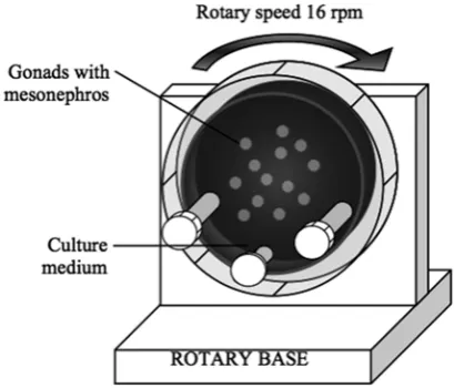
Three different culture conditions were used: 1) Culture on agar blocks (standard in vitro 2D culture). Testes were cultured on agar 3–4 mm cubes of 2% agar (dissolved in phosphate buffered saline, PBS) prepared in sterile conditions and left in the culture medium for 2–3 h at 37°C in a hu-midified incubator with 5% CO2. Four blocks were transferred into a Petri dish (35 mm). The dish was filled with fresh culture medium up to the surface of the blocks. Testes were cultured (one per block) on the top of each cube; 2) Immersion culture. Testes were cultured onto a Petri dish pre-coat-ed with agar to avoid attachment and completely immersed into the culture medium. This culture condition was employed as an additional 2D cul-ture control since in the RWV, testes are complete-ly immersed in the culture medium; 3) Culture in RWV. Testes were placed into a 10 mL RWV dish (not more than 5 testes in each dish). Rota-ry speed 14.7 rpm (rotations per min) was used. The obtained acceleration of gravity was calculat-ed to be about 6.0 × mginstead of Earth’s gravity 1 × g (Fig. 1). In all culture conditions, the testes were incubated for 48 h in a humidified incubator at 37°C with 5% CO2 in air.
Histology of the Gonads
Fixation
The testes were placed in Eppendorf tubes containing freshly prepared Bouin solution (71% picric acid, 24% formaldehyde and 5% acetic acid). After 2–3 h they were washed 3 times with 80% al-cohol and stored in 70% alal-cohol at 4°C.
group, the data was obtained from at least three independent experiments. Comparisons were made by one-way ANOVA, using ISTAT software, P values < 0.05 were considered as significant and p < 0.001 as highly significant.
Results
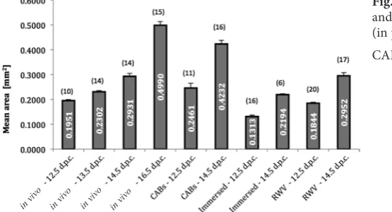
A summary of the measurements of testis size in the freshly collected 12.5–16.5 d.p.c. and 12.5 and 14.5 d.p.c. testis cultured for 2 days under dif-ferent conditions are shown in Fig. 2. We observed a progressive increase in testis size in vivo from 12.5 to 16.5 d.p.c. paralleled by a significant decrease in the number of testis cords. Both 12.5 d.p.c. and 14.5 d.p.c. testes cultured in 2D immersed condi-tions showed frequent necrotic areas and a high-ly significant smaller size in comparison to the freshly collected 14.5 and 16.5 d.p.c. testes, respec-tively. On the other hand, the 12.5 d.p.c. and 14.5 d.p.c. testes cultured onto agar blocks (CAB tes-tes), showed sizes slightly smaller than those of the comparable age, freshly collected 14.5 d.p.c. and 16.5 d.p.c. testes. Testes cultured in RWV reached intermediate size between those cultured under 2D immersed conditions and onto agar blocks. Differ-ences were, however, statistically significant only in comparison to the 2D cultured immersed testes, thus indicating that while 2D CAB and immersed were the best and worse in vitro culture condition for testes growth, respectively, the culture in RWV allowed the testes to reach a size similar to the 2D CAB and in vivo conditions despite complete me-dium immersion.
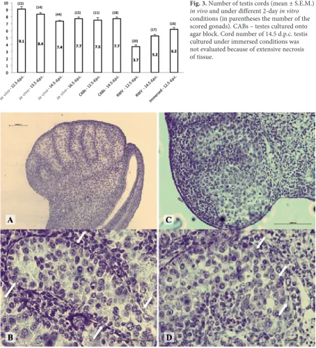
As reported in previous studies, we observed that during the fetal period, testis cords undergo a substantial remodeling resulting in a reduction of their number between 12.5 and 16.5 d.p.c. We found that such changes were not significantly affected by either 2D culture conditions, but appeared severely affected in RWV, in which a significant reduction of the testis cord number occurred (Fig. 3).
Fig. 2. Testis size (mean area ± S.E.M.) in vivo
and under different 2-day in vitro conditions (in parentheses the number of the scored gonads) CABs – testes cultured onto agar block.
Tissue Processing
From 70% alcohol, the samples were trans-ferred to 80% alcohol for 30 min and then in xy-lene (AlanaR) for the next 30 min. Finally, they were embedded in paraffin at 58°C. Samples were sectioned at 9–10 µm. The sections were placed in a warm water bath to reduce wrinkles and then transferred onto microscopic glass. Paraffin was re-moved from the samples using Histo-Clear (Nation-al Diagnostics), twice for 5 min. The samples were re-hydrated and finally rinsed twice in water for 5 min before hematoxylin and eosin (HE) staining.
Testis Size and Cord
Number Count
The length (L) and width (W) of each testis were measured under phase contrast microscope equipped with a micrometric ocular. Since a testis resembles an ellipsoid, the testis size was calculat-ed using the ellipse area formula:
S = π × (L/2) × (W/2).
The number of cords in each testis was also counted.
Counting of Germ Cells
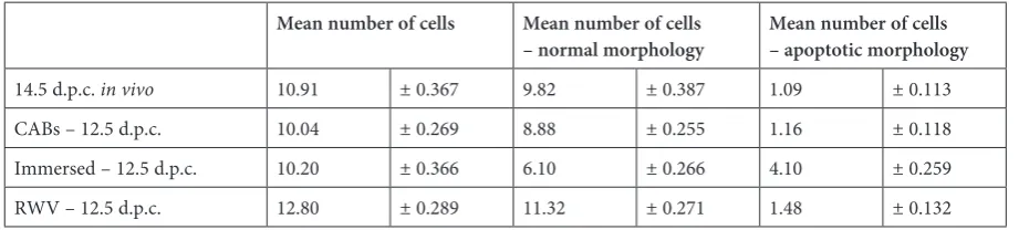
Longitudinal sections of the testis central part (at least 5 for each gonad analyzed in every treat-ment) were examined. Germ cells, identified on the basis of morphology, were counted in ran-dom squares of 2400 µm2; only cells in which the nucleus was clearly visible were scored. Germ cells showing degenerative morphology (reduced size and highly condensed chromatin) were also scored.
Statistical Analysis
All statistical analyses are expressed as mean ± standard error (S.E.M.). For each culture
Histological analyses of the CAB 12.5 d.p.c. testes showed a well-preserved morphology re-sembling that of testes of comparable in vivo age (14.5 d.p.c.). The cords were well outlined and filled with germ cells, showing typical charac-teristics of prospermatogonia with relatively large, spherical nuclei and a few prominent nu-cleoli. A significant number of prospermatogo-nia showed coarse flakes of chromatin associated with the nuclear membrane typical for post-mitot-ic germ cells.Immature Sertoli cells were located at the periphery of the cords, whereas Leydig cells
and other interstitial cells were located between the cords (Fig. 4). Sections of testes cultured under 2D immersion showed clear signs of tissue degener-ation. In some sections, defined cords were diffi-cult to identify; although the cords were well con-served in regions where the tissue appeared better preserved (not shown). In testis cultured in RWV, cord disorganization was evident, whereas the cell number and morphology did not appear gross-ly abnormal (Fig. 4). The mean number of pros-permatogonia in the analyzed areas and their ap-parent normal or degenerating morphologies are
Fig. 3. Number of testis cords (mean ± S.E.M.)
in vivo and under different 2-day in vitro
conditions (in parentheses the number of the scored gonads). CABs – testes cultured onto agar block. Cord number of 14.5 d.p.c. testis cultured under immersed conditions was not evaluated because of extensive necrosis of tissue.
Fig. 4. Representative sections of male gonads. Testis cultured for 2 days under 2D CAB conditions showing normal cord structure: arrows indicate basal lamina (A, B). Sections of testes cultured for 2 days in RWV showing (arrows) disorganized cords and discontinuous basal lamina (C, D).
shown in Table 1. No significant alteration in germ cell number or increase in the number of degen-erating cells were observed in testes cultured on-to agar blocks or in RWV in comparison on-to the
in vivo conditions. On the contrary, in testes cul-tured in 2D immersed conditions, a significant re-duction in the number of prospermatogonia and increasing numbers of cells showing degenerating morphologies in comparison to the in vivo and the other in vitro conditions were found.
Discussion
The RWV was designed to provide continuous sedimentation of particles through a culture me-dium, which is rotating essentially as a solid mass with minimal induced cellular shear and turbu-lence. RWV largely solves the challenges of sus-pension culture: to suspend cells and small organs without inducing turbulence or large degrees of shear, while providing adequate nutrition and ox-ygenation. RWV has been successful in culturing prostate organoids, liver, colon carcinoma and car-tilage, among other tissues [10].
In the present paper, for the first time, we used the RWV bioreactor for in vitro culturing of mouse fetal testes during a crucial period of their mor-phogenesis from 12.5 d.p.c. to 16.5 d.p.c. In the mouse embryo, the sex differentiation of the go-nadal anlage begins around 10.5–11.5 d.p.c., when
Sry activation leads to Sertoli cell differentiation. Soon after, testis cords develop and are visible by 12.5 d.p.c. under a dissecting microscope; the cords consist of Sertoli cells surrounding the germ cells and separate them from the mesenchymal inter-stitium [11]. During the subsequent and up to the perinatal period, testes undergo a considerable size increase paralleled by cell differentiation and very dynamic cord changes [5, 12]. Sertoli cell prolifer-ation, migration of mesenchymal cells into the tes-tis and congregation of interstitial cells around the forming cords, are thought to be the main causes for the increase in testis size [12]. Our results sug-gest that, in testes cultured both under RWV and standard 2D onto agar block conditions, these
processes are likely to be well maintained. Actually, throughout the culture period we observed a size increase in cultured testes analogous to in vivo. At the same time, the cell morphology and germ cell number resembled that of testes of comparable
in vivo age. In this regard, it is worth noting that the culture in RWV was able to markedly alleviate the adverse effects of static 2D immersed condi-tions on testis development, likely providing bet-ter cell nutrition and oxygenation.
Certainly, however, the more intriguing effect of RWV culture conditions which we observed was the striking disorganization of the testis cords. Previous studies have shown that the expression of several genes, such as Inba, Wt1, Sox9 and Sox8
in Sertoli and Leydig cells, are necessary for the maintenance of the testis cords after their initial formation [13–15]. Unpaired expression of these genes could result in defects in molecular signal-ing controllsignal-ing the correct differentiation and ac-tivity of testicular cells. This in turn might down-regulate basal lamina components, which results in a breakdown of the basal lamina and testicu-lar cord disorganization. In fact, interaction be-tween Sertoli cells and peritubular myoid cells leads to the polarized secretion of various extracel-lular matrix (ECM) proteins, such as laminin, type IV and IXa3 collagen and heparan sulfate. These ECM molecules are required for formation of the basal lamina, which surrounds the testis cords and maintains their structural integrity [16–18]. Our morphological observations indicate that in tes-tes cultured in RWV, the basal lamina surround-ing the cords was intermittently disrupted in com-parison to the continuous lamina in the control. Thus, the question arises as to which mechanism is responsible for the loss of testis cord basal lam-ina seen in RWV culture. As the RWV was devel-oped by NASA to simulate culture conditions pre-dicted to occur during experiments in space, these conditions were defined as simulated microgravi-ty. We hypothesize that such microgravity condi-tions could be responsible for altered production of basal lamina molecules and/or of their assem-bly. Although it is known that the altered me-chanical culture conditions of spaceflight might
Table 1. Mean number + SEM of prospermatogonia in testis sections
Mean number of cells Mean number of cells
– normal morphology Mean number of cells – apoptotic morphology
have dramatic effects on cell growth and metabo-lism, as well as the production of bioproducts from growth hormones to hybridomas [10], the mech-anisms by which mechanical culture conditions, including microgravity, could affect gene expres-sion, protein synthesis and the structure and func-tion of diverse cell types is only beginning to be
investigated. Postulated mechanisms include cyto-skeletal changes, electrical and chemical signaling and channel activation [10]. The results presented in the present paper offer the possibility to inves-tigate such mechanisms in the model of fetal testis development that is also relevant for future repro-duction in space.
References
[1] Combes AN, Wilhelm D, Davidson T, Dejana E, Harley V, Sinclair A, Koopman P: Endothelial cell migration directs testis cord formation. Dev Biol 2009, 326, 112–120.
[2] Yao HH, Capel B: Disruption of testis cords by cyclopamine or forskolin reveals independent cellular pathways in testis organogenesis. Dev Biol 2002, 246, 356–365.
[3] Bott RC, McFee RM, Clopton DT, Toombs C, Cupp AS: Vascular endothelial growth factor and kinase domain region receptor are involved in both seminiferous cord formation and vascular development during testis morpho-genesis in the rat. Biol Reprod 2006, 75, 56–67.
[4] Tilmann C, Capel B: Mesonephric cell migration induces testis cord formation and Sertoli cell differentiation in the mammalian gonad. Development 1999, 126, 2883–2890.
[5] Combes AN, Lesieur E, Harley VR, Sinclair AH, Little MH, Wilhelm D, Koopman P: Three-dimensional visualiza-tion of testis cord morphogenesis, a novel tubulogenic mechanism in development. Dev Dyn 2009, 238, 1033–1041.
[6] Pampaloni F, Reynaud EG, Stelzer EH: The third dimension bridges the gap between cell culture and live tissue. Nat Rev Mol Cell Biol 2007, 8, 839–845.
[7] Nickerson CA, Ott CM, Wilson JW, Ramamurthy R, Pierson DL: Microbial responses to microgravity and other low-shear environments. Microbiol Mol Biol Rev 2004, 68, 345–361.
[8] Zwezdaryk KJ, Warner JA, Machado HL, Morris CA, Höner zu Bentrup K: Rotating cell culture systems for human cell culture: human trophoblast cells as a model. J Vis Exp 2012, 18, DOI: 10.3791/3367.
[9] Wolgemuth DJ, Grills GS: Early mammalian development under condition of reorientation relative to the gravity vector. Physiologist 1985, 28, 75–76.
[10] Hammond TG, Hammond JM: Optimized suspension culture: the rotating-wall vessel. Am J Physiol Renal Physiol 2001, 281, F12–F25.
[11] Dym M: The male reproductive system. In: Weiss L, Greep LO, editors. Histology 1997 New York: McGraw-Hill, 979–1038.
[12] Nel-Themaat L, Vadakkan TJ, Wang Y, Dickinson ME, Akiyama H, Behringer RR: Morphometric analysis of testis cord formation in Sox9-EGFP mice. Dev Dyn 2009, 238, 1100–1110.
[13] Liu CF, Archambeault DR, Yao HC: Testis cord morphogenesis: determination, establishment, and maintenance. Anim Reprod 2006, 3, 92–97.
[14] Georg I, Barrionuevo F, Wiech T, Scherer G: Sox9 and Sox8 are required for basal lamina integrity of testis cords and for suppression of FOXL2 during embryonic testis development in mice. Biol Reprod 2012, 87, 99.
[15] Chen SR, Chen M, Wang XN, Zhang J, Wen Q, Ji SY, Zheng QS, Gao F, Liu YX: The Wilms tumor gene, Wt1, maintains testicular cord integrity by regulating the expression of Col4a1 and Col4a2. Biol Reprod 2013, 88, 56, DOI: 10.1095/biolreprod.112.105379.
[16] Perera EM, Martin H, Seeherunvong T, Kos L, Hughes IA, Hawkins JR, Berkovitz G: Tescalcin, a novel gene encoding a putative EF-hand Ca(2+)-binding protein, Col9a3, and renin are expressed in the mouse testis during the early stages of gonadal differentiation. Endocrinology 2001, 142, 455–463.
[17] McClive PJ, Sinclair AH: Type II and type IX collagen transcript isoforms are expressed during mouse testis development. Biol Reprod 2003, 68, 1742–1747.
[18] Jeanes A, Wilhelm D, Wilson MJ, Bowles J, McClive PJ, Sinclair AH, Koopman P: Evaluation of candidate markers for the peritubular myoid cell lineage in the developing mouse testis. Reproduction 2005, 130, 509–516.
Address for correspondence:
Dorian NowackiDepartment of Human Nutrition
Wrocław University of Environmental and Life Sciences Faculty of Food Science
Chełmońskiego 37/41 51-630 Wrocław Poland
E-mail: [email protected]
Conflict of interest: None declared