Akram Beheshti
A, B, e, Younes Shafigh
B, e, Amir-Abdollah Zangivand
C–F,
Fatemeh Samiee-Rad
C–F, Gholamreza Hassanzadeh
B, C, Navid Shafigh
B, GComparison of Topical Sucralfate and Silver
Sulfadiazine Cream in Second Degree Burns in Rats*
Porównanie skuteczności miejscowego sukralfatu i soli srebrowej
sulfadiazyny w kremie w leczeniu oparzeń drugiego stopnia u szczurów
Qazvin University of Medical Sciences, Iran
A – research concept and design; B – collection and/or assembly of data; C – data analysis and interpretation;
D – writing the article; E – critical revision of the article; F – final approval of article; G – other
Abstract
Background. The most prevalent topical treatment for partial thickness burns is silver sulfadiazine 1% (SSD). Recent studies have shown that the healing of partial thickness burns is delayed with the use of SSD. One of the potential burn dressings is sucralfate.
Objectives. With this study the authors have aimed to analyze comparatively the effects of sucralfate and SSD on second degree burn wounds in rats.
Material and Methods. Forty-eight male rats were divided into three equal groups. A burn model was constituted on the back of all rats. The burned areas in the first, second and third groups were covered daily with sucralfate, SSD and cold cream (control), respectively. At the end of the 7th, 14th, 21st and 28th day, the rats were anesthetized
and the burned skin tissue samples were collected for histopathological examination.
Results. At the end of the study, the epidermis and horny layer was completely formed in the SSD and sucralfate group; however the appendix of skin was just formed in the sucralfate group. Also the percentage of wound healing was calculated at 76%, 91% and 100% respectively in the control, silver sulfadiazine and sucralfate groups.
Conclusions. Sucralfate is known to have multiple beneficial effects on wound healing. Using topical sucralfate accelerates the burn wound healing process in comparison with both the control and SSD groups and can be used as an adjunctive or alternative agent in the future (Adv Clin Exp Med 2013, 22, 4, 481–487).
Key words: sucralfate, silver sulfadiazine, second degree burns, rat.
Streszczenie
Wprowadzenie. Najbardziej rozpowszechnionym sposobem miejscowego leczenia oparzeń II stopnia jest sól sre-browa sulfadiazyny 1% (SSD). Ostatnie badania wykazały, że leczenie oparzeń II stopnia jest opóźnione z wykorzy-staniem SSD. Jednym z możliwych opatrunków oparzeniowych jest sukralfat.
Cel pracy. Analiza porównawcza wpływu sukralfatu i SSD na oparzenia drugiego stopnia u szczurów.
Materiał i metody. Czterdzieści osiem samców szczurów podzielono na trzy równe grupy. Model oparzenia utwo-rzono na grzbiecie wszystkich szczurów. Spalone obszary w grupach pierwszej, drugiej i trzeciej codziennie pokry-wano odpowiednio sukralfatem, SSD i zimną śmietaną (grupa kontrolna). Na koniec dnia 7, 14, 21 i 28 szczury usypiano i spalone próbki tkanki skóry zebrano do badań histopatologicznych.
Wyniki. Na koniec badania naskórek i warstwa rogowa skóry były całkowicie uformowane w grupie SSD i sukral-fatu, jednak skóra właściwa została wytworzona w grupie z sukralfatem. Skuteczność gojenia ran obliczono na 76%, 91% i 100% w grupie kontrolnej, z solą srebrową sulfadiazyny i z sukralfatem.
Wnioski. Sukralfat wywiera korzystny wpływ na gojenie ran. Przy zastosowaniu miejscowym sukralfat przyspieszał
Adv Clin exp Med 2013, 22, 4, 481–487 ISSN 1899–5276
ORIGINAl PAPeRS
© Copyright by Wroclaw Medical University
Burns are one of the most widespread injuries all over the world. In the United States, more than 1 million burn victims need medical attention ev-ery year, but only 45,000 of them require hospital-ization [1]. A similar situation exists in the United Kingdom, where burns comprise 1% of the work-load in emergency wards as well as 0.014% of hos-pital admissions [2]. Thus, most burns are not se-vere and could be managed outside the hospital.
The most prevalent topical treatment for partial thickness burns is silver sulfadiazine 1% (SSD) [1, 3]. SSD is the topical agent of choice for severe burns and is used almost universally to-day in preference to compounds such as silver ni-trate and mafenide acetate. SSD cream, in spite of being effective, causes some systemic side effects consisting of neutropenia, erythema multiforme, crystalluria and methemoglobinemia [4–6]. Top-ical agents which are used only as antimicrobials include silver nitrate, sulfamylon and a combina-tion of a sulfonamide and SSD. Sulfamylon has broad spectrum activities, but it is easily absorbed systemically and can lead to toxic complications. SSD has become the standard topical treatment for burn wounds [4]. More recent studies have shown that the healing of partial thickness burns is de-layed with the use of SSD [7, 8], indicating the need for a better burn dressing.
One of the potential burn dressings is sucral-fate. Sucralfate is a basic aluminum complex of sucrose sulfate and a cytoprotective agent. The sporadic studies and case reports available in the literature are all consistent, indicating the favor-able effect of topical sucralfate in wound repair and skin protection. Almost all studies have indi-cated the safe and effective behavior of this com-pound [9–17]. Sucralfate has also been shown to have antibacterial activity [18, 19] and has been successfully studied in decreasing pain and im-proving healing after hemorrhoidectomy[20],in peristomal and perineal dermatoses, in moist des-quamation during radiotherapy, in erosion and ul-ceration of the perineal area, in vaginal ulul-ceration, in dystrophic epidermolysis bullosa, in second and third degree burns, and in a pilot trial with non-healing, full-thickness venous stasis ulcers refrac-tory to 8 weeks of conventional therapy [9–17].
With this study the authors aimed to analyze comparatively the effects of sucralfate and SSD on wound healing in a burn wound that has been made in rats.
Material and Methods
Animals
Forty-eight male Wistar rats (200 to 250 g each) were obtained from the Razi Institute (Karaj, Iran) and housed in groups of three per cage under stan-dard laboratory conditions. All animals were kept at a constant room temperature (21 ± 2ºC) under a normal 12-h light/12-h dark cycle with free ac-cess to food and water. All animal experiments were carried out in accordance with the european Union’s Council Directive of November 24th, 1986 (86/609/eeC) to minimize their suffering.
Chemicals
Sucralfate cream (Avene Cicalfate Restorative Skin Cream, purchased from Avene Co., France), silver sulfadiazine 1% (topical cream, purchased from Sobhan Pharmaceutical Co., Tehran, Iran), cold cream (Botafarma, 12.5% spermaceti + 12% white wax + 56% liquid paraffin + 0.5% borate of soda + 19% distilled water) [21], ketamine (Rotex-medica, GmbH, Germany), and xylazine (loughrea Co., Galway, Ireland) were used in this study.
Method
All animals were administered general anesthe-sia through peritoneal injection of xylazine (5 mg/kg) and ketamine (40 mg/kg) and the back hair of them was shaved. Then a round iron plate (heated to 80°C) was placed on each animal’s skin for 1 second in order to create a 1.75 cm2 second-degree burn injury [22]. Then the animals received an intraperitoneal injec-tion of saline (0.1 l/kg) and were placed in individu-al cages for recovery. To confirm the degree of burns, histopathological samples were taken randomly from eight rats. Then all animals were randomly divided into three groups (n = 16 in each group). The first group was treated with sucralfate cream, the second group was treated with the silver sulfadiazine 1% and the third group was treated with cold cream.
By considering the day that the authors caused burn in rats as zero, the punch biopsies with dispos-able punch were taken of the large diameter wound burn and surrounding skin of 12 animals (n = 4 in each group) at the end of the 7th, 14th, 21st and 28th day (needle size = 8 mm). Skin sections were fixed in 10% formaldehyde, dehydrated in ethanol (50% to 100%), cleared in xylene and embedded in
proces gojenia ran po oparzeniu w porównaniu z grupą kontrolną i SSD oraz może być stosowany jako alternatywa lub środek wspomagający leczenie w przyszłości (Adv Clin Exp Med 2013, 22, 4, 481–487).
paraffin. Sections (4 to 5 µm thick) were prepared and stained with hematoxylin and eosin (He) dyed for histopathological examination and observed under a microscope at a high magnification. The thickness of granulation tissue at the center of each wound was examined and recorded. Also, to deter-mine the percentage of wound healing, the wound surface was measured by tracing the margins of the open wound on digital images of the wounds that were scanned into the computer. The open wound area was then later calculated using Scion Image version beta 4.0.2 (Scion, Frederick, Md) on the aforementioned days (7th, 14th, 21st and 28th). Af-ter that, the following formulas were used to deAf-ter- deter-mine the percentage of wound and healing:
Area of wound = Open wound area was calcu-lated using Scion Image (details above)
Percentage of wound = (Area of wound on particular day × 100) ÷ the area of wound on the first day
Percentage of healing = (100 – percentage of wound)
Ethical Approval
The study was approved by the ethics commit-tee of the Qazvin University of Medical Science before its initiation, and the protocols used con-formed to the ethical guidelines of the 1975 Hel-sinki Declaration.
Statistical Analysis
The collected data was analyzed using SPSS software (Statistical Package for the Social Scienc-es, version 11.0, SPSS Inc, Chicago, Ill, USA). One-way ANOVA tests were used, where appropriate, for comparing data between all groups. Continuous da-ta was demonstrated as mean ± sda-tandard deviation. P – values less than 0.05 were considered significant.
Results
At the End of the First Week
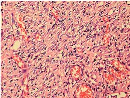
In all groups, no epidermis existed in the burn site and fibrino-leukocytic exudate was seen with edema and transudates in the wound and surround-ing tissue. A necrotic layer with predominant in-flammatory PMN cells and granulation tissue with angiogenesis were seen in all groups (Fig. 1).
At the End of the Second Week
In the sucralfate group, the thickness of the fi-brino-leukocytic layer was decreased in comparison
with the control and SSD groups, although granu-lation tissue had increased. Fibroblast density was increased in the dermal layer in all groups, in addi-tion to this increase; angiogenesis was also greatest in the sucralfate group. The percentage of inflam-matory PMN cells was reduced while the number of mononuclear inflammatory cells had started to increase (Fig. 2).
Fig. 1. The first week in the third group (high mag-nification ×400): exudative and necrotic layer at first week. The predominant inflammatory cells are PMN
Ryc. 1. Stan w pierwszym tygodniu w trzeciej grupie. (duże powiększenie 400×): wysiękowe i martwicze warstwy w pierwszym tygodniu. Dominujące komórki zapalne to granulocyty obojętnochłonne
Fig. 2. The second week in the first group (high magni-fication ×400): The granulation tissue is present. There is more vascular proliferation than in the first week. There is a decrease in the percentage of PMN inflam-matory cells and the mononuclear inflaminflam-matory cells begin to increase in numbers
At the End of the Third Week
In the control group, the cells in the wound bed were more organized. The epidermis layer was not formed and fibroblast density had increased in this layer. In the silver sulfadiazine group, de-spite forming an epidermis layer, the horny layer hadn’t formed and in the sucralfate group, the epi-dermis layer as well as a thin horny layer were seen. like new parallel blood vessels in the silver sulfa-diazine group were also seen on the wound surface in this group. The granulation tissue was more or-ganized than other groups. In this week, the per-centage of inflammatory PMN cells was reduced and density of fibroblast and macrophage was in-creased (Fig. 3).
At the End of the Forth Week
In the control group, the epidermis layer had formed in spite of the fact that some areas showed lack of epidermis in the center of the wound. The horny layer was created in the middle of the wound, otherwise there wasn’t any in the center of the wound. In this week in the silver sulfadiazine group, the epidermis and horny layer was com-pletely formed, however the appendix of skin was not formed.
In the sucralfate group, the epidermis and horny layer was completely formed at the end of the 4th week. There was fibrous tissue in the central
part of the lesion. The inflammatory cells’ content was reduced and a low number of mononuclear in-flammatory cells was seen (Fig. 4).
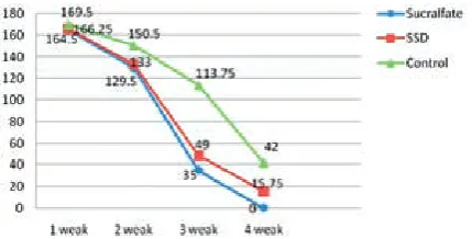
At the end of the fourth week, the percentage of wound healing was calculated at 76%, 91% and 100%, respectively, in the control, silver sulfadia-zine and sucralfate groups. This result was signifi-cantly different between all groups (p < 0.05). The percentage of wound healing in this study is sum-marized in Table 1 and Fig. 5. Also, present re-sults in this study have shown that the thickness of granulation tissue was significantly different between each group (p < 0.05). The mean values of thickness of granulation tissue in the center of the wounds for sucralfate cream, SSD and control groups are shown in Table 2.
Fig. 3. The third week in the first group (High mag-nification ×400): There is decrease in the number of inflammatory cells, particularly PMNs, and an increase in the number of macrophages and fibroblasts. A fine deposit of collagen matrix is also present
Ryc. 3. Stan w trzecim tygodniu w pierwszej grupie (duże powiększenie 400×). Zmniejszyła się liczba komórek zapalnych, w szczególności granulocytów obojętnochłonnych i zwiększyła się liczba makrofagów i fibroblastów. Cienka warstwa macierzy kolagenowej jest również obecna
Fig. 4. The fourth week in the first group (high mag-nification ×400): The inflammatory cells are scat-tered and are mostly mononuclear. Vascularity is also decreased. There is a higher number of fibroblasts than normal and more deposits of collagenous matrix (scar tissue formation)
Ryc. 4. Stan w czwartym tygodniu w pierwszej grupie (duże powiększenie 400×): komórki zapalne są roz-proszone, w większości jednojądrzaste. Unaczynienie również zmniejszyło się. Istnieje większa liczba fibro-blastów niż normalnie i więcej warstw macierzy kola-genowej (powstawanie blizn)
Fig. 5. Wound area (mm2) over 4-week treatment Ryc. 5. Obszar rany(mm2) podczas 4-tygodniowego
Discussion
Wound repair depends on neoangiogenesis, the activation of local immune response, and in the presence of growth factors including epider-mal growth factor (eGF), transforming growth factor b (TGF-b), and basic fibroblast growth fac-tor (bFGF) [23–27].
Sucralfate is known to have multiple benefi-cial effects on wound healing. This drug induces the proliferation of dermal fibroblasts and kera-tinocytes in vitro, and inhibits the release of in-terleukin-2 and interferon-γ from damaged skin cells [28]. The physical barrier feature of sucral-fate is to diminish inflammatory reaction and im-prove mucosal healing [17, 29–31]. limiting the inflammation might decrease fibrosis and stric-ture formation and eGF expression as well as the expression of other factors involved in tissue re-pair processes [32]. Stimulating effects on vascular factors, such as angiogenesis, which play impor-tant roles in tissue repair, have been demonstrat-ed by sucralfate [33, 34]. Sucralfate does not have any adverse effects [35] thus it is widely employed in clinical practice to prevent or treat recurrent aphthous stomatitis and several gastrointestinal diseases [36, 37].
Unfortunately not many in vivo or in vitro his-topathological studies about this drug have been carried out in the world. Because of that, the main objective of this study was to determine the effec-tiveness of the local treatment of burn wounds with sucralfate and compare it to SSD.
According to the results that were obtained in this study, using topical sucralfate accelerates the
burn wound healing process in comparison to both the control and SSD groups, so that at the end of the fourth week, thickness of granulation tissue was significantly higher in the sucralfate group. Also, the percentage of wound healing was calculated at 76%, 91% and 100%, respectively, in the control, sil-ver sulfadiazine and sucralfate groups. These results confirm another study about the role of granulation tissue in wound healing [38] and demonstrate the power of sucralfate in neoangiogenesis, prolifera-tion of dermal fibroblasts, eGF expression and the expression of other factors involved in tissue repair processes. This is in concurrence with other studies, which have shown that sucralfate stimulates epithe-lial cell proliferation by causing accumulation of epidermal growth factor in the ulcerated areas [39, 40]. Burch et al. [28] showed, in animal studies, that sucralfate cream accelerates cell proliferation in the superficial skin layer, leading to a clear thickening of the epidermis the same as in dermis experimen-tal studies by Szabo et al. [34] that have shown that sucralfate stimulates angiogenesis, which increas-es granulation tissue. Moreover, sucralfate induced PGe2 synthesis and Il-6 release from cultured skin cells in the study by Burch et al. [28], which was re-sponsible for the healing process.
Limitations
Unfortunately present study has several limi-tations. The small sample size may have led to an important error. Furthermore, the current study was limited to 28 days only, during which time, all the wounds did not heal completely. On the other
Table 2. Thickness (mm) of granulation tissue in the center of the wound
Tabela 2. Grubość (mm) tkanki ziarninowej na środku rany
1st week (Tydzień 1) 2nd week (Tydzień 2) 3rd week (Tydzień 3) 4th week (Tydzień 4) Sucralfate group 0.985 ± 0.01 1.856 ± 0.03 2.580 ± 0.01 3.200 ± 0.02 SSD group 0.736 ± 0.02 1.214 ± 0.03 2.010 ± 0.03 2.765 ± 0.01 Control group 0.380 ± 0.05 0.896 ± 0.01 1.520 ± 0.03 2.000 ± 0.01 P-Value P < 0.05* P < 0.05* P < 0.05* P < 0.05*
Table 1. The percentages of wound healing
Tabela 1. Odsetek wyleczonych ran
1st week (Tydzień 1) 2nd week (Tydzień 2) 3rd week (Tydzień 3) 4th week (Tydzień 4)
Sucralfate group 6 26 80 100
SSD group 5 24 72 91
Control group 3 14 35 76
hand, the burned areas in this study were covered daily, while 2 or 3 dressing per day probably would have a better outcome in wound healing. Finally, this study is limited to rats with second degree burns and may not generalize to humans.
The authors concluded thatin a burn wound model in rats, sucralfate was found to expedite the healing process both histopathologically and statis-tically as compared to SSD and the control group. Sucralfate powder is a very cheap, safe and well-tolerated drug in addition having a lack of side ef-fects. In the usage of oral medication, aluminum is poorly absorbed and this drug is excreted through the kidneys [41] and in patients with renal disease
should be prescribed with caution. However topi-cal medication, due to its low concentration and lack of absorption through the skin, doesn’t have any such complications [42]. SSD ointment in pa-tients with allergy to sulfonamides can have se-vere complications. Another advantage of these drugs is the ability to be used in children and in all parts of the skin. Through its antimicrobial, anti-oxidant, anti-inflammatory and immunomodula-tory effects, sucralfate can be used as an adjunctive or alternative agent in wound healing therapies in the future. However, further studies are certainly needed to shed more light on the healing mecha-nism of sucralfate.
References
[1] Demling RH: Burns and other thermal injuries. In: Current surgical diagnosis and treatment. eds.: lawrence W, Gerard M. lang Medical Books/McGraw-Hill, 2002, 267–281.
[2] Wilkinson E: The epidemiology of burns in secondary care, in a population of 2.6 million people. Burns 1998, 24, 139–143.
[3] Taddonio TE, Thomson PD, Smith DJ, Prasad JK: A survey of wound monitoring and topical antimicrobial therapy practices in the treatment of burn injury. J Burn Care Rehabil 1990, 11, 423–427.
[4] Gracia CG: An open study comparing topical silver sulfadiazine and topical silver sulfadiazine-cerium nitrate in the treatment of moderate and severe burns. Burns 2001, 27, 67–74.
[5] Gregory RS, Piccolo N, Piccolo MT, Piccolo MS, Heggers JP: Comparison of propolis skin cream to silver sulfa-diazine: A naturopathic alternative to antibiotics in treatment of minor burns. J Altern Complementary Med 2002, 8, 77–83.
[6] Hosnuter M, Gurel A, Bauccu O, Armutcu F, Kargi E, Isikdemir A: The effect of CAPe on lipid peroxidation and nitric oxide levels in the plasma of rats following thermal injury. Burns 2004, 30, 121–125.
[7] Lee AR, Moon HK: effect of topically applied silver sulfadiazine on fibroblast cell proliferation and biomechanical properties of the wound. Arch Pharm Res 2003, 26, 855–860.
[8] Stern HS: Silver sulphadiazine and the healing of partial thickness burns: a prospective clinical trail. Br J Plast Surg 1989, 42, 581–585.
[9] Hayashi AH, Lau HY and Gillis DA: Topical sucralfate: effective therapy for the management of resistant peris-tomal and perineal excoriation. J Pediatr Surg 1991, 26, 1279–1281.
[10] Lyon CC, Stapleton M, Smith AJ, Griffiths CE, Beck MH: Topical sucralfate in the management of peristomal skin disease: an open study. Clin exp Dermatol 2000, 25, 584–588.
[11] Delaney G, Fisher R, Hook C, Barton M: Sucralfate cream in the management of moist desquamation during radiotherapy. Australas Radiol 1997, 41, 270–275.
[12] Maiche A, Isokangas OP, Grohn P: Skin protection by sucralfate cream during electron beam therapy. Acta Oncol 1994, 33, 201–203.
[13] Markham T, Kennedy F, Collins P: Topical sucralfate for erosive irritant diaper dermatitis. Arch Dermatol 2000, 136, 1199–1200.
[14] Lentz SS, Barrett RJ, Homesley HD: Topical sucralfate in the treatment of vaginal ulceration. Obstet Gynecol 1993, 81, 869–871.
[15] Marini I, Vecchiet F: Sucralfate: a help during oral management in patients with epidermolysis bullosa. J Periodontol 2001, 72, 691–695.
[16] Banati A, Chowdhury SR, Mazumder S: Topical use of Sucralfate Cream in second and third degree burns. Burns 2001, 27, 465–469.
[17] Tumino G, Masuelli L, Bei R, Simonelli L, Santoro A, Francipane S: Topical treatment of chronic venous ulcers with sucralfate: a placebo controlled randomized study. Int J Mol Med 2008, 22, 17–23.
[18] Tryba M, Mantey-Stiers F: Antibacterial activity of sucralfate in human gastric juice. Am J Med 1987, 83, 125–127.
[19] Kawazoe, H, Takaoka K., Shibata H, Arakaki N, Higuti T, Negayama K, Houchi H, Tsuchiya K, Takiguchi Y:
Comparison of antibacterial activity of fluoroquinolones with their sucralfate-complexes against clinically-isolated bacteria. J Health Sci 2009, 55, 790–795.
[20] Gupta PJ, Heda PS, Kalaskar S, Tamaskar VP: Topical sucralfate decreases pain after hemorrhoidectomy and improves healing: a randomized, blinded, controlled study. Dis Colon Rectum 2008, 51, 231–234.
[21] Durmus AS, Han MC, Yaman I: Comparative evaluation of Collagenase and Silver Sulfadiazine on Burned Wound Healing in Rats. F Ü Sağ Bil Vet Derg 2009, 23, 135–139.
[23] Eming SA, Krieg T, Davidson JM: Inflammation in wound repair: molecular and cellular mechanisms. J Invest Dermatol 2007, 127, 514–525.
[24] Werner S, Grose R: Regulation of wound healing by growth factors and cytokines. Physiol Rev 2003, 83, 835–870.
[25] Roy H, Bhardwaj S, Yla-Herttuala S: Biology of vascular endothelial growth factors. FeBS lett 2006, 580, 2879–2887.
[26] Neal MS: Angiogenesis: is it the key to controlling the healing process? J Wound Care 2001, 10, 281–287.
[27] Hu YL, Guo SZ, Yan PS: effect of local application of basic fibroblast growth factor and sucralfate on skin tissue structure after expansion. Zhongguo Xiu Fu Chong Jian Wai Ke Za Zhi 2002, 16, 340–344.
[28] Burch RM, McMillan BA: Sucralfate induces proliferation of dermal fibroblasts and keratinocytes in culture and granulation tissue formation in full-thickness skin wounds. Agents Actions 1991, 34, 229–231.
[29] Candelli M, Carloni E, Armuzzi A, Cammarota G, Ojetti V, Pignataro G, Santoliquido A, Pola R, Pola E, Gasbarrini G, Gasbarrini A: Role of sucralfate in gastrointestinal diseases. Panminerva Med 2000, 42, 55–59.
[30] Hollander D, Tarnawski A, Krause WJ, Gergely H: Protective effect of sucralfate against alcohol-induced gas-tric mucosal injury in the rat. Macroscopic, histologic, ultrastructural, and functional time sequence analysis. Gastroenterology 1985, 88, 366–374.
[31] Gupta PJ, Heda PS, Shrirao SA, Kalaskar SS: Topical sucralfate treatment of anal fistulotomy wounds: a random-ized placebo-controlled trial. Dis Colon Rectum 2011, 54, 699–704.
[32] Konturek SJ, Konturek JE, Brzozowski T, et al.: effect of sucralfate on growth factor availability. In: Sucralfate: From Basic Science to the Bedside. Hollander D and Tytgat GNJ (eds). Plenum Medical Book Co., New York 1993, 175–189.
[33] Folkmann J, Szabo S, Stovroff M: Duodenal ulcer. Discovery of a new mechanism and development of angiogenic therapy that accelerates healing. Ann Surg 1991, 214, 414–426.
[34] Szabo S, Vattay P, Scarbrough E, Folkman J: Role of vascular factors, including angiogenesis, in the mechanisms of action of sucralfate. Am J Med 1991, 91, 158–160.
[35] McCarthy DM: Sucralfate. N engl J Med 1991, 325, 1017–1025.
[36] Da Costa RM, Ribeiro Jesus FM, Aniceto C, Mendes M: Randomized, double-blind, placebo-controlled, dose-ranging study of granulocyte-macrophage colony stimulating factor in patients with chronic venous leg ulcers. Wound Rep Reg 1999, 7, 17–25.
[37] Sweetman SC (ed): Sucralfate. In: Martindale: The Complete Drug Reference. 33rd edition, The Pharmaceutical Press, london 2002, 1250–1251.
[38] Wang X, Kimble RM: A review on porcine burn and scar models and their relevance to humans. Wound Pract Res 2010, 18, 41–49.
[39] Nexø E, Poulsen SS: Does epidermal growth factor play a role in the action of sucralfate? Scand J Gasteroenterol 1987,125, 45–49.
[40] Mannari C, Santi S, Migliori M, Filippi C, Origlia N, Sansò M, Boldrini E, Giovannini L: Sucralfate modulates uPAR and eGFR expression in an experimental rat model of cervicitis. Int J Immunopathol Pharmacol 2008, 21, 651–658.
[41] Allain P, Mauras Y, Krari N, Duchier J, Cournot A, Larcheveque J: Plasma and urine aluminium concentrations in healthy subjects after administration of sucralfate. Br J Clin Pharm 1990, 29, 391–395.
[42] Fischer RS: Sucralfate a review of drug tolerance and safety, J Clin Gastroenterol 1981, Suppl. 2, 181–184.
Address for correspondence:
Fatemeh Samiee Rad
Qazvin University of Medical Sciences Clinical Research Development Unit Shahid Rajaie University Hospital Qazvin
Iran
Tel.: +98 2813347496
e-mail: [email protected]
Conflict of interest: None declared