Vanja Petrović
1, A–F, Nataša Pejčić
1, A–F, Saša Čakić
2, A–FThe Influence of Different Therapeutic Modalities
and Platelet Rich Plasma on Apexogenesis
– a Preliminary Study in Monkeys*
Wpływ modyfikacji różnych metod terapeutycznych i osocza
bogatopłytkowego na apeksogenezę – wstępne badanie u małp
1 Department of Preventive and Pediatric Dentistry, School of Dental Medicine, University of Belgrade, Serbia 2 Department of Periodontology and Oral Medicine, School of Dental Medicine, University of Belgrade, Serbia
A – research concept and design; B – collection and/or assembly of data; C – data analysis and interpretation;
D – writing the article; E – critical revision of the article; F – final approval of article; G – other
Abstract
Background. Traumatic injuries of permanent teeth with incomplete root formation are frequent during child-hood. Adequate therapy is important for the further destiny of teeth with damaged pulp.
Objectives. To evaluate the effect of pulpotomy and high pulpotomy on the pulp and on root development, and the effect of platelet rich plasma (PRP) with hydroxyapatite (HAP) as a carrier and hydroxyapatite alone on apexo-genesis.
Material and Methods. The study included eight monkeys (Cercopithecus aethiops) in which high pulpotomies were performed on the mandibular lateral incisors and canines, and pulpotomies were performed on the mandibu-lar central incisors and premomandibu-lars. The materials used in the study were commercial HAP (Apatec®, Stomygen) and
PRP (prepared at the Torlak Institute of Immunology and Virology, Belgrade, Serbia). Histological and radiological evaluations were done six months after the treatment.
Results. Considering the differences between HAP+PRP treated teeth in the pulpotomy group and teeth in the high pulpotomy group, two times more root growth retardation was observed in the high pulpotomy group. In the high pulpotomy group, root growth retardation was less common in HAP+PRP treated teeth (42.9%) than in HAP treated teeth (50%). In the pulpotomy group, retardation of root development was also less common in HAP+PRP treated teeth (25%) compared to HAP treated teeth (50%). There were differences between the pulpotomy and high pulpotomy groups, but without statistical significance.
Conclusions. The application of endogenous growth factors in conjunction with the preservation of dental pulp vitality can result in a good outcome for pulp therapy of injured teeth, which means successfully completed apexo-genesis (Adv Clin Exp Med 2013, 22, 4, 469–479).
Key words: platelet rich plasma, apexogenesis, pulpotomy, monkeys.
Streszczenie
Wprowadzenie. Pourazowe uszkodzenia zębów stałych z niepełną formacją korzenia są częste w dzieciństwie. Odpowiednia terapia jest ważna dla dalszych losów zębów z uszkodzeniem miazgi.
Cel pracy. Ocena wpływu pulpotomii i wysokiej pulpotomii na miazgę i rozwój korzeni, a także wpływ osocza bogatopłytkowego (PRP) z hydroksyapatytem (HAP) jako nośnikiem i samego hydroksyapatytu na apekso-genezę.
Materiał i metody. Badaniem objęto osiem małp (Cercopithecus aethiops), u których wysokie pulpotomie przepro-wadzono na bocznych zębach siecznych żuchwy i kłach, a pulpotomie przeproprzepro-wadzono na zębach siecznych żuchwy i zębach przedtrzonowych środkowych. Materiały użyte w badaniu były to dostępne w handlu HAP (Apatec®,
Adv Clin Exp Med 2013, 22, 4, 469–479 ISSN 1899–5276
ORIgINAl PAPERS
© Copyright by Wroclaw Medical University
During childhood, traumatic injuries of young permanent teeth are very frequent [1]. These in-juries often happen before root formation is completed, and influence future root develop-ment [2]. Pulp inflammation or necrosis are un-favorable outcomes [3], while positive outcomes result in successful continuation of the process of apexogenesis.
Apexogenesis is the process of root devel-opment under physiological conditions, and the main aim of therapy for affected dental pulp in teeth with immature roots. Since this process de-termines the further destiny of an injured tooth, regenerative apexogenesis is a subject of great in-terest in current dentistry. The main aim in re-generative apexogenesis is preservation of pulp vi-tality, since dental pulp is the source of cells with regenerative potential specific to the dentin-pulp complex [4]. Post-neonatal stem cells, along with growth factors, play a central role in regenerative apexogenesis [5, 6].
Platelet rich plasma (PRP) is an autologous source of highly concentrated growth factors, hence it has high inductive potential on stem cells [7–9]. The process of sequestering and concentrating platelets by gradient density concentration will yield PRP [10], so its composition is a modifica-tion of fibrin glue containing a platelet concen-tration 338% higher than untreated blood [11]. growth factors are polypeptides that regulate cell functions and participate in active tissue regener-ation [12]. Considering all these facts, PRP could be a beneficial agent in regenerative apexogene-sis [13]. The aim of this study was to evaluate the effect of pulpotomy and high pulpotomy on the pulp and also on root development, the effect of PRP with hydroxyapatite (HAP) as a carrier and the effect of HAP alone. Histological analyses were performed to evaluate the tissue reaction, and ra-diological analyses were used to evaluate apexo-genesis as a whole.
Material and Methods
Experimental Animals
Eight monkeys (Cercopithecus aethiops) were included in the study. Their average age was three years and the formation of the roots of their per-manent dentition was incomplete. The study was carried out at the Torlak Institute of Immunology and Virology in Belgrade, Serbia, which was also where the animals were bred. The Ethical Commit-tee of the Dentistry School of Belgrade Universi-ty and Animal Ethical Screening Committee ap-proved the experiment, and it was carried out in accordance with the European Communities Di-rective (86/609/EEC) [14].
Material
– Commercial hydroxylapatite (Apatec®,
Stomygen),
– PRP (prepared at the Torlak Institute of Im-munology and Virology, Belgrade, Serbia).
Study Design
Before the procedure, the animals were pre-medicated with atropine sulphate (0.5 mg/kg) and the animals were anesthetized with an intramus-cular injection (0.005 mg/kg body weight) of Con-bern® (Bayne, germany) and an intravenous
in-jection (25 mg/kg) of Nembutal (Abbott labs, Chicago, USA). The Nembutal® was given by IV
for 15 minutes to ensure the animals were uncon-scious during the surgery.
Only teeth from the lower jaw were treated. High pulpotomies were carried out on the later-al incisors and canines, and pulpotomies on cen-tral incisors and premolars. Cencen-tral incisors and canines were treated with HAP+PRP; lateral in-cisors and first premolars were treated only with HAP. A contralateral untreated tooth from each animal was used as a control. Four teeth for the
Stomygen) i PRP (przygotowane przez Instytut Immunologii i Wirusologii Torlak, Belgrad, Serbia). Oceny histolo-gicznej i radiolohistolo-gicznej dokonano sześć miesięcy po leczeniu.
Wyniki. Biorąc pod uwagę różnice między zębami leczonymi HAP + PRP w grupie, w której przeprowadzono pulpotomię i zębami w grupie, w której przeprowadzono wysoką pulpotomię, zahamowanie wzrostu korzeni obser-wowano 2 razy częściej w grupie, w której przeprowadzono wysoką pulpotomię. W grupie, w której przeprowa-dzono wysoką pulpotomię opóźnienie wzrostu korzenia występowało rzadziej w zębach leczonych HAP + PRP (42,9%) niż zębach leczonych HAP (50%). W grupie, w której przeprowadzono pulpotomię opóźnienie rozwoju korzeni występowało również rzadziej w zębach leczonych HAP + PRP (25%) w porównaniu z zębami leczonymi HAP (50%). Wystąpiły różnice między grupami, w której przeprowadzono pulpotomię i w której przeprowadzono wysoką pulpotomię, ale bez istotności statystycznej.
Wnioski. Zastosowanie endogennych czynników wzrostu w połączeniu z zachowaniem żywotności miazgi zębów może prowadzić do dobrych wyników terapii miazgi uszkodzonych zębów, co pozwala pomyślnie zakończyć apek-sogenezę (Adv Clin Exp Med 2013, 22, 4, 469–479).
study and four control teeth were removed from each animal. All the treated teeth were submitted to the same preparatory procedure. The teeth were isolated with a cofferdam and washed with 0.5% chlorhexidine. After that, the study teeth were giv-en differgiv-ent treatmgiv-ent depgiv-ending on which group they were assigned to. In the teeth chosen for pul-potomy, cavity preparation was done with an air turbine and the pulp was removed with a carbide round bur at the cement-enamel junction; after that the wound left by the pulp was covered with ma-terial (HAP or HAP+PRP), and glass ionomer ce-ment and amalgam were used for sealing. The high pulpotomies entailed the removal of the pulp close to the radiographically visible end of the immature root, the application of gutta percha along with se-lected material (HAP or HAP+PRP) and sealing with the same materials used in the pulpotomies. Radiographs and histological specimens were per-formed after six months. The animals were sacri-ficed with an overdose of Nembutal® at the end of
the experiment.
Preparation of PRP
PRP was prepared using Weibrichs et al. 2002 method [15]. Whole blood in 2.6 ml volumes was centrifuged for 20 minutes in a centrifuge (MSE, England) at 1200 rpm with Na-citrate. Subquently, two fractions appeared: yellow (blood se-rum) and red (containing blood elements). The red fraction consisted of two areas: The highest platelet concentration was in the upper 6–7 mm, and be-low that was the area of blood elements. Resuspen-sion of the extracted platelet concentrate was then carried out, and after that the platelet concentrate was centrifuged for 15 minutes at 2000 rpm in or-der to further concentrate the PRP. The platelets were ready for application when the serum was re-moved from the upper parts.
Immediately before application, the PRP was mixed with hydroxyapatite powder to a paste con-sistency suitable for application with a lentulo spiral.
The radiological analysis was done as described in one of the authors’ earlier publications [16]: The long cone parallel technique was used for the radio-graphs, and the recording conditions were 35 KV, 8 mA, for 0.02 second. The recorded parameters were: the dentin bridge, which could be observed only in the pulpotomy group; root development according to Demirjian’s system, which represents eight stages of tooth maturation, from A to H [17]; pathological formations in canals (canal oblitera-tion and denticles), which could be observed only in the pulpotomy group; deformities in the peria-pical region.
The histological analysis was done as described in the authors’ earlier publication [16]:
Histological specimens were taken en bloc, in-cluding the experimental tooth, according to ISO usage guidelines (Technical Report 7405) [18]. Buffered formalin 10% (pH = 2) was used for sam-ple fixation, then demineralization was done in 5% trichloracetic acid for 40–50 days and the samples were put into paraffin. Sections with a thickness of 6µm were cut in a mesio-distal direction. Staining was done with hematoxylin and eosin, and with gram to identify bacteria, and then the sections were analyzed under a microscope (Carl Zeiss Inc., Oberkochen, germany).
In the pulpotomy group the following para-metrs were recorded:
– The presence and quality of a dentin bridge: the formation of tertiary dentin at the site of pulp exposure by newly differentiated odontoblast-like cells [19]. During the examination of the sections, reactionary and reparative dentin were differenti-ated as follows: 1) a newly forming tissue barrier behind the applied material; 2) a newly forming tissue barrier far away from the material; and 3) no tissue barrier.
– The reaction of the pulp to therapy: 1) tissue with normal morphology around the exposed pulp; 2) the exposed pulp is changed, but tissue under the exposed zone is normal; 3) a loss of pulp mor-phology and altered tissue under the exposed zone; 4) necrosis in a minimum of 1/3 of the pulp.
– Inflammatory reaction: 1) no inflammato-ry cells or only a few inflammatoinflammato-ry cells below the exposed zone; 2) polimorphonuclears (PMNs) or mononuclears (MNs) in the inflammatory zone; 3) an enhanced inflammatory lesion overtaking a third or more of the pulp; 4) necrotic pulp.
In the high pulpotomy group the following pa-rameters were recorded:
– The presence of an apical barrier; – Inflammatory reaction;
– Deformities in the periapical region.
Statistical Analysis
Results
Radiological Evaluation
Comparisons of treated teeth and their con-trols in the pulpotomy group are presented in Table 1. HAP treated teeth showed a statistical-ly significant decrease in dentin bridge formation (p = 0.046) and a statistically significant increase in root growth retardation (p = 0.046).
Comparisons of treated teeth and their con-trols in the high pulpotomy group are presented in Table 2. Root growth retardation was significantly higher in HAP treated teeth in this group as well (p = 0.046).
Radiological parameters obtained after six months are listed in Table 3. In the pulpotomy group the formation of a dentin bridge was evi-dent in 50% of HAP treated teeth and in 62.5% of HAP+PRP treated teeth. Root growth retarda-tion was detected in 50% of HAP treated teeth and in 25% of HAP+PRP treated teeth. No root canal obliteration, denticles or deformities in the peri-apical region were detected in the treated teeth.
In the high pulpotomy group, one tooth treat-ed with HAP+PRP was excludtreat-ed because of the loss of the amalgam filling. Root development re-tardation was evident in 50% of HAP treated teeth and in 42.9% of the HAP+PRP treated teeth. De-formities in the periapical region were not noted in any of the treated teeth. Dentin bridges, denti-cles and obliteration of root canal could not be ob-served in the high pulpotomy group (Fig. 1).
Considering the differences between HAP+PRP treated teeth in the pulpotomy group and in high pulpotomy group, two times more root growth retardation was observed in the high pulpotomy group, but that was not statistically significant (Ta-ble 4.) In terms of the characteristics of HAP treat-ed teeth, no differences were observtreat-ed between the pulpotomy and high pulpotomy groups.
Histological Evaluation
The histological evaluation of the treatments is presented in Table 5 for the pulpotomy group and in Table 6 for the high pulpotomy group. Inflam-matory reaction in the pulp-periodontal complex
Table 1. Statistical analysis of the pulpotomy groups vs. the control group
Tabela 1. Analiza statystyczna grupy, w której przeprowadzono pulpotomię vs grupy kontrolnej
Observed parameters
(Badane wskaźniki) gROUPS presence absence gROUPS presence absence
Presence of dentin bridge (Obecność mostku dentystycz-nego)
HAP 4/8 (50%) 4/8(50%) HAP+PRP 5/8(62.5%) 3/8(37.5%)
CONTROl 8/8(100%) 0/8 (0%) CONTROl 8/8 (100%) 0/8 (0%)
Wilcoxon’s test; significance
(Istotność statystyczna, test Wilcoxona) p = 0.046 Wilcoxon’s test; signifi-cance
p = 0.083
Retardation of root growth
(Opóźnienie rozwoju korzenia) HAP 4/8 (50%) 4/8(50%) HAP+PRP 2/8 (25%) 6/8 (75%)
CONTROl 0/8 (0%) 8/8(100%) CONTROl 0/8 (0%) 8/8 (100%)
Wilcoxon’s test; significance
(Istotność statystyczna, test Wilcoxona) p = 0.046 Wilcoxon’s test; signifi-cance
p = 0.157
Obliteration of root canal and presence of denticles
(Obliteracja kanału korzeniowego i obecność wyrostków)
HAP 0/8 (0%) 8/8(100%) HAP+PRP 0/8 (0%) 8/8 (100%)
CONTROl 0/8 (0%) 8/8(100%) CONTROl 0/8 (0%) 8/8 (100%)
Wilcoxon’s test; significance
(Istotność statystyczna, test Wilcoxona) ns. Wilcoxon’s test; signifi-cance
ns.
Presence of deformities in peri-apical region
(Obecność deformacji w obszarze okołowierzchołkowym)
HAP 0/8 (0%) 8/8(100%) HAP+PRP 0/8 (0%) 8/8 (100%)
CONTROl 0/8 (0%) 8/8(100%) CONTROl 0/8 (0%) 8/8 (100%)
Wilcoxon’s test; significance
(Istotność statystyczna, test Wilcoxona) ns. Wilcoxon’s test signifi-cance
Table 2. Statistical analysis of the high pulpotomy groups vs. the control group
Tabela 2. Analiza statystyczna grupy, w której przeprowadzono wysoką pulpotomię vs. grupy kontrolnej
Observed parameters
(Badane wskaźniki) gROUPS presence absence gROUPS presence absence
Retardation of root growth
(Opóźnienie rozwoju korzenia) HAP 4/8 (50%) 4/8 (50%) HAP+PRP 3/7(42,9%) 4/7(57.1%)
CONTROl 0/8 (0%) 8/8 (100%) CONTROl 0/7 (0%) 7/7 (100%)
Wilcoxon’s test; significance
(Istotność statystyczna, test Wilcoxona) p = 0.046 Wilcoxon’s test signifi-cance
p = 0.083
Presence of deformities in periapi-cal region
(Obecność deformacji w obszarze okołowierzchołkowym)
HAP 0/8 (0%) 8/8(100%) HAP+PRP 0/7 (0%) 7/7 (100%)
CONTROl 0/8 (0%) 8/8(100%) CONTROl 0/7 (0%) 7/7 (100%)
Wilcoxon’s test; significance
(Istotność statystyczna, test Wilcoxona) ns. Wilcoxon’s test signifi-cance
ns.
Table 3. Radiological evaluation of apexogenesis
Tabela 3. Radiologiczna ocean apeksogenezy
Pulpotomy (Pulpotomia) High pulpotomy (Wysoka pulpotomia)
Clinical parameters (Wskaźniki kliniczne)
stage of develop-ment before the treatment
observed
parame-ters after 6 months stage of develop-ment after 6 months
clinical param-eters
stage of develop-ment before the treatment
observed parameters after 6 months
stage of develop-ment after 6 months
a b c d a b c d
HAP F – – – – g HAP F + – F
F + – – – g g + – g
F – + – – F g + – g
F – + – – F g – – H
F + – – – g F + – F
g – + – – g F – – g
g + + – – g F – – g
g + – – – H F – – g
HAP
+ PRP g + – – – H HAP + PRP F – – g
F + – – – g F + – F
g – + – – g F + – F
g – + – – g g – – g
g + – – – g X X X X
F + – – – g g – – H
F + – – – g F – – g
F – – – – g g + – g
a – presence of dentin bridge; b – retardation of root growth; c – obliteration of root canal and presence of denticles; d – presence of deformities in the periapical region.
+ present; – absent; X – failed.
a – obecność mostu dentystycznego, b – opóźnienie wzrostu korzenia, c – obliteracja kanału korzeniowego i obecność wyros tków, d – obecność deformacji w obszarze okołowierzchołkowym.
of both HAP and HAP+PRP teeth was moderate, where the infiltrate predominantly consisted of lymphocytes, plasma cells and macrophages. No signs of severe inflammation, massive infiltration of neutrophils or abscess formation were noted in the specimens. In the pulpotomy group inflamma-tory reaction was more expressed in HAP treated teeth than in HAP+PRP treated teeth. In the high
Table 4. Statistical analysis of radiological parameters of treated teeth
Tabela 4. Analiza statystyczna wskaźników radiologicznych leczonych zębów
Observed parameters N (%)
(Badane wskaźniki) Pulpotomy (Pulpotomia) High pulpotomy (Wysoka pulpotomia)
presence absence
HAP 4/8 (50%) presence absence
Retardation of root growth (Opóźnienie
rozwoju korzenia) HAP+PRP 2/8 (25%) 4/8 (50%) 4/8 (50%) 4/8 (50%)
p = 0.608 p = 0.879 6/8 (75%) 4/7 (57.1%) 3/7(42.9%) Fisher’s test significance (Istotność statystyczna, test Fishera) HAP 0/8 (0%) Presence of deformities in periapical region
(Obecność deformacji w obszarze okołowierzchołkowym)
HAP+PRP 0/8 (0%) 8/8 (100%) 0/8 (0%) 8/8 (100%)
ns. ns. 8/8 (100%) 0/7 (0%) 7/7 (100%)
Fisher’s test significance (Istotność statystyczna, test Fishera) HAP 4/8 (50%) Presence of dentin bridge (Obecność
most-ka dentystycznego) HAP+PRP 5/8 (62.5%) 4/8 (50%)
p = 0.989 3/8 (37.5%)
Fisher’s test significance (Istotność statystyczna, test Fishera) HAP 0/8 (0%) Obliteration of root canal and presence of
denticles
(Obliteracja kanału korzeniowego i obecność wyrostków)
HAP+PRP 0/8 (0%) 8/8 (100%)
ns. 0/8 (0%) 8/8 (100%)
Fisher’s test significance (Istotność statystyczna, test Fishera)
Fig. 1A. Mandibular right incisors, canine and first premolar in stage F of apex formation before the treat-ment
Ryc. 1A. Prawe zęby sieczne, kły i pierwsze przedtrzono-we w stadium F formacji wierzchołka przed zabiegiem
Fig. 1B. Central incisor in stage g of apex formation 6 months after pulpotomy and HAP+PRP application; a dentin bridge was observed. lateral incisor in stage H of apex formation after high pulpotomy and HAP applica-tion. Canine in stage H of apex formation after high pulp-otomy and HAP+PRP application; slight deformities of the root were observed. First premolar in stage g of apex formation after high pulpotomy and HAP application
pulpotomy group, no such differences were noted between the applied materials. Changes in tissue morphology were present in all samples, including reorganization of morphologically altered fibro-blasts, immune cells and odontofibro-blasts, which were elongated. Considering pulp tissue stratums in the pulpotomy group, the deeper tissue parts were in-tact, and in HAP+PRP treated teeth the cell infil-trate was in more superficial stratums than in HAP treated teeth. In the pulpotomy group, dentin for-mation was equally present in HAP and HAP+PRP treated teeth, covering the area of pulp exposure (Figure 2). The newly formed dentin had the char-acteristics of reparative dentin, and it was observed that a greater density of odontoblast-like cells was present in HAP+PRP treated teeth than in HAP treated teeth. Before the treatment, the apical fo-ramen was wide open and in contact with the sur-rounding periodontium in all teeth. In the high pulpotomy group an apical barrier was observed in 50% of the HAP treated teeth and in 57.1% of the HAP+PRP treated teeth. Deformities in the peri-apical region were present in the same percentage (12.5%) in both HAP treated teeth and HAP+PRP treated teeth.
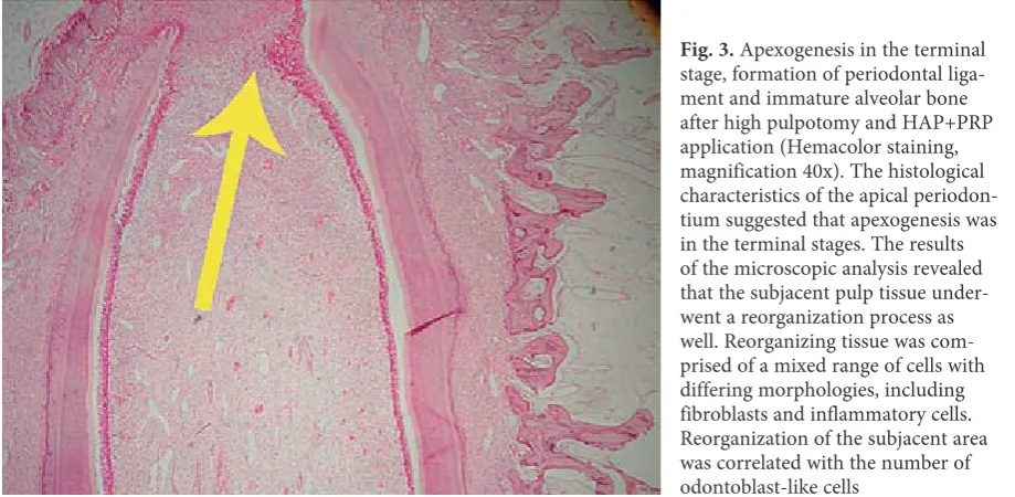
Completion of apexogenesis was associated with the formation of a periodontal ligament and surrounding alveolar bone in all teeth (Figure 3).
It was known in advance that pulpotomy is a more beneficial treatment for the further devel-opment of the root than high pulpotomy. Adding platelet rich plasma to hydroxyapatite only inten-sified the decrease in retardation of root develop-ment, especially after pulpotomy.
Table 5. Analysis of histological data in the pulpotomy group
Tabela 5. Analiza danych histologicznych w grupie, w której przeprowadzono pulpotomię
Observed parameters
(Badane wskaźniki) HAP HAP+PRP
Inflammatory reac-tion
(Reakcja zapalna)
1 2/8 (25%) 4/8 (50%) 2 6/8 (75%) 4/8 (50%)
3 0 0
4 0 0
Fisher’s test significance (Istotność statystyczna, test Fishera)
p = 0.608
Pulp response to applied therapy (Odpowiedź miazgi na zastosowane leczenie)
1 0 0
2 6/8 (75%) 8/8 (100%) 3 2/8 (25%) 0/8 (0%)
4 0 0
Fisher’s test significance (Istotność statystyczna, test Fishera)
p = 0.467
Presence and qual-ity of dentin bridge (Obecność i jakość mostka dentystycz-nego)
1 5/8
(62.5%) 5/8 (62.5%)
2 0 0
3 3/8
(37.5%) 3/8 (37.5%) Fisher’s test significance
(Istotność statystyczna, test Fishera)
p = 0.989
Table 6. Analysis of histological data in the high pulpotomy group
Tabela 6. Analiza danych histologicznych w grupie, w której przeprowadzono wysoką pulpotomię
Observed parameters (Badane wskaźniki) HAP HAP+PRP
Presence of apical barrier (Obecność bariery wierzchołkowej) presence 4/8 (50%) 4/7(57.1%) absence 4/8 (50%) 3/7(42.9%) Fisher’s test significance (Istotność statystyczna, test Fishera) p = 0.782 Presence of Inflammatory reaction (Obecność reakcji zapalnej) presence 1/8 (12.5%) 1/7 (14.3%)
absence 7/8 (87.5%) 6/7 (85.7%) Fisher’s test significance (Istotność statystyczna, test Fishera) p = 0.919
Presence of deformities in periapical region (Obecność deformacji w obszarze
okołowierzchołkowym) presence 1/8 (12.5%) 1/7 (14.3%)
Fig. 3. Apexogenesis in the terminal stage, formation of periodontal liga-ment and immature alveolar bone after high pulpotomy and HAP+PRP application (Hemacolor staining, magnification 40x). The histological characteristics of the apical periodon-tium suggested that apexogenesis was in the terminal stages. The results of the microscopic analysis revealed that the subjacent pulp tissue under-went a reorganization process as well. Reorganizing tissue was com-prised of a mixed range of cells with differing morphologies, including fibroblasts and inflammatory cells. Reorganization of the subjacent area was correlated with the number of odontoblast-like cells
Fig. 2. Compact formation of dentin bridge after pulpotomy and HA + PRP treatment; the pulp was closed (Hemacolor staining, magnification 40x). The dentin bridge covered the area of pulp exposure, preventing microleakage and bacterial invasion, which was crucial for maintain-ing pulp integrity and providmaintain-ing the conditions for root formation. Beneath the dentin bridge formation, odontoblast-like cells were identified. A great variation in the number of these cells was observed. These newly differentiated cells have a similar morphology to odontoblast cells. The original odontoblasts were positioned peripherally to the site of the expo-sure
Ryc. 2. Zwarta formacja mostka dentystycznego po pulpotomii i leczeniu HA + PRP; miazga została zamknięta (barwienie Hemacolor, powiększenie 40x). Mostek dentystyczny zajmował powierzchnię eksponowania miazgi, zapobiegając mikroprzeciekom i inwazji bakterii, co było kluczowe dla utrzymania integralności miazgi oraz zapewnienia warunków do formacji korzenia. Pod mostkiem dentystycznym zidentyfikowano komórki podobne do odontoblastów. Zaobserwowano wielką różnorodność liczby tych komórek. Te nowo zróżnicowane komórki mają podobną morfologię do komórek odontoblastów. Pierwotne odontoblasty zostały umieszczone obwodowo na miejscu ekspozycji
Ryc. 3. Apeksogeneza w fazie końcowej, tworzenie więzadła przyzębia i niedojrzałych kości wyrostka zębodołowego po wysokiej pulpotomii i podaniu HAP + PRP (barwienie Hemacolor, powiększenie 40x). Histologiczne cechy wierzchołkowego przyzębia sugerują fazę końcową apeksogenezy. Wyniki analizy mikroskopowej ujawniły, że spodnia tkanka miazgi także przeszła proces przebudowy. Przebudowana tkanki składała się z mieszanych komórek o różnej morfologii, w tym fibroblastów oraz komórek zapalnych. Przebudowa tkanki spodniej była skorelowana z liczbą komórek podobnych do odontoblastów
Discussion
Regenerative medicine seems to be the most appropriate and successful approach in therapeutic
its complete biocompatibility, the low invasiveness of the protocol for obtaining it, and its compatibil-ity with the holistic biological approach.
The present authors chose to use PRP with the aim of stimulating and enhancing local de-velopmental processes, with the high concentra-tion of growth factors as the main regulators of stem cells as the key links responsible for devel-opment [20]. The biocompatibility of HAP is well documented [21, 22], which is the main reason it was chosen as the PRP carrier. A secondary rea-son for choosing hydroxyapatite was the fact that it acts beneficially on apexogenesis and produces new calcification tissue without the necrosis char-acteristic of Ca[OH] 2 materials [23, 24]. The au-thors know of no possible chemical reaction be-tween HAP and PRP. The results of the present study confirmed this concept, since there was two times greater success in root development using HAP+PRP than HAP alone. In both therapeutic protocols, no statistically significant differences in root development were found in HAP+PRP treat-ed teeth as compartreat-ed to the controls; in contrast, in HAP treated teeth statistically significant retar-dation of root development was observed. These findings are in compliance with results of lee et al. [25], who that showed that PRP has sufficient potential to promote apexogenesis, comparable to physiological apexogenesis. The differences be-tween the HAP and HAP+PRP groups could be additionally explained by the previously report-ed role of PRP in the early phase of wound heal-ing [26] based on the mechanism of fast regenera-tion [10]. With regard to the formaregenera-tion of a dentin bridge, a statistical decrease was evidenced in HAP treated teeth compared to the control teeth, whereas in HAP+PRP treated teeth there was no difference compared to the control teeth. Den-tin bridges were present in the HAP+PRP in one more sample than in the HAP group. Considering the histological characteristics, the dentin bridge was characterized by a greater cell density in the HAP+PRP treated teeth. This indicated that PRP treatment led to histologically better quality com-position of the bridge. The effects of PRP were in-vestigated under the condition of preserved pulp tissue (pulpotomy) and with reduced pulp tissue (high pulpotomy), and it was found that gener-ally more beneficial effects were obtained in the pulpotomy group. In the pulpotomy group, PRP contributed to the preservation of pulp vitality by bringing about a compact dentin bridge and by stimulating pulp regeneration, and simultaneous-ly by stimulating development process. In terms of root development, there was twice as much re-tardation in the high pulpotomy group. A poten-tial explanation could be that in the pulpotomy
teeth the dental pulp-derived stem cells – the ma-jor ones in dentin-pulp complex development – were stimulated. In contrast, in the high pulpo-tomy group where pulp tissue was reduced (and therefore the capacity for pulp-derived stem cells was proportionately reduced) it was predominant-ly stem cells originating from the periodontal lig-ament that were stimulated, and since they have a lower regenerative potential for the dentin-ce-ment complex, the final outcome of the treatdentin-ce-ment was lower. These findings confirmed that den-tal pulp is the main source of stem cells responsi-ble for the development of the dentin-pulp com-plex, which corresponds to findings of Tziafas and Kodonas [4].
Sonoyama et al. [27] found that pulp contains more cellular and vascular components than the apical papilla does. Although maintenance of the dental pulp is the most important for healing pro-cess, the role of the mesenchymal stem cells of the apical papilla cannot be disputed [28] because it seems that these stem cells could be the source of odontoblasts that are responsible for the forma-tion of root dentin. It is known that the capabili-ties of these cells are increased by treatment with appropriate stimuli [29], so PRP probably has ben-eficial effects on this as well. That could be the ex-planation for such a beneficial effect in the high pulpotomy group with significantly reduced pulp regenerative capacity. Overall, PRP has dem-onstrated beneficial effects on apexogenesis in both the pulpotomy and high pulpotomy proto-cols [30]. Conventional treatment of immature teeth with open apices rarely has a good outcome, due to the high possibility of recontamination and increased risk of fracture. Recent dental research is therefore focused on the development of new treatment modalities such as the use of growth factors from the platelets in reestablishing pulp vi-tality, continuing root development [31], and pre-venting complications.
References
[1] Andreasen J: Apexogenesis. In: Textbook and color atlas of traumatic injuries to the teeth. Eds.: Andreasen JO, Andreasen FM, 3rd ed 1994. Munksgaard, Copenhagen, 63–65.
[2] Rafter M: Apexification: a review. Dent Traumatol 2005, 21, 1–8.
[3] Andreasen JO, Borum MK, Andreasen: Replantation of 400 avulsed permanent incisors, Endod Dent Traumatol 1992, 8, 45–55.
[4] Tziafas D, Kodonas K: Differentiation potential of dental papilla, dental pulp, and apical papilla progenitor cells. J Endod 2010, 36, 781–789.
[5] Friedlander LT, Cullinan MP, Love RM: Dental stem cells and their potential role in apexogenesis and apexifica-tion. Int Endod J 2009, 42, 955–962.
[6] Huang GT, Gronthos S, Shi S: Mesenchymal stem cells derived from dental tissues vs. those from other sources: their biology and role in regenerative medicine. J Dent Res 2009, 88, 792–806.
[7] Kotsovilis S, Markou N, Pepelassi E, Nikolidakis D: The adjunctive use of platelet-rich plasma in the therapy of periodontal intraosseous defects: a systematic review. J Periodontal Res 2010, 45, 428–443.
[8] Camargo PM, Lekovic V, Weinlaender M, Vasilic N, Madzarevic M, Kenney EB: Platelet-rich plasma and bovine porous bone mineral combined with guided tissue regeneration in the treatment of intrabony defects in human. J Periodont Res 2002, 37, 300–306.
[9] Whitman DH, Berry RL, Green DM: Platelet gel: an autologous alternative to fibrin glue with applications in oral and maxillofacial surgery. J Oral Maxillofac Surg 1997, 55, 1294–1299.
[10] Lekovic V, Camargo PM, Weinlaender M, Vasilic N, Aleksic Z, Kenney EB: Effectiveness of a combination of platelet-rich plasma, bovine porous bone mineral and guided tissue regeneration in the treatment of mandibular grade II molar furcations in humans. J Clin Periodontol 2003, 30, 746–751.
[11] Marx RE, Carlson ER, Eichstaedt RM, Schimmele SR, Strauss JE, Georgeff KR: Platelet-rich plasma: growth factor enhancement for bone grafts. Oral Surg Oral Med Oral Pathol Oral Radiol Endod 1998, 85, 638–646.
[12] Camargo PM, Lekovic V, Weinlaender M, Divnic-Resnik T, Pavlovic M, Kenney EB: A surgical reentry study on the influence of platelet-rich plasma in enhancing the regenerative effects of bovine porous bone mineral and guided tissue regeneration in the treatment of intrabony defects in humans. J Periodontol 2009, 80, 915–923.
[13] Arora NS, Ramanayake T, Ren YF, Romanos GE: Platelet-rich plasma: a literature review. Implant Dent 2009, 18, 303–310.
[14] EEC: Council Directive 86/609/EEC of 24November 1986 on the approximation of laws, regulations and admin-istrative provisions of the Member States regarding the protection of animals used for experimental and other scientific purposes. Official J Eur Communities 1986, l358, 1–29.
[15] Weibrich G, Kleis WK, Hafner G, Hitzler WE: growth factor levels in platelet-rich plasma and correlations with donor age, sex, and platelet count. J Craniomaxillofac Surg 2002, 30, 97–102.
[16] Petrović V, Pejčić N, Rakić M, Leković V, Vasić U, Stojić Ž: Effects of the platelet rich plasma on apexogenesis in young monkeys: radiological and histological evaluation. Acta Vet 2012, 62, 39–52.
[17] Demirjian A, Goldstein H, Tanner JM: A new system of dental age assessment. Human Biol 1973, 45, 211–227.
[18] ISO-7405. Preclinical evaluation of biocompatibility of medical devices used in dentistry, test methods for dental materials. International Organization for Standardization – ISO E, 1997, 1–18.
[19] Murray PE, Hafez AA, Smith AJ, Windsor LJ, Cox CF: Histomorphometric analysis of odontoblast-like cell numbers and dentin bridge secretory activity following pulp exposure. Int Endod J 2003, 36, 106–116.
[20] Huang GT: Pulp and dentin tissue engineering and regeneration: current progress. Regen Med 2009, 4, 697–707.
[21] Jin HH, Kim DH, Kim TW, Shin KK, Jung JS, Park HC, Yoon SY: In vivo evaluation of porous hydroxyapatite/ chitosan-alginate composite scaffolds for bone tissue engineering. Int J Biol Macromol 2012, 51(5), 1079–1085.
[22] Zandi M, Mirzadeh H, Mayer C, Urch H, Eslaminejad MB, Bagheri F, Mivehchi H: Biocompatibility evalua-tion of nano-rod hydroxyapatite/gelatin coated with nano-HAp as a novel scaffold using mesenchymal stem cells. J Biomed Mater Res A 2010, 92(4), 1244–1255.
[23] Lee UL, Jeon SH, Park JY, Choung PH: Effect of platelet-rich plasma on dental stem cells derived from human impacted third molars. Regen Med 2011, 6, 67–79.
[24] Strayhorn CL, Garrett JS, Dunn RL, Benedict JJ, Somerman MJ: growth factors regulate expression of osteo-blastassociatedgenes. J Periodontol 1999, 70, 1345–1354.
[25] Alliot-Licht B, Jean A, M.Gregoire M: Comparative effect of calcium hydroxide and hydroxyapatite on the cel-lular activity of human pulp fibroblasts in vitro. Arch Oral Biol 1994, 39, 481–489.
[26] Tjäderhane L: The mechanism of pulpal wound healing. Aust Endod J 2002, 28, 68–74.
[27] Sonoyama W, Liu Y, Yamaza T, et al.: Characterization of the apical papilla and its residing stem cells from human immature permanent teeth: a pilot study. J Endod 2008, 34, 166–171.
[28] Huang GT-J, Sonoyama W, Liu Y, Liu H, Wang S, Shi S: The hidden treasure in apical papilla: the potential role in pulp/dentin regeneration and bioroot engineering. J Endod 2008, 34, 645–651.
[29] Sonoyama W, Liu Y, Fang D, et al.: Mesenchymal stem cell-mediated functional tooth regeneration in swine. PloS ONE 2006, 1, e79.
[30] Fréchette JP, Martineau I, Gagnot G: Platelet-rich plasmas: growth factor content and roles in wound healing. J Dent Res 2005, 84, 434–439.
Address for correspondence:
Nataša Pejčić
Department of Preventive and Pediatric Dentistry School of Dentistry
University of Belgrade Doktora Subotića 2 11000 Belgrade Serbia
E-mail: [email protected] Conflict of interest: None declared Received: 23.08.2012