Turkish Journal of Fisheries and Aquatic Sciences 16: 251-258 (2016)
www.trjfas.org ISSN 1303-2712 DOI: 10.4194/1303-2712-v16_2_04
RESEARCH PAPER
© Published by Central Fisheries Research Institute (CFRI) Trabzon, Turkey in cooperation with Japan International Cooperation Agency (JICA), Japan
Histological Profile and Fatty Acid Composition in Hepatopancreas of Blue
Swimming Crab,
Portunus pelagicus
(Linnaeus, 1758) at Different Ovarian
Maturation Stages
Introduction
Blue swimming crab, Portunus pelagicus (Linnaeus, 1758) is a commercially important species for both fisheries and aquaculture in South-east Asia. Great market demands, especially for human consumption, have put this species in attention. The high harvests of this species are due to delicious taste, good market price and supplied nutrients. In addition, it is also relatively expensive in comparisons to sea fishes consumed locally. This has led to increasing demand on this species. However, the interests towards aquaculture of this species have been growing. This species is suitable for the aquaculture industry because it has special abilities to withstand against changes of physical parameters and activity like temperature, salinity, oxygen, pH and starvation alongside with fast growth and high reproductive rate. Broodstock nutrition determines the reproductive physiology of crustacean larvae. If the nutritional requirements of the broodstock are met, the larvae of P. pelagicus will have high survival rate. Some of the components in broodstock diet like fatty acids (FA) can influence the fecundity, larval quality and
hatching ability of several fishes and crustaceans (Racotta et al., 2003; Azra and Ikhwanuddin, 2016).
In brachyuran crabs, the hepatopancreas is an important organ for storage of organic matter and plays main roles in nutrition metabolism and ovarian development (Wang et al., 2014). Currently, there is a lack of study related to histological characteristics and FA composition of the hepatopancreas in P. pelagicus since most of the studies related to P. pelagicus were only focused on its natural diets, growth and survival and their size at maturity (Xiao and Kumar, 2004; Romano et al., 2012; Zainal, 2013). Information about the FA composition in female crabs during maturation is also lacking although it is related to reproductive success and influential towards ovarian maturation. Histology study of hepatopancreas can be used as a reference for the study related to the other species of crabs. The concentration of FA by female crabs at the different ovarian maturation stages may be able to be useful for the creation of formulated diet instead of using natural diet.
The objectives of this study are to analyze the histological character of hepatopancreas and to determine the FA composition in hepatopancreas at
Ambok Bolong Abol-Munafi
1, Mohd Syafiq Mukrim
2, Roswati Md Amin
2, Mohamad N Azra
1,
Ghazali Azmie
3, Mhd Ikhwanuddin
3,*
1
Universiti Malaysia Terengganu,School of Fisheries and Aquaculture Sciences, 21030, Kuala Terengganu, Terengganu, Malaysia.
2
Universiti Malaysia Terengganu, School of Marine Science and Environment, 21030, Kuala Terengganu, Terengganu, Malaysia
3
Universiti Malaysia Terengganu, Institute of Tropical Aquaculture, 21030, Kuala Terengganu, Terengganu, Malaysia.
* Corresponding Author: Tel.: +609.668 3638; Fax: +609.668 3502; E-mail: ikhwanuddin@umt.edu.my
Received 27 December 2015 Accepted March 2016
Abstract
The study of the hepatopancreas structure is important to provide the morphological and molecular information for
future research involving the nutrition requirements of Portunus pelagicus culture. Thus, the study of histological
characteristics and fatty acid (FA) composition in hepatopancreas of different ovarian maturation stages of the P. pelagicus
were investigated. There are five stages in ovarian maturation of P. pelagicus which are: Stage I (immature), Stage II (early
maturing), Stage III (advance maturing), Stage IV (mature) and Stage V (spawned/re-maturation/spent stage). The histological study of hepatopancreas showed that the lumens of most of the tubules were irregular-like shape in Stage I, circle-like shape
in Stage III and ovul-like shape in Stage IV. A total of 29 types of FA were found in the hepatopancreas of P. pelagicus. The
most dominant FA was C16:0 (Palmitic acid) with concentration of 456.10±266.23 mg/g(38.73±4.12%) at stage II. Second most dominant FA was C18:0 (Stearic acid) with concentration of 203.99±120.28 mg/g (13.97±1.62%) at stage III and followed by C20:5n3 (Eicosapentanoic acid) with concentration of 131.19±84.70 mg/g (8.98±1.13%) at stage III. In conclusion, the tubules structure and FA concentration of hepatopancreas play an important role in reproduction and diet formulation for portunid crab broodstock.
different ovarian maturation stages of P. pelagicus.
Materials
and
Methods
Study Area and Sample Collection
The study was conducted at Pendas Jetty, Gelang Patah situated south of the state of Johor, and Setiu Wetland, Terengganu, Coastal water of Malaysia. The samples were collected on June 2013 and fifty female crabs’ samples were obtained, ten female crabs for each ovarian maturation stages (Stage 1-5). The female crab samples were obtained from the market and placed in a container equipped with an aeration system. Female crabs were randomly picked. During the selection of the crabs, several obvious criteria that differentiate female and male crabs were taken into account (shape of the abdomen; female crab with wider and more globular abdomen meanwhile male crab with narrow and straight abdomen). Then, two morphometric characteristics of the crab, carapace width (CW) and body weight (BW) were measured. Subsequently, the crabs were dissected by using dissecting tools and the stages of ovarian maturation were determined based on the study by Efrizal et al., (2015) which divided it in five stages; immature stage (Stage I), early maturing (Stage II), advance maturing (Stage III), mature (Stage IV) and spent stage (Stage V). Stage differentiation was done through histological color and structure. Hepatopancreas sample in a crab was carefully collected by using forceps and placed in sample bottles. Hepatopancreas were divided into two, histology analysis and another one for FA analysis.
Sample Storage and Preparation
The hepatopancreas for histology sample were fixed with 5% formalin for 12 hours. After that, hepatopancreas in sample bottles were transferred into histological cassettes and immersed in 70% ethanol. The FA samples were stored in an ice chest. In the laboratory, the samples were stored in freezer with -80 ºC according to the study by Abdulkadir and Tsuchiya (2008). Then, samples were transferred into smaller sample bottles. The purpose of transfer the samples are to safe some space during drying in freeze dry instrument. After that, samples were freeze dried to dry it. After drying process, the samples were crushed by using mortar and pestle. The samples were weighted by using analytical balance.
Histological Analysis
By referring to standard procedure with Haematoxylin and Eosin staining method, samples in histological cassettes were processed by using automated tissue processor. Tissue samples were processed with the following sequence- 70% alcohol,
90% alcohol, 95% alcohol twice, alcohol 100% twice, xylene I, xylene II, xylene III and paraffin wax. After processing, samples were mounted onto their cassettes using paraffin wax, sectioned into 5 μm films using a microtome, transferred into water bath (at 45 °C) for expansion before mounting onto slides using glycerol and egg white (used as adhesive). Every section on slide were dried on hot plate (60°C) and stained by Haematoxylin-Eosin staining method. Samples were mounted by using DPX and observed under compound microscope model Leica DME. For tubules characterization, the hepatopancreas tubules at each ovarian maturation stage were observed by using DinoEye model AM4023X. For tubules measurement selection (calculated as largest distance of the tubule), the heights in unit micrometer (µm) of the hepatopancreas tubules at each ovarian maturation stages were measured by using DinoEye model AM4023X . The mean tubules heights were calculated after calibration of the DinoEye.
Fatty acid Composition Analysis
Fatty acid analysis was conducted by referring to one step method by Abdulkadir and Tsuchiya (2008). Preparation of internal standard which contains known amounts of FAs is important to quantify the amounts of FA in samples. The preparations of internal standard solution were started by dissolving 100 mg of 19:0 (Nonadecanoic acid) in 100ml hexane until the 1 mg/ml of C 19:0 achieved. For each sample, the weights required were around 200-300 mg. The sample from glass vial were weighted and transferred into a centrifuge tube. Then, it was mixed with 4 ml of hexane and 1 ml of internal standard solution. The mixture in the tube was mixed with 2 ml of 14% BF3 in methanol. A magnetic stirrer was placed in the centrifuge tube carefully. After that, the nitrogen gas was used to flush head spaces of tubes. The tube was closed tightly by using a Teflon-lined screw-cap. The tube was heated on a hot plate at 100 ºC for 120 min under continuous stirring. Then, the tube was cooled down to room temperature. Another 1 ml of hexane was added and 2 ml of distilled water as follow up. The tube was shaken vigorously for 1 min and centrifuged for 3 min at 2500 rpm. After that, two phases were formed. The upper phase, hexane layer were taken for further analysis as it contains FA Methyl Esters (FAMEs). The layer was transferred by using micropipette into the sample vial that was injected into the Gas Chromatography (GC). One µL of FAME was injected into gas chromatography (GC –FID) by using syringe. 37-Component FAME Mix was used to be standard for FA content in sample. Hydrogen gas was used as the carrier gas. Full peak of FA could be observed on chromatogram after about 20 min.
Mean height of hepatopancreas tubules were compared among the samples based on ovarian maturation stages by using one way ANOVA. Fisher’s exact tests were used to know the difference between mean in XLSTAT software (Addinsoft, New York, USA). The mean concentration of individual and classes of FA in hepatopancreas were compared among the samples based on ovarian maturation stages by using one way ANOVA. Fisher test were used to know the difference between mean in XLSTAT software. In order to check whether the data is normally distributed or not, Kolmogorov-Smirnov test were used. In this study, the data is normally distributed after checking it by using the test in SPSS 16.0 software.
Results
Histological Characteristics of Hepatopancreas Tubules at Different Ovarian Maturation Stages
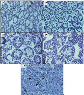
Generally, cells had an irregular-like shape with
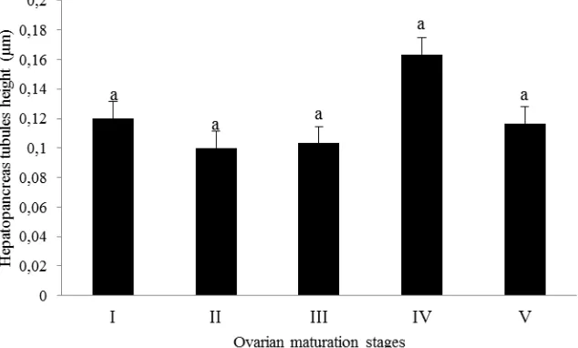
clear bluish nuclei, and the lumen of most tubules was small (Figure 1a). Tubules in ovarian stage II crabs were of circle-like shape and with smaller lumens when compared to ovarian stage I crabs (Figure 1b). The nuclei were non observable. Meanwhile, tubules in ovarian stage III were circle-like shape (Figure 1c) compared to the ovarian stage IV which in ovul-like shape (Figure 1d). Tubule lumens of stage III was smaller than those from stage IV. On the other hand, most tubules in Stage V (Figure 1e) were in various shapes (ovul+irregular shape) with smaller lumen compared to Stage IV. Figure 2 showed the pattern of mean hepatopancreas tubules height through the increasing ovarian maturation stages was fluctuate. The tubules were decreasing in height from Stage I to Stage II, while decreased in Stage III, increased back in Stage IV and decreased at Stage V.
Fatty Acid Composition at Different Ovarian Maturation Stages
In total, 29 individual FAs belonging to three
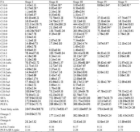
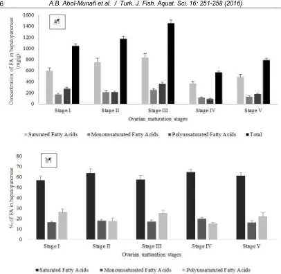
major classes (saturated FAs – SFA; monounsaturated FAs – MUFA and polyunsaturated FAs – PUFA) were identified in hepatopancreas of P. pelagicus (Table 1). Concentrations of total FA in hepatopancreas were increased from Stage I to Stage III (Figure 3a). The most dominant FA was C16:0 (Palmitic acid) with mean concentration of 456.10±266.23 mg/g (38.73±4.12%) at stage II of the ovarian maturation stage followed by C18:0 (Stearic acid) with mean concentration of 203.99±120.28 mg/g (13.97±1.62%) at stage III of the ovarian maturation stage and C20:5n3 (Eicosapentanoic acid) with mean concentration of 131.19±84.70 mg/g (8.98±1.13%) at stage III of the ovarian maturation stage. Based on Figure 4, percentage of SFA was the highest compared to two other classes of fatty acid and put SFA as dominant fatty acid classes. SFA has highest percentage of FA, ranging from 57.33 – 73.29% which followed by PUFA, 15.18 – 26.33%. Least dominant FA was MUFA, with range of 16.28 – 19.75%. All the data were normally distributed after checked with Kolmogorov-Smirnov test. Then, concentrations were decrease dramatically at Stage IV and increased back at Stage V. Same goes as the concentration of SFA and MUFA at Stage I to Stage V. The pattern of PUFA was different than for other two classes (Figure 3b). There were significant differences in concentration of FA at different ovarian maturation stages. There were significantly different of percentage at different maturation stages in MUFA and PUFA classes and vice versa in SFA. Based on the results from Table 1, ratio of n-6/n-3 has maximum value of 0.17 and minimum value of 0.10 while ratio of PUFA/SFA has maximum value of 4.24 and minimum value of 2.16.
Discussion
Histological Characteristics of Hepatopancreas
Tubules
To clearly understand the physiological significance of the hepatopancreas, it is important to understand its histological structure. In this study, mean tubules heights at immature stage (Stage I) to early maturing stage (Stage II) of the ovary were increasing. Mean concentration of tubules height was decreased at late maturing stage (Stage III). It may be influenced by the decrease size in lumen of tubules from Stage II to Stage III and presence of vacuole at the tubules at Stage III. Mean height of hepatopancreas was increased back at maturing stage (Stage IV) as the size of lumen were increased from Stage III to Stage IV. Spent stage (Stage V) showed decreasing of hepatopancreas tubules height from Stage IV. The decrease of cell height was caused by decreased size of lumen of tubules at Stage V. There was significant different of tubules height at different ovarian maturation stages. Hepatopancreas tubules did not have clear relationship with the total FA concentration at different ovarian maturation stages. The trends were different. At stage IV, the hepatopancreas tubules height was highest when compared to other stages, while for total FA the concentration at same stage was the lowest compared to other stages. This showed that not only tubules contained FA, the space between tubules also stored FA.
Fatty Acid Composition
SFA was dominant FA. The dominant individual FA of SFA that have the highest concentration were firstly C16:0, C18:0 and followed by C14:0 (Myristic acid). The results were similar to the study of Soundarapadian et al., (2013) for FA composition of crustacean, Macrobrachiumidae. In addition, palmitic acids have high content of metabolic energy during
growth of female marine animal (Huynh et al., 2007). PUFA was second most dominant FA. The dominant individual FA of PUFA were firstly C20:5n3, C20:3n3 (Eicosatrienoic acid), and followed by C22:6n3 (Docosahexaenoic acid). Least dominant FA was MUFA, with concentration of 175.90 mg/g (17.56%).
The most dominant individual FA of MUFA were C16:1 (Palmotelic acid), C18:1n9c (oleic acid) and followed by C20:1 (eicosenoic acid). The main function of MUFA is to supply energy source for spawning (Rosa and Nunes, 2002). Total FA were increased steadily from Stage I to Stage III. Then, total FA decreased dramatically at Stage IV and bounce back to increase at Stage V. There was no significant different of total fatty acids at different ovarian maturation stages. In previous study by
Raviet al., (2013) on FA changes in hepatopancreas of P. pelagicus at different ovarian maturation stages, the output from the study is different. The different results suggested that content of fatty acids may caused by different sampling area. At different geographical area, the diet may distinct. From the past study, the lipid concentration was increased from immature to early maturing stage and then decreased at spawning stage. In addition, present study also relatively similar to study done by Wen et al., (2001) on chinese-mitten crab, Eriocheir sinensis. The crab’s lipid was at peak at Stage III and decreasing during spawning stage, similar like in present study.
There was an increase in total FA from Stage I to Stage III. The possible reason why this happen was accumulation of FA from other tissues to oocytes of ovary occured. The tissue that responsible for the
Table 1. Fatty acid (FA) concentration in hepatopancreas of Portunus pelagicus at different ovarian maturation stages
FA Stage I Stage II Stage III Stage IV Stage V
C10:0 1.45±1.11 5.82±4.50* 3.05±3.62 0.68±1.04* 0.52±0.91*
C11:0 0.27±0.28* 0.92±0.10* 0.50±0.68 - -
C12:0 1.24±0.94 2.14±1.56 2.69±2.94 - -
C13:0 0.35±0.37 0.35±0.61 1.31±1.57 - -
C14:0 63.03±46.21 72.70±43.28 75.02±48.36 37.01±6.81 47.74±6.77
C15:0 16.05±4.80 24.70±13.57 18.52±9.81 12.38±0.84 16.53±3.08
C16:0 321.31±150.71 456.10±266.23 444.42±255.51 226.69±12.83 269.84±50.05
C17:0 21.08±9.45 21.18±8.49 35.45±23.68* 10.28±0.94* 18.13±4.49
C18:0 140.29±70.07 138.73±62.26 203.99±120.28 75.30±9.62 115.54±23.61
C20:0 8.54±7.78 9.19±4.89 15.01±10.77 1.76±2.69 3.76±3.26
C21:0 2.84±3.41 - 5.05±4.37 - -
C22:0 5.21±6.18 2.07±3.59 7.54±6.64 - -
C23:0 14.69±6.73 17.19±8.80 23.28±14.74 5.67±3.97 12.18±2.16
C24:0 1.60±2.78 - 1.63±2.83 - -
C14:1 0.68±0.35 0.82±0.84 1.40±0.82 - -
C16:1 76.44±37.01 103.73±63.84 104.41±58.98 64.91±3.33 62.42±4.98
C17:1 4.16±2.90 8.24±3.88 10.12±7.19* 1.33±2.02* 3.98±3.96
C18:1n9t 3.10±2.90 3.14±5.44 6.22±5.66 - -
C18:1n9c 70.87±32.72 81.39±41.37 111.98±68.89* 39.62±3.46* 51.97±18.14
C20:1 12.71±5.53 12.72±7.38 12.02±7.67 7.60±0.63 10.59±2.28
C22:1n9 4.99±2.84 2.37±4.11 5.61±7.62 - -
C18:2n6c 14.94±8.07 15.05±7.16 24.88±16.98 8.38±1.02 12.83±2.75
C18:3n6 5.58±6.69 8.43±7.42 13.06±10.00 - 3.06±5.30
C20:2 4.68±5.174 3.66±3.17 12.00±9.96 - -
C20:3n3 57.40±22.50 61.66±31.53 88.17±52.56* 31.08±2.76* 52.09±8.60
C20:3n6 4.10±2.51 3.64±3.21 8.57±6.14 - -
C20:4n6 3.62±2.34 1.78±3.08 6.10±4.12 - -
C20:5n3 108.64±72.01 73.21±39.33 131.19±84.70 47.76±13.07 79.55±12.45
C22:6n3 78.04±51.12 42.23±20.85 82.70±57.54 - 29.80±26.75
SFA 597.98±310.82 751.09±417.87 837.45±505.80 369.77±38.73 484.25±94.33
MUFA 172.94±84.24 212.41±126.85 251.75±156.84 113.45±9.45 128.96±29.36
PUFA 277.02±171.71 209.66±115.76 366.68±244.90 87.22±16.85 177.34±55.85
Total fatty acids 1047.94±566.78 1173.17±638.47 1455.88±907.54 570.43±49.18 790.55±124.26
Omega 3 (n-3)
244.08±25.78 177.11±15.66 302.06±26.55 78.84±24.24 161.45±24.92
Omega 6
(n-6) 28.2±5.32 28.90±5.92 52.61±8.33 8.38±4.19 15.89±6.08
n-6/n-3 ratio 0.12 0.16 0.17 0.11 0.10
PUFA/SFA ratio 2.16 3.58 2.28 4.24 2.73
accumulation was muscle tissue. Accumulation of FA happened in muscle during inactivity of the ovary. Then, the FA in muscles were transferred into maturing ovary. A crustacean, female shrimp were proved to double its consumption of food to accumulate lipid at ovaries. Besides, hepatopancreas itself was the place where digestion and absorption happened. Based on the findings from present study suggested that the digestion and absorption may occur at high rate at Stage I to Stage III with low transfer out of FA from hepatopancreas to other part of the crab system like ovary, muscle and other tissue. The thing was the ovaries were taken the lipids directly from gut during this stage. Maybe the ovaries had sufficient requirement of fatty acids from the supply from muscle and gut. Instead of being the lipid storage site, hepatopancreas also involved in vitellogenesis of ovary. Vitellogenesis is the formation of yolk at oocyte in the ovary. Study by Ravi et al., (2013) showed that there were mobilization of FA from hepatopancreas to the ovary at vitellogenesis I and II (from Stage I to Stage III).
Great changes between these stages were also observed in marine shrimp, Penaeus kerathurus (Mourente and Rodriguez,1991). In present study, there is a possibility of high gonadal activity during later stages of ovarian maturation which could have caused the great decreases. Beside the high gonadal activity, transfer period of lipid store in hepatopancreas could also be the reason why the declination of FA happened during Stage IV. In present study, reduction of FA content from Stage III to Stage IV proposed that high leaching of fatty acid sources from hepatopancreas to ovaries were triggered by huge amount energy supply needed by ovaries for reproduction. This suggested that the lipid transport from hepatopancreas to the ovary was maximum at Stage IV.
Result from present study also were supported with increment of lipid content in hepatopancreas of spiny lobster at spent stage. After spawning, the energy reserves in ovary were drained and decreased. Instead of that, rise in total FA in hepatopancreas in present study also due to inactivity of ovary during
Figure 3. (a): Concentration of fatty acids (FA) in hepatopancreas of P. pelagicus at different ovarian maturation stages;
spent stage. Although it is classified as resting stage, some activities in ovary like preparation of next maturation cycles were occured inthe study by Raviet al., (2013)but the activity seems not necessary at the stage. Inactivity of ovary aid in low rate of leaching of FA from hepatopancreas thus the FA content in hepatopancreas were increased at Stage V. SFA and MUFA followed pattern of total FA but PUFA concentration at Stage 1 was higher than Stage II, shown different pattern from two other classes of FA, SFA and MUFA. This was due to natural diets consumption by the crabs, as it contributes to the composition of FA. Diets that contained high level of PUFA may consume more by P. pelagicus during Stage I. The example of high PUFA containing diet is squid (Turner and Rooker, 2005). There was no significant difference of SFA, MUFA and PUFA at different ovarian maturation stages.
As hepatopancreas is the organ that is responsible to absorb digested materials (Wang et al., 2014), it is essential that the biomarkers of FA could be indicator for diet for P. pelagicus. Based on past study, there was a presence of brown algae, green algae, animal tissues, and highly digested materials in the foregut of P. pelagicus (Zainal et al., 2013). In addition, domination of palmitic acid and eicosapentanoic acid in foregut content of P. pelagicus was noted by Ikhwanuddinet al., (2014). Some fatty acids could be indicator for requirement of human diet and health hazard. For example, n-6/n-3 and PUFA/SFA ratio. From present study, it showed that the ratio of n-6/n-3 at different ovarian maturation stages is less than 4. PUFA/SFA ratio was above 0.45 (minimum ratio recommended). The ratio of present study is above minimum ratio. C16:0 (Palmitic acid) are biomarker for microalgae, mostly from Cyanophyceae and Chlorophyceae. It was also the biomarkers for plants and marine animal sources (Sahu et al., 2013). C18:0 (Stearic acid) was the second most abundant and it is the biomarker for marine animals and fungi and followed by C20:5n3 (Eicosapentanoic acid), as the third most abundance which is biomarker for microalgae and marine bacteria (Yazawa, 1996).
Conclusion
In conclusion, the study shows important results on tubules structure and FA composition of hepatopancreas of P. pelagicus in various ovarian maturation stages. The results were essential for further study on reproduction and diet formulation for portunid crab broodstock in the future.
Acknowledgements
This study was supported by research grant from Malaysian Ministry of Education under Niche Research Grant Scheme (NRGS, 2013–2018) –
Improving the Health of Setiu Wetlands Ecosystem and Productivity of Crustacean Resources for Livelihood Enhancement (Vote No. 53131) to MI. The authors wish to thank all the staff at Institute of Tropical Aquaculture, the School of Marine Science and Environment and the Marine Hatchery of School of Fisheries and Aquaculture Sciences, Universiti Malaysia Terengganu for technical assistance rendered. We would like to thank anonymous reviewer (s) for their constructive criticism.
References
Abdul kadir, S. and Tsuchiya, M. 2008.One-step method for quantitative and qualitative analysis of fatty acids in marine animal samples.Journal of Experimental
Marine Biology and Ecology, 354: 1-8.
doi:10.1016/j.jembe.2007.08.024
Azra, M.N. and Ikhwanuddin, M. 2016. A review of
maturation diets for mud crab genus Scylla
broodstock: Present research, problems and future perspective. Saudi Journal of Biological Sciences, 23: 257-267. doi:10.1016/j.sjbs.2015.03.011
Efrizal, Arshad, A., Kamarudin, M.S., Saad, C.R. and Amin, S.M.N. 2015.Some reproductive biology of
blue swimming crab (Portunuspelagicus (Linnaeus,
1758)) under laboratory conditions. Journal of
Fisheries and Aquatic Science, 10: 77-91.
doi:10.3923/jfas.2015.77.91
Huynh, M.D., Kitts, D.D., Hu, C. and Trites, A.W. 2007. Comparison of fatty acid profiles of spawning and
non-spawning Pacific herring,
Clupeaharenguspallasi. Comparative Biochemistry
and Physiology: Part B, 146: 504-511.
doi:10.1016/j.cbpb.2006.11.023
Mourente, G. and Rodriguez, A. 1991.Variation in the lipid content of wild-caught females of the marine shrimp
Penaeus kerathurus during sexual maturation.Marine Biology, 110: 21-28. doi:10.1007/BF01313088 Patil, C., Paul, R. and Malkanna. 2008. Neuroendocrine
regulation and pesticidal impact on freshwater crab,
Barytelphusaguerini (H. Milne Edwards). Journal of
Environmental Biology, 29: 887-892.
http://www.jeb.co.in/journal_issues/200811_nov08/pa per_15.pdf
Racotta, I.S., Palacios, E. and Ibarra, A.M. 2003. Shrimp larval quality in relation to broodstock condition. Aquaculture, 227: 107-130. doi:10.1016/S0044-8486(03)00498-8
Ravi, R., Manisseri, M.K. and Sanil, N.K. 2013.Ovarian maturation and oogenesis in the blue swimmer crab,
Portunus pelagicus (Decapoda: Portunidae). Acta
Zoologica, 94: 291-299.
doi:10.1111/j.1463-6395.2011.00555.x
Romano, N., Zeng, C., Noordin, N.M. and Ng, W.K. 2012.Improving the survival, growth and hemolymph ion maintenance of early juvenile blue swimmer
crabs, Portunus pelagicus, at hypo- and
hyper-osmotic conditions through dietary long chain PUFA
supplementation. Aquaculture, 342-343: 24-30.
doi:10.1016/j.aquaculture.2012.02.013
Rosa, R. and Nunes, M.L. 2002.Biochemical changes during the reproductive cycle of the deep-sea decapod
Nephropsnorvegicus on the south coast of Portugal.
doi:10.1007/s00227-002-0911-9
Sahu, A., Pancha, I., Jain, D., Paliwal, C., Ghosh, T., Patidar, S., Bhattacharya, S. and Mishra, S. 2013.Fatty acids as biomarkers of microalgae.Phytochemistry, 89: 53-58. doi:10.1016/j.phytochem.2013.02.001 Soundarapandian, P., Sudhakar, S., Varadharajan, D. and
Dinakaran, G.K. 2013.Biochemical composition during the embryonic development and freshly
hatched zoea of Macrobrachiumidae (Heller,
1862).Earth Science and Climatic Change, 5: 171. doi:10.4172/2157-7617.1000171
Turner, J.P. and Rooker, J.R. 2005.Effect of diet on fatty
acid compositions in Sciaenopsocellatus.Journal of
Fish Biology, 67: 1119-1138. doi:10.1111/j.0022-1112.2005.00816.x
Wen, X., Chen, L., Ai, C., Zhou, Z. and Jiang, H. 2001.Variation in lipid composition of Chinese
mitten-handed crab, Eriocheirsinensis during ovarian
maturation.Comparative Biochemistry and Physiology Part B: Biochemistry and Molecular Biology, 130: 95-104. doi:10.1016/S1096-4959(01)00411-0
Wang, W., Wu, X., Liu, Z., Zheng, H. and Cheng, Y. 2014.Insights into hepatopancreatic functions for nutrition metabolism and ovarian development in the
crab Portunustrituberculatus: Gene discovery in the
comparative transcriptome of different
hepatopancreas stages. Plos One, 9:
e84921.doi:10.1371/journal.pone.0084921
Xiao, Y. and Kumar, M. 2004. Sex ratio, and probability of sexual maturity of females atsize, of the blue
swimmer crab, Portunus pelagicus Linneaus, off
southern Australia. Fisheries Research, 68: 271-282. doi:10.1016/j.fishres.2003.11.012
Yazawa, K. 1996. Production of eicosapentaenoic acid from
marine bacteria.Lipids, 31: S297-S300.
doi:10.1007/BF02637095
Zainal, K.A.Y. 2013.Natural food and feeding of the
commercial blue swimmer crab, Portunus pelagicus