Introduction
Cell therapy for diabetes mellitus will be advanced by the availability of additional donor cells. Islet cells may
originate from pancreatic ductal or other epithelial cells, especially after damage in the adult pancreas [1, 2]. However, the replication potential of human pancreatic islet cells is limited, which generally restricts expansion of these cells under culture conditions [3]. Although cell lines have been generated by oncogenetic transformation of islet cells [4, 5] tumorigenic cells will obviously be unsuitable for basic studies under various contexts, as well as for cell therapy. This problem of genetic trans-formation afflicts other sources of cells, for example, inducible pluripotential stem cells (iPS), which are Abstract
Introduction:The ability to expand organ-specific stem/progenitor cells is critical for translational applications, although uncertainties often arise in identifying the lineage of expanded cells. Therefore, superior insights into lineage maintenance mechanisms will be helpful for cell/gene therapy.
Methods: We studied epithelial cells isolated from fetal human pancreas to assess their proliferation potential, changes in lineage markers during culture, and capacity for generating insulin-expressing beta cells. Cells were isolated by immunomagnetic sorting for epithelial cell adhesion molecule (EpCAM), and characterized for islet-associated transcription factors, hormones, and ductal markers. Further studies were performed after modification of cells with the catalytic subunit of human telomerase reverse transcriptase (hTERT).
Results: Fetal pancreatic progenitor cells efficiently formed primary cultures, although their replication capacity was limited. This was overcome by introduction and expression of hTERT with a retroviral vector, which greatly enhanced cellular replication in vitro. However, we found that during culture hTERT-modified pancreatic progenitor cells switched their phenotype with gain of additional mesodermal properties. This phenotypic switching was inhibited when a pancreas-duodenal homeobox (Pdx)-1 transgene was expressed in hTERT-modified cells with a lentiviral vector, along with inductive signaling through activin A and serum deprivation. This restored endocrine properties of hTERT-modified cellsin vitro. Moreover, transplantation studies in immunodeficient mice verified the capacity of these cells for expressing insulin in vivo.
Conclusions: Limited replication capacity of pancreatic endocrine progenitor cells was overcome by the hTERT mechanism, which should facilitate further studies of such cells, although mechanisms regulating switches between meso-endodermal fates of expanded cells will need to be controlled for developing specific applications. The availability of hTERT-expanded fetal pancreatic endocrine progenitor cells will be helpful for studying and recapitulating stage-specific beta lineage advancement in pluripotent stem cells.
© 2010 BioMed Central Ltd
Switching of mesodermal and endodermal
properties in hTERT-modified and expanded fetal
human pancreatic progenitor cells
Kang Cheng
1, Antonia Follenzi
2, Manju Surana
3, Norman Fleischer
3, Sanjeev Gupta*
4R E S E A R C H
Open Access
*Correspondence: [email protected]
4Hepatology Division, Department of Medicine, Cancer Research Center, Diabetes Research Center, Center for Human Embryonic Stem Cell Research, Marion Bessin Liver Research Center, Ruth L. and David S. Gottesman Institute for Stem Cell and Regenerative Medicine Research, and Institute for Clinical and Translational Research, Albert Einstein College of Medicine, Ullmann Bldg., Rm 625, 1300 Morris Park Avenue, Bronx, NY 10461, USA
Full list of author information is available at the end of the article
teratogenic [6]. Whereas reprogramming of cells through transcription factor modifications was recently found to be effective for transdifferentiating pancreatic exocrine cells to endocrine beta cells in vivo [7], generalization of this observation in developing treatments for diabetes mellitus requires much more work.
We consider fetal tissues to be of considerable interest as cell donors because these are particularly enriched in lineagecommitted stem/progenitor cells. However, in comparison with pluripotent human embryonic stem cells (hESC) or iPS, the replication potential of fetal cells was limited [8, 9], possibly due to telomere shortening [3, 10]. Modification of fetal human liver cells with the catalytic subunit of human telomerase reverse trans criptase (hTERT) enhanced cell replication without loss of stem/progenitor cell properties and additional expression of pancreatic duodenal homeobox (Pdx)1 in these cells induced regulated insulin expression [10, 11]. On the other hand, we recently determined that epithelial fetal human liver cells became altered under continuous culture, with development of a novel conjoint meso endodermal phenotype under control of transcriptional regulation mechanisms [9]. As nuclear transcription factors are of fundamental significance in directing embry onic development of the foregut endoderm, which originates both hepatic and pancreatic stem/progenitor cells [12], we considered that pancreatic fetal stem/pro genitor cells may share this property of lineage switching. Among lineagespecific mechanisms, some trans crip tion factors are of particular significance in pancreatic lineage development, for example, Pdx1, Neurogenin 3 (NGN3), and others, while cytokine networks represent another level of endocrine regulation in stem cells, for example, activin A a member of the transforming growth factor (TGF)β superfamily inducts beta cell differentiation in hESC [2, 12, 13].
To generate further insights into fetal human endocrine stem/progenitor cells, we isolated epithelial cells charac terized by the display of epithelial cell adhesion molecule (EpCAM), and studied mechanisms of proliferation and differentiation. This led us to define phenotypic changes in cells during culture, including after increased cell replication with hTERT expression. We found that the initial epithelial/endodermal phenotype of fetal endocrine cells was altered with gain of mesenchymal/ mesodermal phenotype, which was similar to changes in stem/progenitor cells isolated from fetal human livers, further emphasizing sharing of mechanisms in cells originating from the foregut endoderm [9]. The ability to control the phenotype of these cells through additional manipulations offers potential ways to regulate cell differentiation for superior β cell functions and to understand mechanisms in stagespecific β lineage advancement in other types of stem/progenitor cells.
Materials and methods Human tissues
Fetal tissues of 17 to 24 week gestation were from Human Fetal Tissue Repository of Albert Einstein College of Medicine (Bronx, NY, USA). A total of 20 fetal pancreata were used. Mature pancreatic islets were from Islet Distribution Program of Juvenile Diabetes Research Foundation New York, NY, USA. Procedures for fetal tissue collection, including informed consent in writing from donors, as well as this research were approved by the Committee on Clinical Investigations (Institutional Review Board) at Einstein.
Cell isolation and culture
Fetal pancreases were rinsed in Leffert’s buffer (10 mM HEPES, 3 mM KCl, 130 mM NaCl, 1 mM NaH2PO4 and 10 mM glucose, pH 7.4) with 5 mM CaCl2, 100 U/ml DNase (Worthington Biochemical Corp., Lakewood, NJ, USA), and 0.03% collagenase P (Roche Applied Science, Indianapolis, IN, USA). Tissue was repeatedly passed through 5 ml syringe and gently agitated in this buffer for 20 to 30 minutes at 37°C. Dissociated cells were passed through 80 μm dacron (Sefar Filtration Inc. Depew, NY, USA) and washed twice in phosphate buffered saline (PBS, pH 7.2), 2 mM EDTA, 0.5% BSA (wash buffer) under 800 x g for five minutes at 4°C. After resuspending 5x10⁷ cells in 0.3 ml wash buffer, FcR blocking reagent was added, and cells were incubated with 100 μl micro beads conjugated with antibody against human EpCAM (Miltenyi Biotec Inc, Bergisch Gladbach, Germany) for 30 minutes at 4°C. To verify cell separation, aliquots were incubated with FITCconjugated antiEpCAM (Miltenyi Biotec) for 10 minutes at room temperature and examined under epifluorescence. Remaining cells were resuspended in 1 ml PBS with pelleting of EpCAM positive and negative fractions in wash buffer under 300 x g for 10 minutes. Cell viability was determined by exclusion of 0.2% trypan blue dye.
4 nM human recombinant activin A (R&D Systems, Minneapolis, MN, USA) for another three days.
hTERT retrovirus
The vector encoding hTERT and puromycin selection marker was previously described [10]. EpCAMpositive cells were transduced with 2 to 4 x 10⁴ units/ml of hTERT retrovirus. Fresh DMEM was added after 24 h and cells were cultured to 7080% confluency before adding 0.75 μM puromycin (Sigma Biochemical Co., St. Louis, MO, USA). A clone of EpCAMpositive cells transduced with hTERT was designated hTERTFPC.
Pdx1 lentivirus vector (LV)
The pONY4Pdx1 plasmid containing rat Pdx1 cDNA was provided by Dr. S. Efrat (Tel Aviv University, Tel Aviv, Israel). Pdx1 cDNA was excised by BamHI and SalI and 850 base pair fragment was subcloned into pCCLsinPPT. hPGK.IRES.GFP.Wpre plasmid to obtain transfer plasmid designated pCCLsinPPT.hPGK.Pdx1.IRES.GFP.Wpre Pdx1 LV was produced by calcium phosphate transfec tion of 293T cells as described [14]. pMDLg/pRRE was the HIV derived packaging construct, pRSVRev construct expressed Rev regulatory protein and pMD2.G construct expressed VSVG envelope protein. To obtain hightiter LV, 293T medium was concentrated under 50,000 x g for 140 minutes with tittering of LV in HeLa cells. To transduce cells, LV under 10 multiplicities of infection (MOI) was added to cells overnight at 37°C. Cell trans duction was analyzed after 72 h with flow cytometry for green fluorescent protein (GFP).
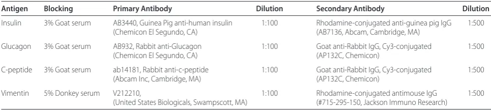
Cytostainings
A total of 4 x 10³ cells were cultured on glass cover slips and fixed in cold ethanol for 10 minutes. Histochemical staining for glycogen, dipeptidyl peptidase IV (DPPIV) and γglutamyl transpeptidase (GGT) was as previously described [9]. For immunostaining, cells were fixed in 4% paraformaldehyde, blocked in goat or donkey serum with 0.1% Triton X100 in PBS for 10 minutes, and incubated for one hour with antibodies at room temperature (Table 1). To measure intensity of immunostaining,
microphotographs were analyzed with ImageJ software (NIH, Bethesda, MD, USA).
Insulin radioimmunoassay
Cells were lysed for measuring insulin and cpeptide as previously described [4]. Data were normalized to cell numbers.
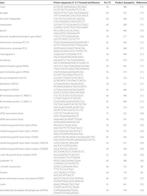
Reverse-transcription polymerase chain reaction (RT-PCR) Total RNA was extracted with Trizol Reagent (Invitrogen Life Technologies, Carlsbad, CA, USA). After incubation with Amplification Grade DNase I (Invitrogen), RNAs were reverse transcribed with Omniscript RT system (Qiagen Inc., Valencia, CA, USA). Equal amounts of cDNAs were amplified with Platinum PCR Supermix (Invitrogen) as follows: 94°C x 5 minutes and 35 cycles at 94°C x 30 sec, annealing at various temperatures for 30 sec, and 72°C for one minute, with final extension at 72°C for seven minutes. PCR products were resolved in 1.5% agarose containing ethidium bromide. Oligo nucleo tide primers, annealing conditions and expected product sizes are listed in Table 2, including previously published conditions [911, 1517].
Cell transplantation studies
The Animal Care and Use Committee of Albert Einstein College of Medicine approved animal use in conformity with National Research Council’s Guide for the Care and Use of Laboratory Animals (United States Public Health Service publication, revised 1996). All animals were maintained in the Institute of Animal Studies at Albert Einstein College of Medicine. We transplanted 2 x 10⁶ hTERTFPC modified by Pdx1 LV in NODCB17prkdc SCID mice (Jackson Labs., Bar Harbor, ME, USA) into portal vein as described previously [18]. Animals were sacrificed in groups (n = 3 each) 24 hours and one, two or four weeks after transplantation to analyze cell engraft ment and gene expression.To verify presence of trans planted cells in the liver,genomic DNA was isolated by Trizol Reagent. A primatespecific sequence from the CharcotMarieTooth (CMT)1A element was amplified by PCR and in situ hybridization was performed with a
Table 1. Immunostaining protocols
Antigen Blocking Primary Antibody Dilution Secondary Antibody Dilution
Insulin 3% Goat serum AB3440, Guinea Pig anti-human insulin 1:100 Rhodamine-conjugated anti-guinea pig IgG 1:500
(Chemicon El Segundo, CA) (AB7136, Abcam, Cambridge, MA)
Glucagon 3% Goat serum AB932, Rabbit anti-Glucagon 1:100 Goat anti-Rabbit IgG, Cy3-conjugated 1:500
(Chemicon El Segundo, CA) (AP132C, Chemicon)
C-peptide 3% Goat serum ab14181, Rabbit anti-c-peptide 1:100 Goat anti-Rabbit IgG, Cy3-conjugated 1:500
(Abcam Inc, Cambridge, MA) (AP132C, Chemicon)
Vimentin 5% Donkey serum V212210, 1:100 Rhodamine-conjugated antimouse IgG 1:500
(United States Biologicals, Swampscott, MA) (#715-295-150, Jackson Immuno Research)
Table 2. RT-PCR Primers
Gene Primer sequences (5’-3’): Forward and Reverse Tm (°C) Product (basepairs) References
Insulin GCTGCATCAGAAGAGGCCATCAGGC 58 380 [11]
GCGTCTAGTTGCAGTAGTTCTCCAG
Glucagon GAATTCATTGCTTGGCTGGTGAAAGGC 60 255 [11]
CATTTCAAACATCCCACGTGGCATGCA
Pancreatic Polypeptide CTGCTGCTGCTGTCCACCTGCGTG 60 206 [11]
CTCCGAGAAGGCCAGCGTGTCCTC
Somatostatin CGTCAGTTTCTGCAGAAGTCCCTGGCT 60 206 [11]
CCATAGCCGGGTTTGAGTTAGCAGATC
NeuroD ATCCCAACCCACCACCAACC 60 440 [15]
CAGCGGTGCCTGAGAAGATT
Pancreatic duodenal homeobox1 gene (Pdx1) CTGCCTTTCCCATGGATGAA 58 277 [15]
CGCTTCTTGTCCTCCTCCTTT
Prohormone convertase (PC1/3) TTGGCTGAAAGAGAACGGGATACATCT 65 456 [11]
ACTTCTTTGGTCATTGCTTTGGCGGTG
Prohormone convertase (PC2) GCATCAAGCACAGACCTACACTGG 60 308 [11]
GAGACACAAACCACCCTTCATCCTTC
Chromogranin-A CGGACAGTTCCATGAAGCTCTC 58 444 [11]
GAGTCAGGAGTAGGAGACAAGG
Glucokinase GACGAGTTCCTGCTGGAGTATGAC 65 523 [11]
GACTCGATGAAGGTGATCTCGCAGCTG
Paired homeobox gene4 (PAX4) CACCTCTCTGCCTGAGGACACGGTGAG 64 443 [11]
CTGCCTCATTCCAAGCCATACAGTAGTG
Paired homeobox gene 6 (PAX6) CAGTCACAGCGGAGTGAATCAGC 58 519 [11]
GCCATCTTGCGTAGGTTGCCCTG
Glucose transporter2 (GLUT2) GCCATCCTTCAGTCTCTGCTACTC 65 523 [11]
GCTATCATGCTCACATAACTCATCCA
Betacellulin2 (BETA2) CCTGAGCAGAACCAGGACATGCC 58 221 [11]
ATCAAAGGAAGGGCTGGTGCAATCA
Neurogenin3 (NGN3) ACTGAGCAAGCAGCGACGGAGTC 65 448 [11]
GCACCCACAGCCGAGCGACAGAC
NK homeobox protein 6.1 (NKX6 1) CTCCTCCTCGTCCTCGTCGTCGTC 60 699 [11]
CTTGACCTGACTCTCTGTCATC
NK homeobox protein 2.2 (NKX2 2) CGGACAATGACAAGGAGACCCCG 65 490 [11]
CGCTCACCAAGTCCACTGCTGCTGG
Islet1 (ISL1) GTGCGGAGTGTAATCAGTATTTGG 58 519 [11]
GTCATCTCTACCAGTTGCTCCTTC
GATA2 transcription factor CGTCTTCTTCAATCACCTCG 55 225 [10]
CGTCTTGGAGAAGGGGCTC
GATA6 transcription factor GAAGAAGCACATGATTTTGGAC 58 181 [10]
GATGGAAGGGAAGGGCCAG
Transforming growth factor alpha (TGFα) ATGGTCCCCTCGGCTGGA 58 297 [10]
GGCCTGCTTCTTCTGGCTGGCA
Transforming growth factor beta1 (TGFβ1) GCCCTGGACACCAACTATTGCT 58 161 [10]
AGGCTCCAAATGTAGGGGCAGG
Transforming growth factor beta2 (TGFβ2) GATTTCCATCTACAAGACCACGAGGGACTTGC 65 503 [10]
CAGCATCAGTTACATCGAAGGAGAGCCATTCG
Transforming growth factor beta1 receptor (TGFβ1R) CGTGCTGACATCTATGCAAT 54 251 [10]
AGCTGCTCCATTGGCATAC
Transforming growth factor beta2 receptor (TGFβ2R) TGCACATCGTCCTGTGGAC 58 784 [10]
GTCTCAAACTGCTCTGAAGTGTTC
Insulin-like growth factor receptor (IGFR) ACCCGGAGTACTTCAGCGCT 55 229 [10]
CACAGAAGCTTCGTTGAGAA
Cytokeratin-19 ATGGCCGAGCAGAACCGGAA 60 308 [16]
CCATGAGCCGCTGGTACTCC
α-Smooth muscle actin AGTACCCGATAGAACATGG 60 153 [9]
TTTTCTCCCGGTTGGC
Vimentin CACCTACAGCCTCTACG 60 170 [9]
AGCGGTCATTCAGCTC
Human telomerase reverse transcriptase (hTERT) AAGTTCCTGCACTGGCTGATGAG 60 378 [17]
TCGTAGTTGAGCACGCTGAACAG
β-actin AGAGCTATGAGCTGCCTGAC 55 361 [15]
CTGATCCACATCTGCTGGAA
Glyceraldehyde phosphate dehydrogenase (GAPDH) CCATGGAGAAGGCTGGGG 58 194 [10]
pancentromere probe to identify human cells as des cribed [19]. GFP immunostaining was performed as described [18]. In some studies, tissues were additionally stained for insulin as described above. After washing with PBS, tissues were counterstained with DAPI (4’,6diamidino2phenylindole)antifade (Molecular Probes, Invitrogen Life Technologies Corporation, Carlsbad, CA, USA) and examined under epifluorescence.
Statistical analysis
Data are shown as means ± SD. Significances were exam ined by ttest. P values <0.05 were considered significant.
Results
Characterization of pancreatic epithelial cells
After immunomagnetic sorting, we obtained 13 x 107 EpCAMpositive epithelial cells per fetal pancreas with viability of 85 to 90%. Intact fetal pancreas showed primitive acini, as well as islets with ductal and vascular structures (Figure 1a). Cells in ductal, periductal and acinar regions expressed the ductal markers, DPPIV and GGT. EpCAM staining verified the presence of epithelial cells with DPPIV and GGT expression in those areas. Similarly, EpCAMpositive isolated cells expressed DPPIV and GGT, whereas cells with glycogen, which is a characteristic of pancreatic acinar cells and not beta cells, were not observed in this fraction (Figure 1b). By
contrast, the EpCAMnegative cell fraction showed only infrequent EpCAM, DPPIV or GGTpositive cells, whereas cells with glycogen were frequent (Figure 1c).
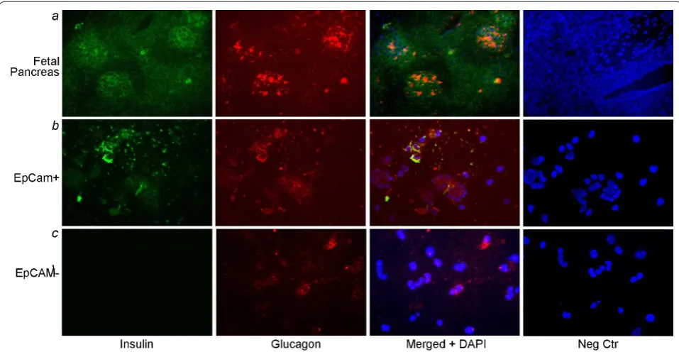
Cells in the EpCAMpositive fraction expressed insulin or glucagon, often both, whereas in the EpCAMnegative fraction, cells expressing insulin or glucagon were rare (Figure 2). Taken together, these findings indicated that EpCAM selected epithelial cells and that endocrine progenitor cells were present within epithelial cell niches in the fetal pancreas.
After culture, differences in EpCAMpositive and – negative fractions were apparent, as the former retained epithelial morphology, whereas the latter showed spindle shaped morphology (Figure 3a and 3b). In primary (P0) culture, EpCAMpositive cells proliferated within several days to form confluent monolayers of cells, although replication of cells declined over 10 to 12 passages, and cells required greater times to form confluent cultures. It was noteworthy that cells continued to express EpCAM, as shown by immunostaining, which simultaneously verified the absence of nonepithelial cells in cultures. Also, EpCAMpositive cells expressed pancreatic polypeptide (PP), somatostatin, glucagon and insulin in shortterm culture (1 to 2 d), whereas after longerterm culture (1014 d), PP was no longer expressed (Figure 3C). By contrast, in EpCAMnegative cells, insulin, glucagon and somatostatin were expressed at lower levels and only
in the shortterm, while PP was not expressed at all. Pdx1 was expressed in EpCAMpositive cells in short and longterm cultures. BETA2 and NeuroD transcription factors, which regulate βcell differentiation, were expressed in EpCAMpositive cells, as were transcriptional regula tors, GATA2, GATA4 and GATA6, which govern differentiation of additional mesodermal lineages. NGN3 was expressed in EpCAMpositive cells. Other regulatory transcription factors, for example, Pax4, Pax6, Nkx2.2 and Nkx6.1, were either not expressed, or were expressed less well in EpCAMpositive cells, although Isl1, which promotes islet cell development, was expressed during shortterm cell culture. The glucose trans porter, GLUT2, and glucokinase (GK), which partici pate in signalsecretion coupling in βcells, were expressed in only EpCAMpositive cells. These cells expressed proinsulin processing enzymes, prohormone convertases, PC1/3 and PC2, and chromogranin A (CGA), the major component of densecore secretory granules. Finally, growth factor genes regulating islet and βcell development, for example, TGFα, TGFβ1, TGF β2, and their receptors, as well as insulinlike growth factor1 receptor (IGF1R), were expressed.
Proliferation of EpCAM-positive cells after telomerase reconstitution
In mature pancreatic islets or in fetal pancreas, hTERT mRNA was absent (Figure 4a). After hTERT introduction,
RTPCR showed hTERT mRNA in transduced cells, similar to hTERTFHB fetal human liver cells [10]. Serial cultures of hTERTFPC demonstrated robust prolifera tion despite >50 passages, during which >170 estimated cell population doublings occurred, which was beyond the boun daries of replicative senescence in somatic cells. Comparison of cell doubling rates showed proliferation was significantly increased in FPC after expression of hTERT (Figure 4b). The human identity of hTERTFPC was verified by PCR of DNA with humanspecific CMT1A probe, as well as in situ hybridization for primatespecific alphoid satellite sequences in centro meres [19]. Morpho logically, hTERTFPC resembled unmanipulated primary EpCAMpositive fetal cells. To address whether hTERT FPC maintained endocrine functions, we deter mined insulin expression (Figure 4c). Primary EpCAMpositive fetal cells expressed insulin during culture over up to two weeks and insulin was expressed in cultured hTERTFPC after up to four passages (P4). Subsequently, insulin expression declined in hTERTFPC, and was undetectable after 10 passages. This was asso ciated with a parallel decline, and eventual loss, of Pdx1 expres sion in hTERT FPC, indicating that the βcell pheno type of hTERTFPC was perturbed during cell culture.
Restoration of endocrine phenotype in hTERT-FPC
We studied whether restoring Pdx1 expression will reconstitute the endocrine phenotype of hTERTFPC.
Therefore, we expressed rat Pdx1 and green fluorescent protein (GFP) under phosphogylcerate kinase (PGK) promoter, which is ubiquitously active (Figure 5a). After transduction of hTERTFPC with Pdx1 LV >90% cells were GFPpositive (Figure 5b and 5c). GFP expression was maintained over multiple cell passages. Rat Pdx1 induced glucagon and ISL1 expression in hTERTFPC, without expression of endogenous hPdx1 or insulin under basal conditions (Figure 5d). However, when hTERTFPC were cultured in serumfree medium with
activin A, hPdx1 and insulin mRNA were expressed and hTERTFPCPdx1 also contained immunostainable insulin and cpeptide. Neither hPdx1 nor insulin was detected in hTERTFPC without transduction with Pdx1 LV. No insulin was detected by immunoassay in un differ entiated hTERTFPC, whereas hTERTFPCPdx1 cells cultured without serum and with activin A contained 5 ± 3 ng insulin per 1 x 10⁶ cells. This indicated that Pdx1 restored endocrine progenitor phenotype in hTERTFPC, although activin A was required. Detailed studies of regulated insulin secretion were not attempted in view of relatively limited insulin expression in hTERTFPCPdx1 cells and requirement of further differentiation in cells.
Endodermal-mesenchymal phenotype conversions
To examine the phenotyperegulating mechanism of activin A, we determined whether hTERTFPC transi tioned to an epithelialmesenchymal state, which was recently recognized in endodermal cells [9]. We found that EpCAMpositive fetal pancreatic cells expressed the intermediate filament characteristic of epithelial cells, cytokeratin (CK)19, with minimal expression of the mesenchymal filament, vimentin (Figure 6a). By contrast, in Pdx1LVtransduced hTERTFPC, expression of vimentin increased, while CK19 was also expressed, indicating a mixed epithelialmesenchymal phenotype. After activin A, expression of vimentin declined without affecting CK19 expression. It was noteworthy that expression of TGFβ1, TGFβ2 and their receptors was unchanged. This was associated with changes in cell morphology, such that cultured hTERTFPC were larger and flatter in the presence of serum, whereas hTERT FPCPdx1 cells became smaller and rounder when cultured without serum and in the presence of activin A (Figure 6b). Moreover, these morphological changes were accompanied by decreased expression of vimentin, which was verified by immunostaining of cultured cells (Figure 6c).
Transplantation of hTERT-FPC cells in NOD/SCID mice The fate of hTERTFPC was established in vivo by transplantation studies in mice. Transplanted hTERT FPC were present in the liver throughout the fourweek period of studies (Figure 7a). Transplanted cells were localized in the liver by in situ hybridization (Figure 7b and 7f). Moreover, presence of GFP expression, as well as simultaneous expression of insulin in GFPpositive hTERTFPC that had been modified by Pdx1LV was observed (Figure 7g and 7i). No tumors were detected in animals after cell transplantation over the fourweek period of the study.
Discussion
We successfully isolated epithelial progenitor cells from the fetal human pancreas and isolated cells were greatly
expanded after genetic modification with hTERT in vitro. These cells exhibited endocrine progenitor phenotype with multiple pancreatic hormones, although this pheno type was altered in culture conditions with acquisition of additional mesenchymal/mesodermal properties. As this phenotype change occurred through spontaneous pro cesses, we believe the availability of hTERTmodified pan creatic cells will offer additional substrates for
dissect ing regulatory mechanisms in beta cell differen tiation. In particular, our findings should be of interest in determining whether other stem/progenitor cells will transition through similar fetal stages during generation of β or betalike cells.
During development, pancreatic progenitor cells arise from the primitive foregut endoderm, with cellspecifi cation requiring Pdx1 and other transcription factors, for
example, HNF homeobox B, NGN3, pancreasspecific transcription factor 1a, GATA4, and HNF6 [12]. We were able to isolate pancreatic epithelial progenitor cells characterized by the epithelialspecific adhesion molecule, EpCAM, which is a feature of endodermal stem/progenitor cells [8, 9], although these cells demonstrated limited proliferation capacity in culture conditions. The replication potential of fetal human liver stem/progenitor cells expressing EpCAM was similarly limited and was accompanied by telomere shortening, which could be circumvented by hTERT expression [10]. In the same way, modification of pancreatic progenitor cells with hTERT greatly expanded their capacity to proliferate. The ability of other somatic cells, and even iPS cells, to assume greater proliferation capacity after modification with hTERT has been established [6, 10]. This role of hTERT should be noteworthy because of the absence of tumorigenicity in hTERTmodified cells [10, 20], as shown previously, and again in our cell trans plantation studies here, where transplanted cells did not generate foci of proliferating cells and no tumors were observed over at least four weeks.
We do not propose that geneticallymodified cells with hTERT expression will be appropriate for immediate clinical applications, as more information will be needed in regards to their biological properties, regulation of differentiation through nonintegrating vectors in case of gene therapytype approaches or of alternative small
molecule approaches for this purpose, as recently identified during β lineage derivation in hESC [21]. The availability of fetal cells described here will permit comparative studies of differentiation mechanisms in pluripotent stem cells versus stem/progenitor cells with specific lineage commitment. Our preliminary studies indicated that ()indolactam V, which was effective for inducing beta cell maturation in hESCderived cells [21], failed to enhance beta cell phenotype in hTERTFPC, suggesting fundamental differences in regulation of beta cell differentiation in various types of stem/progenitor cells.
The loss of insulin expression in cultured FPC was in agreement with previous studies, for example, down regulation of hormone expression was a feature of immorta lized human pancreatic cell lines, as well as of growthstimulated primary human pancreatic cells [22, 23]. Whether loss of gene expression was due to transcriptional downregulation, independent of cell differentiation, or switching of progenitor cells to more primitive states, were both possibilities. In our studies, EpCAMpositive fetal epithelial cells displayed properties of islet progenitor cells with expression of multiple hormones and relevant transcription factors in early cell culture conditions. We found that loss of insulin expression was coupled with simultaneous loss of Pdx1 expression in hTERTFPC. Subsequently, we found that although this initial epithelial/endodermal phenotype
was maintained in cells, additional properties typically encountered in mesenchymal/mesodermal cells, for example, vimentin expression, were displayed. This was strongly reminiscent of changes in EpCAMpositive fetal human liver stem/progenitor cells, where downregulation of the epithelial/endodermal phenotype was due to the onset of conjoint mesoendoderm (epithelial plus mesen chymal) phenotype under culture conditions [9]. Also, these fetal human liver cells regained expression of epithelial genes after culture under serumfree condi tions, which strengthened sharing of mechanisms observed in fetal pancreatic cells here.
Therefore, our findings of increased insulin expression in hTERTFPC following culture under serumfree conditions with activin A were in agreement with the role
of pathwayspecific soluble signals in cell differentiation. The role of activin A in enhancing insulin expression in hTERTFPC resembled the role of inductive signaling in generating βlike cells from hESC [13], and of extrinsic signaling in inducing hepatic properties in fetal stem/ progenitor cells [9]. These shared mechanisms will offer new opportunities for exploring genetic and epigenetic processes in pancreatic lineage regulation, including induction of the beta cell phenotype. The availability of hTERTFPC and hTERTFHB cells derived from fetal liver and pancreas, respectively, should provide impor tant opportunities for comparing hESC or iPSderived cells in understanding transitions of embryonictofetal and fetaltoadult stages in the β cell lineage. For instance, it should be relevant to determine whether candidate
approaches capable of advancing β cell maturation in hESCderived cells, could be equally effective in hTERT FPC or hTERTFHB cells.
As hTERTFPC were capable of expressing insulin in vivo, this was similar to the expression of hepatic functions in fetal human liver epithelial cells following transplantation in xenotolerant mice [9]. In other studies, transplantation of hTERTFHB fetal liver cells modified by Pdx1, was successful in correcting hyperglycemia in mice [11, 24]. Here, our cell transplantation studies were limited to establishing differentiation of hTERTFPC in the liver. Whether these cells could correct hyperglycemia in animals will require detailed studies in the future, particularly after further differentiation and maturation of cells along the β lineage.
Conclusions
Insights into lineageswitching mechanisms during expan sion of pancreatic stem/progenitor cells will be critical for understanding the fundamental nature of expanded cells. The availability of hTERTFPC should facilitate efforts to identify effective mechanisms to maintain stem/progenitor cells under suitable states for cell expansion without extinction of differentiation capacity. This will be useful for translational applications, including cell therapy in type1 diabetes mellitus.
Abbreviations
BETA2 = betacellulin 2; BSA = bovine serum albumin; CaCl2 = calcium chloride;
cDNA = complementary deoxyribonucleic acid; CGA = chromogranin A;
CK = cytokeratin; CMT =Charcot-Marie-Tooth; DMEM = Dulbecco’s Minimal
Eagle Medium; Dnase = deoxyribosenuclease; DPPIV = dipeptidyl peptidase IV; EDTA = ethylenediaminetetraacetic acid; EpCAM = epithelial cell adhesion molecule; FBS = fetal bovine serum; FITC = fluorescein isothiocyanate; GATA = GATA family of transcription factors; GFP = green fluorescent protein; GGT = γ-glutamyltranspeptidase; GK = glucokinase; GLUT = glucose transporter; HEPES = 4-(2-hydroxyethyl)-1-piperazineethanesulfonic acid; hESC = human embryonic stem cells; HNF = hepatic nuclear factor; hTERT = human telomerase reverse transcriptase; hTERT-FPC = human telomerase reverse transcriptase containing fetal pancreatic cells; IGF-1R = insulin-like growth factor-1 receptor; iPS = inducible pluripotential stem cells; Isl = islet; KCl: = potassium chloride; LV = lentivirus vector; NaCl = sodium chloride;
NaH2PO4 = sodium dihydrogen phosphate; NGN = Neurogenin; Nkx = NK
homeobox protein; NOD-SCID = natural onset diabetes-severe combined immunodeficiency; Pax = paired homeobox gene; PBS = phosphate buffered saline; PC = prohormone convertase; pCCLsinPPT.hPGK.IRES.GFP.Wpre = third generation lentivirus plasmid; Pdx = pancreatic duodenal homeobox gene; PGK = phosphogylcerate kinase; pMD2.G = plasmid construct expressing vesicular stomatitis virus G envelope protein; pMDLg/pRRE = plasmid containing human immunodeficiency virus-derived packaging construct; PP = pancreatic polypeptide; pRSV-Rev = plasmid construct expressing Rev regulatory protein; RT-PCR = reverse-transcription polymerase chain reaction; TGF = transforming growth factor.
Competing interests
The authors declare that they have no competing interests.
Authors’ contributions
KC performed studies, interpreted data, and prepared the manuscript. AF performed studies, interpreted data, and reviewed the manuscript. MS performed studies, interpreted data, and reviewed the manuscript. NF interpreted data, reviewed and approved the manuscript. SG designed studies, interpreted data and prepared the manuscript.
Acknowledgements
This work was supported in part by NIH grants P01 DK52956, P20 GM075037, R01 DK46952, R01 DK071111, P30 DK41296 and P30 CA13330. Mark Zern, MD, University of California, Davis, provided hTERT retrovirus and E. Vigna contributed to the lentiviral vector.
Author details
1Hepatology Division, Department of Medicine, Albert Einstein College of
Medicine, Ullmann Bldg., Rm 625, 1300 Morris Park Avenue, Bronx, NY 10461,
USA. 2Department of Pathology, Albert Einstein College of Medicine, Ullmann
Bldg., Rm 625, 1300 Morris Park Avenue, Bronx, NY 10461, USA. 3Endocrinology
Division, Department of Medicine, Diabetes Research Center, Albert Einstein College of Medicine, Forchheimer Bldg., Rm 505, 1300 Morris Park Avenue,
Bronx, NY 10461, USA. 4Hepatology Division, Department of Medicine, Cancer
Research Center, Diabetes Research Center, Center for Human Embryonic Stem Cell Research, Marion Bessin Liver Research Center, Ruth L. and David S. Gottesman Institute for Stem Cell and Regenerative Medicine Research, and Institute for Clinical and Translational Research, Albert Einstein College of Medicine, Ullmann Bldg., Rm 625, 1300 Morris Park Avenue, Bronx, NY 10461, USA.
Received: 03 Jul 2009 Revised: 14 Sep 2009 Accepted: 15 Mar 2010 Published: 15 Mar 2010
References
1. Hao E, Tyrberg B, Itkin-Ansari P, Lakey JR, Geron I, Monosov EZ, Barcova M,
Mercola M, Levine F: Beta-cell differentiation from nonendocrine epithelial
cells of the adult human pancreas.Nat Med 2006, 12:310-316. 2. Xu X, D’Hoker J, Stangé G, Bonné S, De Leu N, Xiao X, Van de Casteele M,
Mellitzer G, Ling Z, Pipeleers D, Bouwens L, Scharfmann R, Gradwohl G,
Heimberg H: β Cells Can Be Generated from Endogenous Progenitors in
Injured Adult Mouse Pancreas.Cell 2008, 132:197-207.
3. Halvorsen TL, Beattie GM, Lopez AD, Hayek A, Levine F: Accelerated telomere
shortening and senescence in human pancreatic islet cells stimulated to divide in vitro.J Endocrinol 2000, 166:103-109.
4. Fleischer N, Chen C, Surana M, Leiser M, Rossetti L, Pralong W, Efrat S:
Functional analysis of a conditionally transformed pancreatic beta-cell line.Diabetes 1998, 47:1419-1425.
5. Wang S, Beattie GM, Mally MI, Cirulli V, Itlin-Ansari P, Lopez AD, Hayek A,
Levine F: Isolation and characterization of a cell line from the epithelial
cells of the human fetal pancreas.Cell Transplant 1997, 6:59-67 6. Park IH, Zhao R, West JA, Yabuuchi A, Huo H, Ince TA, Lerou PH, Lensch MW,
Daley GQ: Reprogramming of human somatic cells to pluripotency with
defined factors. Nature 2008, 451:141-146.
7. Zhou Q, Brown J, Kanarek A, Rajagopal J, Melton DA: In vivo reprogramming
of adult pancreatic exocrine cells to beta-cells.Nature 2008, 455:627-632. 8. Inada M, Benten D, Cheng K, Joseph B, Berishvili E, Badve S, Logdberg L,
Dabeva M, Gupta S: Stage-specific regulation of adhesion molecule
expression segregates epithelial stem/progenitor cells in fetal and adult human livers.Hepatol Int 2008, 2:50-62.
9. Inada M, Follenzi A, Cheng K, Surana M, Joseph B, Benten D, Bandi S, Qian H,
Gupta S: Phenotype reversion in fetal human liver epithelial cells identifies
the role of an intermediate meso-endodermal stage before hepatic maturation.J Cell Sci 2008, 121:1002-1013.
10. Wege H, Le HT, Chui MS, Liu L, Wu J, Giri RK, Malhi H, Sappal BS, Kumaran V,
Gupta S, Zern MA: Telomerase reconstitution immortalizes human fetal
hepatocytes without disrupting their differentiation potential.
Gastroenterology 2003, 124:432-444.
11. Zalzman M, Gupta S, Giri RK, Berkovich I, Sappal BS, Karnieli O, Zern MA,
Fleischer N, Efrat S: Reversal of hyperglycemia in mice using human
expandable insulin-producing cells differentiated from fetal liver progenitor cells.Proc Natl Acad Sci USA 2003, 100:7253-7258.
12. Zaret KS: Genetic programming of liver and pancreas progenitors: lessons
for stem-cell differentiation.Nat Rev Genet 2008, 9:329-340.
13. Shiraki N, Yoshida T, Araki K, Umezawa A, Higuchi Y, Goto H, Kume K, Kume S:
Guided differentiation of embryonic stem cells into Pdx1-expressing regional-specific definitive endoderm.Stem Cells 2008, 26:874-885.
14. Follenzi A, Ailles LE, Bakovic S, Geuna M, Naldini L: Gene transfer by lentiviral
vectors is limited by nuclear translocation and rescued by HIV-1 pol sequences.Nat Genet 2000, 25:217-222.
Pipeleers D, Heimberg H: Recapitulation of embryonic neuroendocrine differentiation in adult human pancreatic duct cells expressing neurogenin 3.J Cell Biol 2002, 159:303-312.
16. Schwartz RE, Reyes M, Koodie L, Jiang Y, Blackstad M, Lund T, Lenvik T,
Johnson S, Hu WS, Verfaillie CM: Multipotent adult progenitor cells from
bone marrow differentiate into functional hepatocyte-like cells.J Clin
Invest 2002, 109:1291-1302.
17. Piao CQ, Liu L, Zhao YL, Balajee AS, Suzuki M, Hei TK: Immortalization of
human small airway epithelial cells by ectopic expression of telomerase.
Carcinogenesis 2005, 26:725-731.
18. Benten D, Follenzi A, Bhargava KK, Kumaran V, Palestro CJ, Gupta S: Hepatic
targeting of transplanted liver sinusoidal endothelial cells in intact mice.
Hepatology 2005, 42:140-148.
19. Benten D, Cheng K, Gupta S: Identification of transplanted human cells in
animal tissues. Methods Mol Biol 2006, 326:189-201.
20. Jiang XR, Jimenez G, Chang E, Frolkis M, Kusler B, Sage M, Beeche M, Bodnar
AG, Wahl GM, Tlsty TD, Chiu CP: Telomerase expression in human somatic
cells does not induce changes associated with a transformed phenotype.
Nat Genet 1999, 21:111-114.
21. Chen S, Borowiak M, Fox JL, Maehr R, Osafune K, Davidow L, Lam K, Peng LF,
Schreiber SL, Rubin LL, Melton D. A small molecule that directs
differentiation of human ESCs into the pancreatic lineage. Nat Chem Biol
2009, 5:258-265.
22. Itkin-Ansari P, Demeterco C, Bossie S, Tour DD, Beattie GM, Movassat J,
Movassat J, Mally MI, Hayek A, Levine F: Pdx1 and cell-cell contact act in
synergy to promote δ-cell development in a human pancreatic endocrine precursor cell line.Mol Endocrinol 2000, 14:814-822.
23. Ramiya VK, Maraist M, Arfors KE, Schatz DA, Peck AB, Cornelius JG: Reversal of
insulin-dependent diabetes using islets generated in vitro from pancreatic stem cells.Nat Med 2000, 6:278-282.
24. Zalzman M, Anker-Kitai L, Efrat S: Differentiation of human liver-derived,
insulin-producing cells toward the beta-cell phenotype.Diabetes 2005,
54:2568-2575.
doi:10.1186/scrt6