RESEARCH ARTICLE
Micro differential scanning calorimetry
and micro hot table method for quantifying
deterioration of historical leather
Cristina Carsote
1†and Elena Badea
2,3*†Abstract
The aim of this study was to advance the current understanding on the mechanism of deterioration of historical vegetable tanned leathers and establish new criteria for quantifying their deterioration using micro differential scan-ning calorimetry (micro DSC) and micro hot table (MHT) method. Ten historical leather objects were investigated to this purpose. The calorimetric indices of macromolecular change identified for fibrous collagen provided quantitative results on the bulk material and deconvolution of DSC denaturation peaks revealed the dynamics of deterioration in historical leather. The results brought clear evidence that long-term natural ageing of leather induces the destabi-lization of chemically modified collagen thereby promoting its partial de-tanning and allowing micro-unfolding of chemically unmodified collagen. As deterioration continues toward the extreme, collagen molecules become highly unstable allowing for gelatinisation and irreversible denaturation. It is thus explained the coexistence of collagen pop-ulations with distinct thermal stability in historical leather. They are grouped in three main structural domains, namely “leather-like”, “parchment-like” and “gelatine-like”, whose mass percentages determines leather stability against further natural ageing and deterioration. As a result of the MHT and micro DSC parameters correlation, a more comprehen-sive set of criteria, including Tf and Tl values, as well ∆C and ∆T intervals’ lengths, was introduced for better interpreting the shrinking activity of collagen in historical vegetable-tanned leathers and open thus the way for the highly sought in situ evaluation of leather artefacts.
Keywords: Collagen matrix, Vegetable tannage, Deterioration mechanism, Thermal destabilisation, De-tanning, Gelatinisation, Denaturation, Shrinking behaviour, MHT method, Micro DSC
© The Author(s) 2019. This article is distributed under the terms of the Creative Commons Attribution 4.0 International License (http://creat iveco mmons .org/licen ses/by/4.0/), which permits unrestricted use, distribution, and reproduction in any medium, provided you give appropriate credit to the original author(s) and the source, provide a link to the Creative Commons license, and indicate if changes were made. The Creative Commons Public Domain Dedication waiver (http://creat iveco mmons .org/ publi cdoma in/zero/1.0/) applies to the data made available in this article, unless otherwise stated.
Introduction
Historical leathers, in a huge variety of items as foot-wear and garments, bookbinding, wall tapestry, uphol-stery, harnesses, armours, storage vessels, household tools, cases, musical instruments, toys, ritual objects, are important parts of our cultural heritage, from remote time to modernity. It is vital therefore that these objects remain well preserved along with all the knowledge involved, from their material aspects and value of use to historical, cultural and artistic values. However, analysis
and diagnosis of historical objects is especially challeng-ing owchalleng-ing to the fact they are unique and irreplaceable, which demands that analytical investigation must be non-invasive or, at the very least, employ microsamples. The quality of the information obtained (descriptive, explanatory, quantitative) is a critical factor when select-ing the investigatselect-ing technique. Ensurselect-ing the optimum ratio between the invasiveness of investigation and qual-ity of data provided is generally a difficult task, and in the case of leather artefacts the difficulty increases due to their damage non-uniformity which lead to complex composition (old leathers are complex materials consti-tuted of tanned collagen, untanned collagen and gelati-nised collagen) and structural heterogeneity.
Open Access
*Correspondence: [email protected]
†Cristina Carsote and Elena Badea contributed equally to this work 2 Faculty of Sciences, University of Craiova, Calea Bucuresti 107 I, 200512 Craiova, Romania
For rendering the raw animal skin, a more useful and durable material, i.e. non-putrescible under warm and moist conditions, vegetable extracts, fermented materi-als, fats or aluminium salts have been used since the end of Neolithic [1, 2]. Leather can thus be considered as the first biomaterial ever produced. The term “tanning” (tannage in French) was originally reserved only for the leather production with tannins [3, 4], while the term “tawing” was used to indicate the method of tanning with aluminium salts (alum, a double salt of aluminium and potassium phosphate, occurs naturally in many warm cli-mates, hence it’s early use as a tanning agent). The more versatile vegetable-tannage has continuously progressed to become the most used method to produce leather in Western and Mediterranean Europe until the end of the XIXth century [5, 6], when chromium mineral tanning came into use and gradually replaced vegetable tannins. Tannins derive their name from the Latin word tannum, which means “crushed oak bark,” since in early times oak trees served as a major source of tannin for the leather production. Tannins are a class of complex biomolecules of polyphenolic nature synthesized by a large variety of plants that have the property of combining with proteins, cellulose, gelatin, and pectin to form an insoluble com-plex [7]. Depending on the main polyphenolic constitu-ent, they are classified in condensed tannins, also named proanthocyanidins, and hydrolysable tannins, which comprise two subclasses, gallotannins and ellagitannins. Condensed tannins are oligomeric or polymeric flavo-noids whereas hydrolysable tannins consist of a polymer containing a polyol core (d-glucose is the commonest) multi-esterified with gallic acid (gallotannins) or its oxi-dized derivative, ellagic acid (ellagitannins) [8]. Today, the terms tanning and tannage are applied for all differ-ent processes of leather making which include vegetable tanning with plant polyphenols, mineral tanning with metal salts, oil and aldehyde tannages, synthetic tanning agents and organic tannages based on natural polyphe-nols or synthetic organic oligomers [9].
The chemical nature of collagen, the main constituent of animal skin, allows it to react with a variety of tanning agents, resulting in skin conversion to leather, a much more resistant material whose appearance and proper-ties enables a wide range of uses. De facto, the purpose of tannage is primarily to increase the hydrothermal sta-bility of native collagen, secondarily to increase its bio-logical inertness, and finally, to improve the utility of the hide’s physical properties. The hydrothermal stability can be measured by observing the temperature at which a thoroughly wetted leather specimen experiences shrink-age, when it is heated in water (or glycerine-water solu-tion when the shrinkage temperature is above 98 °C) at a rate of 2 °C per minute (Standard Test Method for
Shrinkage Temperature of Leather, ASTM D6076-18). This temperature is called shrinkage temperature, Ts. The
collagen fibres shrinking process occurs as a result of the hydrothermal denaturation of the collagen molecules. Even though the tanning reaction is highly complex on the molecular level, to the extent that there is no clear model of its effect on hydrothermal stability (i.e. what causes the Ts to rise), the value of the shrinkage
tempera-ture of leather is commonly used as an indicator of the type of tannage or degree of tannage, or both. As dena-turation causes collagen to shrink, which is a convenient gross metric, shrinkage has long been used as a measure of heating-induced damage to collagen and collagen-ous tissues [10, 11] and more recently it is considered a fine measure of the deterioration of ancient leathers [12,
13]. A micro-destructive method called Micro Hot Table (MHT) method that requires a few micro-fibres only and can be easily used by a conservator was set up by Larsen et al. [14] for measuring the hydrothermal stability and hence damage level of historical leather. This method has widely spread in the conservation laboratories and the knowledge gained so far allows for a partly quanti-tative assessment of deterioration at the fibre level. The MHT method is thus currently used as a routine method to measure Ts and rank the damage level in ancient
col-lagen-based materials such as leather and parchment [15–19]. It has been improved by incorporating an algo-rithm called imageMHT that allows the automation of the shrinkage intervals determination, with a significant effect on reducing the analysis time, accuracy increas-ing and inter-laboratory comparison [20]. Yet, MHT assessment is based only on intensive parameters (i.e. not depending on the sample mass) such as temperature which strongly limits the quantitative nature of dam-age evaluation. It was shown that the high heterogeneity of composition of historical collagenous materials can lead to the overestimation of the “health” condition of a material if only shrinkage temperature measurement is performed [21]. Besides, we must be aware that leather deterioration generally proceeds from the surface to inner layers and can vary from macroscopic to molecular level. Consequently, a comprehensive and reliable dam-age assessment requires that surface analysis be comple-mented with bulk analysis, and descriptive information be correlated with explanatory and quantitative informa-tion. It is therefore highly advisable that MHT method be always accompanied by another analysis able to provide complementary information on specific alterations of collagen at a different structural level and thus prevent partial or even improper evaluations [22–24].
information on bulk leather [16, 18]. Although DSC is a powerful quantitative technique which provides a measure of protein (i.e. collagen) hydrothermal stability and an indication of its long-term stability [25–27], no quantitative metrics of heat-induced collagen denatura-tion such as changes in the enthalpy or DSC peak asym-metry have been used so far for assessing damage in historical leather [28, 29]. Badea et al. [30, 31] reported a quantitative assessment of damage in ancient parch-ments based on the calorimetric parameters associate to collagen denaturation using micro-samples of only 1–2 mg. We have recently used micro DSC to reveal the accelerated ageing induced-deterioration pattern of collagen in vegetable tanned leather and quantify dam-age in leather after dehydrothermal treatment and vis-ible light exposure [32].
Based on these considerations, in the present study we used both micro DSC technique and MHT method to characterise the deterioration pattern in historical leather at both fibre (shrinkage process) and fibrillar (denaturation process) levels and find quantitative rela-tionships between denaturation/shrinking and dete-rioration. To this end, a comparison with the outcomes of previous studies on artificially aged and historical parchment and leather [21, 30, 32–35] was made. Our results provide clear evidence that long-term natural ageing (i.e. exposure to natural environmental fluctua-tion of temperature, relative humidity and light as well as to the more recent chemical pollutants, and exten-sive use/manipulation) of leather destabilises tanned collagen thereby promoting partial de-tanning and allowing micro-unfolding of chemically unmodified collagen, and thus progressively lead to its gelatinisa-tion and irreversible denaturagelatinisa-tion. A further aim of this work was to compare the results obtained by micro DSC with the results obtained by MHT method a to set up a more comprehensive set of criteria for interpreting
the shrinkage activity of collagen in historical vegetable tanned leathers.
Materials and methods Historical leathers
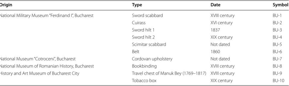
The investigated leather-made objects owned by some Romanian museums are listed in Table 1. All investi-gated historical leathers appear heavily damaged with dry, shrunk and brittle looking, frequent cracking and spotting. Micro-samples were taken from each historical object, generally from the most fragile areas, inherently exposed to material loss during normal use (i.e. edge, bookbinding spine, etc.).
New vegetable tanned leathers from calf, sheep and goat hides were used for comparison. They were prepared according to procedures inspired by traditional reci-pes at the National Research and Development Institute for Textile and Leather, Division Leather and Footwear Research, Bucharest (Romanian patent no.122098/2006:
Piele naturala pentru legatorie carte de patrimoniu si procedeu de realizare a acesteia). Mimosa bark (con-densed tannin) and chestnut wood (hydrolysable tannin) extracts were selected for tanning because both extracts were widely used in the European post-medieval period (the dating period of the investigated historical samples), and remained even nowadays among the mostly used vegetable tanning agents [36]. Chestnut tannin belongs to the ellagitannins [37], while mimosa is a prorobineti-nidin polyphenol [38]. Samples were taken from the butt, where fibres are tightly packed making it the strongest part of the skin/leather.
Micro differential scanning calorimetry
Collagen molecules denaturation is a kinetic process [39] and, as such, it must be treated thermodynamically. Micro DSC measurements were carried out with a high-sensitivity SETARAM Micro-DSC III calorimeter using
Table 1 List of historical leather objects
Origin Type Date Symbol
National Military Museum “Ferdinand I”, Bucharest Sword scabbard XVIII century BU-1
Cuirass XVI century BU-2
Sword hilt 1 1837 BU-3
Sword hilt 2 XIX century BU-4
Scimitar scabbard Not dated BU-5
Belt 1860 BU-6
National Museum “Cotroceni”, Bucharest Cordovan upholstery Not dated BU-7 National Museum of Romanian History, Bucharest Bookbinding XVIII century BU-8 History and Art Museum of Bucharest City Travel chest of Manuk Bey (1769–1817) XVIII century BU-9
850 µl stainless steel (Hastelloy C) sample cells. Meas-urements were performed in the temperature range (5 to 95) °C at 0.5 K min−1 heating rate. Micro-samples of
about 2.0 mg were suspended in 0.5 M acetate buffer (pH = 5.0) directly in the measure cell and left for 2 h at 5 °C to assure fully hydration [40] and thus reproducible calorimetric values since it is known that both denatura-tion temperature and denaturadenatura-tion enthalpy depend on hydration level. It was reported [18] that leather’s thermal characteristics are stable in the pH range 3 to 5. We have previously found that at 5 °C the values of calorimetric parameters of leather thermal denaturation practically remained unchanged for immersion times longer than 2 h indicating that the fibrils had reached their maxi-mum extent in hydration [32]. Depending on the avail-ability, each micro-sample was divided into few very tiny sub-samples and multiple measurements were performed with fresh samples.
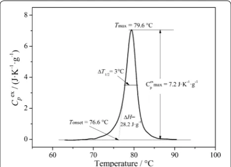
Experimental DSC data acquired with the SETA-RAM SetSoft2000 software were analysed using Peak-Fit 4.1 (Jandel Scientific) to obtain the specific heat capacity values of the sample Cpex(T) (J K−1 g−1) in the scanned temperature interval and derive the DSC peak parameters featuring collagen fibrils denaturation (Fig. 1). Denaturation temperature, Tmax, was
deter-mined as the temperature attained at peak maximum. Temperature span of the transition was reported as peak width at half height, ΔT1/2, and specific
denatura-tion enthalpy, ΔH was calculated as the area under the
peak by integrating Cpex(T) curve across the denatura-tion temperature range [26, 32]. DSC multiple peaks of historical leather were deconvoluted using the PeakFit asymmetric Gaussian fit function to improve the fit of the asymmetry in the peaks.
It is worth noting that the DSC measurements pro-vide quantitative information on bulk leather whereas the non-invasive investigation techniques are qualita-tive and mainly relate to the surface of the sample. DSC micro invasiveness could, however, constitute a critical factor for some historical objects/artefacts even if it requires only a minimal amount of sample.
Micro hot table method
The shrinking measurements were carried out using a Linkam LTS120 stage equipped with a temperature controller and Linksys32 temperature control software which enables full PC programming of temperature. The water circulator ensures a temperature stability and accuracy to 0.1 °C and rapid cooling. Shrinkage motion was digitally recorded with a SMZ 745 Nikon stereomicroscope equipped with a Nikon D90 digital camera. This process can be viewed either through the microscope, or on the PC using the camera and image capture and processing software [41]. Of each sub-sam-ple used for micro DSC measurements, few fibres (0.1– 0.2 mg) were taken and placed on a microscope slide with a concavity, completely immersed in demineral-ised and degassed water to ensure thoroughly wetting. Then, the fibre bundles were well separated using fine needles and the microscope slide, covered with a cover glass, placed on the Peltier element of the stage and heated at a rate of 2 °C min−1 in the range (25–95) °C.
All measurements were performed three times for each sample and the average values are reported with the standard errors.
Collagen fibres shrinkage activity associated with thermal denaturation is described by a sequence of temperature intervals: no activity—A1–B1–C–B2– A2—complete shrinkage as previously reported [14]. In the first two intervals, A1 and B1, shrinkage discretely occurs in individual fibres and displays higher activity (namely higher amount of shrinkage per unit of time) in B1 interval. Most of the fibre mass shrinks in the main interval ΔC. The starting temperature of this interval is the shrinkage temperature, Ts. Generally, the
shrink-age activity levels off through B2 and A2 intervals. Tf
is defined as the temperature at which the very first motion is observed while Tl is the temperature of the
very last observed motion. The total shrinkage interval is thus calculated as ΔT=Tl−Tf.
Results and discussion
Hydrothermal stability of collagen in leather
Collagen type I (the main constituent of skin) is a hier-archically organised protein characterised by an intimate relationship and connectivity between individual struc-tural levels: molecules, fibrils and fibres [42–44]. Colla-gen fibres are composed of fibrils, which in turn are made up of collagen molecules. The collagen molecule is made of three polypeptide chains, called α-chains, which form a unique triple helix structure which is around 1.4 nm in diameter and 300 nm in length. The primary amino acid sequence of collagen is glycine-proline-X or gly-cine-X-hydroxyproline, where X can be any of the other 17 amino acids, and every third amino acid is glycine. Hydroxyproline stabilizes the triple helix via a stereoelec-tronic effect and enables the formation of water-medi-ated hydrogen bonds that stitch together the folded triple helix. The triple helix configuration is 3 left-handed heli-ces twisted into a right-handed coil. The chemical nature of the side chains attached to the methylene group in the centre of each amino acid residue, i.e. ε-amino group of lysine residues, carboxyl groups of glutamic and aspartic acids, or hydroxyl groups of hydroxylysine and hydroxy-proline, ensures intermolecular and interfibrillar cross-linking of collagen with a variety of agents [42] having tanning properties. In addition, the backbone peptide bonds offer different reactions sites that can be exploited during tanning process. The nature and stability of these different chemical bonds produce a significant variation in the properties of the cross-linked product, i.e. appear-ance, biological, mechanical and thermal properties. One of the most important changes induced by tanning is in fact the increased hydrothermal stability of collagen. This would be due to the increase of packing density of colla-gen by increasing the density of crosslinking.
According to the latest theory of tanning based on the concept of link-lock [45, 46], all tanning processes are either single- or multi-component. The reaction between collagen and vegetable tannins is a single component reaction termed “linking” because the tanning agent only links to the collagen structure. It confers the same degree of moderate hydrothermal stability (Ts < 85 °C)
independently of the tannin type because the bound species merely interfere with the shrinking/denatura-tion process. For example, the primary link reacshrinking/denatura-tion between hydrolysable plant polyphenols and collagen occurs through multiple hydrogen bonding [9]. Similarly, the condensed tannins react with collagen via hydrogen bonds. In addition, they can react via quinoid species, resulting in covalent bonding at the collagen lysine amino group [45]. Multi-component processes additionally involve crosslinking the primary reagent and “locking” the molecules together to create a stable matrix within
the collagen structure. This step results in an increase of the shrinkage/denaturation temperature since the colla-gen-tannin matrix becomes less easily displaced. Further stabilisation of collagen within leather involves the sub-stitution of some of the supramolecular water (i.e. dehy-dration) and the interaction with some of the remaining water [46].
It is important to underline that, regardless of the tan-ning mechanism, the denaturation/shrinking process involves the unravelling of the triple helix aided by the disruption of a network of hydrogen-bonded water mol-ecules surrounding the collagen molecule as elegantly explained by Bella et al. [47]. When present as isolated monomers in solution, the triple helix of type I collagen becomes unstable near body temperature, causing mol-ecules to convert into random coils [48]. When packed in a fibril, the proximity of neighbouring molecules restricts the conformational freedom of individual α-chains, sub-stantially increasing the thermal stability of collagen mol-ecules [49, 50]. This mode of stabilisation is known as polymer-in-a-box constraint [51]. Taken together, poly-mer-in-a-box molecular stabilization combined with the tanning-induced stabilisation of collagen matrix provides adequate explanation for the increased stability of colla-gen fibrils in leather.
Micro DSC evidences of new leather hydrothermal stability The parameters of the denaturation process represent a measure of the collagen matrix stability while at the same time provide a measure of the distribution of molecular thermal stabilities [31–33]. Figure 1 illustrates the typi-cal sharp endotherm related to the denaturing process of collagen in mimosa-tanned goat leather. This transition of collagen macromolecules from their native triple heli-cal structure into a random coiled structure via unfold-ing (helix-to-coil transition) is revealed by micro DSC at a defined temperature, Tmax, which has been assumed as
denaturation temperature of collagen fibrils according to a statistical process [10]. Besides the position (Tmax)
of the DSC peak, its height (Cpex,max) , width (∆T1/2), area (∆H) and symmetry provide valuable information about the denaturation process [52]. In addition, the onset tem-perature Tonset of the transition, calculated by using the
tangent method, is explained by conformational changes occurring within the fibrils before the main denaturation transition Tmax, such as partial shrinkage of the fibrils.
This shrinkage phenomenon was also reported by Lin et al. [53], using a second harmonic generation micros-copy approach, and by Bozec and Odlyha [54], using localized thermomechanical analysis. We can there-fore assume that Tonset and Tmax correspond at
macro-scopic level to Tf, and Ts, respectively. This is confirmed
leathers and 48 artificially aged samples [32] by the good linear correlations obtained when Tmax was plotted
ver-sus Ts (R = 0.961) and Tonset versus Tf (R = 0.908) (Fig. 2a,
b).
The experimentally derived enthalpy of denaturation ∆H is the area under the peak and is a measure of the amount of energy required for collagen denaturation, i.e. to disrupt the interactions stabilizing the triple helix. The peak width at half height ΔT1/2 is the full width at half
maximum of the endothermic peak and gives a measure of the breadth of the distribution of molecular thermal stabilities.
The calorimetric parameters associated with collagen denaturation in newly obtained vegetable tanned leather are reported in Table 2 together with the correspond-ing calorimetric parameters of collagen denaturation in parchment. As expected, Tmax in leather clearly depends
on the tannin type and show higher values than that of collagen in parchment. Higher values of Tmax (73–80 °C)
result from collagen reaction with mimosa condensed tannin than from its reaction with chestnut hydrolys-able tannin (66–72 °C), while chemically non-modified collagen in parchments undergoes denaturation at even lower Tmax (55–60 °C). Unlike denaturation
tempera-ture, the specific enthalpy (calculated by dividing the total enthalpy by the sample mass) of the new vegetable tanned leathers seems to not significantly depend on the tannin type [32]. However, the denaturation enthalpy of collagen in vegetable tanned leather is much lower than that of parchment [26, 27] and chrome-tanned leather [55, 56]. One reason for this low enthalpic value is the much higher mass percentage of non-collagenous mate-rial in vegetable tanned leather compared to parchment and chrome-tanned leather. On the other hand, an influ-ence of the tanning mechanism is expected, since colla-gen changes its conformation and hydrothermal stability as a result of the specific interactions with tannin. It is only after the complex tannin–collagen breakdown that the denaturation process is controlled by the breaking of hydrogen bonds within the triple helix [44]. In addition, it is worth mentioning that the few enthalpy data from the literature reported by Chahine [18] and Budrugeac et al. [19] for vegetable tanned leather are very scattered and even lower than those measured by us. This can be ascribed, on one hand, to leather high heterogeneity (i.e. non-uniformity in tannage) induced by the fabrication technology and, on the other hand, to the experimental conditions which resulted in various hydration levels. It is known that both Tmax and ∆H depends on the
hydra-tion level [40]. So, it is very likely that the very long swell-ing time (e.g. 12–24 h) used by Chahine and Budrugeac is the cause of altered DSC denaturation parameters [27]. Consequently, long swelling times, are not recommended for historical damaged leather which are very sensitive to excessive moist.
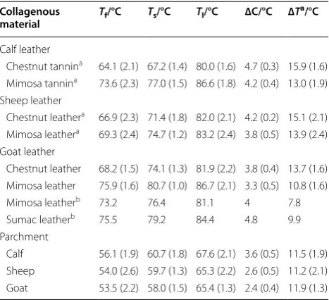
MHT evidences of new leather hydrothermal stability The parameters associated with collagen fibres shrink-ing in vegetable tanned leathers are reported in Table 3
together with the corresponding shrinkage parameters of collagen fibres in parchment. It is apparent from these data the distinct shrinkage behaviour of vegetable tanned leather depending on the tannin type and animal spe-cies. Mimosa tanned leathers show higher Ts values, as
expected, but shorter ΔC and ΔT intervals by compari-son with chestnut tanned leathers. This could be attrib-uted to the further covalent interaction of mimosa tannin with collagen compared to chestnut tannin as explained before. Ts values for the investigated new leathers listed
in Table 3 well compare with values reported by Larsen et al. [14] as well as with those typically observed for collagen stabilisation by polyphenol reactions (e.g.
gallotannin or ellagitannin reaction generally raises Ts to
75–80 °C, whereas flavonoids raise Ts to 80–85 °C). Our
slightly higher ΔT intervals may be attributed to the tan-ning technology.
Deterioration mechanism in historical leather. Quantification of deterioration
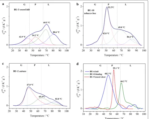
Some representative DSC curves associated with hydro-thermal denaturation of collagen in historical leather are presented in Fig. 3. The multiple character of the curves 3a, 3b and 3c is well resolved and multiple peaks have been singled out by appropriate deconvolution. Such a thermal behaviour indicates that each dena-turation endotherm contains a distribution of collagen molecules with distinct thermal stabilities. Consider-ing that the thermal stabilisation of leather is due to the linking reaction between elements of collagen structure and tannin molecules [46], one can infer that on age-ing this linkage-ing capability gradually decreases until total de-tanning when leather thermal stability equals that of chemically unmodified collagen in parchment. Fur-ther deterioration results in collagen gelatinisation and irreversible denaturation, i.e. the conversion of triple helix into random coils. We can therefore assume that the ageing/deterioration mechanism of leather includes the following key-steps: thermal destabilisation of col-lagen–tannin complex (chemically modified colla-gen), de-tanning, thermal destabilisation of chemically
Table 2 Parameters of thermal denaturation of collagen in new vegetable tanned leathers and new parchment
Literature data for new vegetable leathers are also reported
Calf, sheep and goat leather and parchment were manufactured at INCDTP-ICPI, Bucharest. Uncertainty is the standard deviation. At least three measurements with fresh samples were performed for each leather/parchment type
Literature data [15, 16] were obtained by performing DSC measurements at the rate of 10 °C min−1 a Ref. [29]; bRef. [15]; cRef. [16]
Collagenous material Tonset/°C Tmax/°C ∆H/J g−1 ∆T1/2/°C Cpex,max/J K−1 g−1
Calf leather
Chestnut tannina 63.1 (2.1) 66.3 (0.9) 27.7 (2.2) 3.2 (0.1) 6.7 (0.2) Mimosa tannina 70.8 (1.0) 75.1 (1.1) 29.5 (2.4) 4.4 (0.2) 5.1 (0.2) Sheep leather
Chestnut tannina 66.5 (1.1) 69.1 (1.3) 26.8 (2.2) 2.4 (0.2) 8.6 (0.4) Mimosa tannina 70.5 (1.0) 73.6 (1.1) 29.6 (2.4) 3.4 (0.3) 7.3 (0.2) Goat leather
Chestnut tannin 66.8 (0.5) 71.7 (1.1) 26.5 (2.5) 2.8 (0.2) 6.9 (0.2) Mimosa tannin 76.6 (0.3) 79.6 (1.4) 28.2 (1.5) 2.9 (0.3) 7.2 (0.2)
Vegetable leather 88.0b 90.0b 21.8b – –
76.3c 80.1c 11.5c – –
Parchment
Calf 53.1 (1.3) 55.8 (1.8) 53.4 (5.0) 5.4 (1.2) 5.8 (1.0)
Sheep 55.5 (0.6) 57.4 (1.0) 46.4 (2.4) 4.6 (0.4) 5.8 (0.5)
Goat 55.1 (0.7) 57.6 (1.1) 47.5 (3.1) 3.6 (0.3) 5.6 (0.4)
Table 3 Shrinkage temperatures and intervals of collagen fibres from new vegetable tanned leathers and new parchment measured by MHT method with the aid of imageMHT software for image capture and processing
Literature data for new vegetable leathers are also reported
Uncertainty is the standard deviation. For each tannin type, three leathers obtained from different hides were analysed. For each leather, three measurements with fresh samples were performed
a Ref. [19]; bRef. [11] Collagenous
material Tf/°C Ts/°C Tl/°C ∆C/°C ∆T
a/°C
Calf leather
Chestnut tannina 64.1 (2.1) 67.2 (1.4) 80.0 (1.6) 4.7 (0.3) 15.9 (1.6) Mimosa tannina 73.6 (2.3) 77.0 (1.5) 86.6 (1.8) 4.2 (0.4) 13.0 (1.9) Sheep leather
Chestnut leathera 66.9 (2.3) 71.4 (1.8) 82.0 (2.1) 4.2 (0.2) 15.1 (2.1) Mimosa leathera 69.3 (2.4) 74.7 (1.2) 83.2 (2.4) 3.8 (0.5) 13.9 (2.4) Goat leather
Chestnut leather 68.2 (1.5) 74.1 (1.3) 81.9 (2.2) 3.8 (0.4) 13.7 (1.6) Mimosa leather 75.9 (1.6) 80.7 (1.0) 86.7 (2.1) 3.3 (0.5) 10.8 (1.6) Mimosa leatherb 73.2 76.4 81.1 4 7.8 Sumac leatherb 75.5 79.2 84.4 4.8 9.9 Parchment
unmodified collagen, gelatinisation and denaturation. This assumption is based on our findings concerning the behaviour of both artificially aged leather [32] and parchment [27]. First, thermally destabilised intermedi-ate stintermedi-ates of chemically modified collagen are formed through a progressive impairment of collagen–tannin binding. This allows conformational changes and pro-gressive de-tanning and is expressed by the lowering of denaturation temperature. When the breakdown of the collagen–tannin bonding exceeds a hypothetical criti-cal level, full de-tanning occurs. Then, leather exhibits a parchment-like hydrothermal behaviour as we previ-ously reported [32, 57]. Thermally destabilised interme-diate states of chemically unmodified collagen are formed through peptide bonds cleavage without disorganising
the triple helical structure—this is expressed by further lowering of denaturation temperature [32]. Once enough covalent bonds have been cleaved beyond a hypothetical critical level, shortened peptide segments occur, which contain less than a critical number of hydrogen bonds needed to maintain the coiled-coil, helical structure. As deterioration continues toward the extreme, the stabil-ity of some local areas drops below a lower limit between neighbouring crosslinks. At this point, the collagen mol-ecules become highly unstable allowing for gelatinisation and irreversible denaturation. Once the leather structure contains both chemically modified and unmodified colla-gen, all the steps may occur simultaneously and severely deteriorated collagen progressively accumulates in the leather material. For example, in Fig. 3b we can observe
the coexistence of multiple collagen populations: chemi-cally modified collagen with distinct linking capability showing Tmax at 84.4 °C and 69 °C, chemically
unmodi-fied collagen showing Tmax at 52.2 °C and gelatinised
col-lagen having Tmax at 42.8 °C. Based on these findings we
can characterise the pattern of deterioration of historical leather by simply dividing the temperature range of ther-mal transitions of collagen in three intervals correspond-ing to the followcorrespond-ing structural domains:
• “Leather-like” (L) domain (65 °C < T < 85 °C), where chemically modified collagen shows denaturation; • “Parchment-like” (P) domain (45 °C < T < 65 °C),
where chemically unmodified collagen (fully de-tanned) shows denaturation;
• “Gelatine-like” (G) domain (T≤ 45 °C), where gelati-nised collagen shows thermal transition.
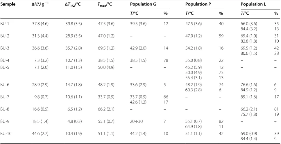
The multiple thermal transitions of the various colla-gen populations identified by deconvolution can be thus allocated to one of these domains depending on the Tmax
value of each DSC peak component. This grouping ena-bles us to also quantify the chemically modified collagen (tanned collagen), chemically unmodified collagen (fully de-tanned collagen) and gelatinised collagen within his-torical leather based on the percentage of enthalpy of
each population (Table 4). It is of outmost importance for conservators to get this information because gelatine-like population may spontaneously denature at room temper-ature when in the presence of enough moisture content, while leather-like and parchment-like collagen popula-tions are stable and can withstand seasonal temperature and humidity excursions.
Additional information on deterioration comes from the half-width of the overall DSC peak, ∆T1/2 listed
in Table 4, too. It is apparent from these data and from Fig. 3a–d that the shape of the overall DSC peak of his-torical leathers reflects their structural heterogeneity. Very large ∆T1/2 values indicate a distribution of
colla-gen populations in all structural domains whereas very low ∆T1/2 values indicate one major collagen population
belonging to a specific structural domain.
It is interesting to note that overall specific enthalpy of collagen denaturation in historical leathers show both lower and higher values than that we measured for collagen in the new vegetable tanned leathers (Table 1). First, it should be stressed that modern tan-ning technologies use commercial tannins and numer-ous chemical auxiliaries that greatly accelerate the tanning process, thus reducing the traditional tanning time from 1 to 2 years to 2 to 3 weeks or even a few days. For this reason, there are still no valid references
Table 4 Thermal stability and proportions of the various collagen populations in historical leathers obtained by deconvolution of the full DSC peak
The overall peak temperature, enthalpy and peak half-width are also reported
Columns 2 to 4: data refer to the full DSC peaks. Columns 5 to 10: data refer to the deconvoluted DSC peaks. Uncertainty is the standard deviation
Sample ∆H/J g−1 ∆T
1/2/°C Tmax/°C Population G Population P Population L
T/°C % T/°C % T/°C %
BU-1 37.8 (4.6) 39.8 (3.5) 47.5 (3.6) 39.5 (3.6) 12 47.5 (3.6) 40 66.0 (3.6)
84.4 (3.2) 3513 BU-2 31.3 (4.4) 28.9 (3.5) 47.0 (1.2) – – 47.0 (1.2) 59 65.4 (1.0)
82.8 (1.8) 3110 BU-3 36.6 (3.6) 35.7 (2.8) 69.5 (1.2) 42.9 (2.0) 14 54.2 (1.8) 16 69.5 (1.2)
80.6 (1.5) 4228
BU-4 7.3 (3.2) 10.7 (1.3) 38.5 (1.5) 38.5 (1.5) 78 55.0 (0.8) 22 – –
BU-5 7.1 (2.0) 11.0 (1.5) 50.0 (4.9) – – 45.2 (5.9) 50.0 (4.9) 55.4 (3.1)
12 75 13
– –
BU-6 28.9 (2.9) 14.7 (1.8) 48.2 (1.9) 33.6 (2.9) 5 48.2 (1.9)
60.3 (2.8) 746 76.6 (1.6)84.9 (1.2) 69 BU-7 9.8 (0.7) 10.6 (1.1) 33.7 (0.9) 33.7 (0.9)
42.6 (1.2) 6617 – – 85.1 (1.6) 17
BU-8 16.6 (0.5) 6.5 (1.2) 66.2 (2.1) – – – – 66.2 (2.1)
75.7 (1.8) 8119 BU-9 18.5 (1.4) 4.8 (0.3) 55.1 (0.7) 20÷30 7 55.1 (0.7)
64.9 (1.8) 8211 – –
BU-10 44.6 (2.7) 10.4 (1.9) 51.1 (1.1) 44.2 (1.4) 10 51.1 (1.1) 42 69.0 (0.9)
for historical vegetable tanned leathers and hence nei-ther for their calorimetric indices such as enthalpy and temperature of denaturation. The lower ∆H values showed by the historical leathers can undoubtedly be attributed to the collagen content decrease over time as a result of its irreversible denaturation induced by deterioration. In fact, severely deteriorated col-lagen that does not contribute to the denaturational change in enthalpy accumulates in the leather over time, resulting in a decrease of the overall denatura-tion enthalpy. Although the higher ∆H values showed by the historical leathers may seem surprisingly at first sight, it is very plausible that the initial enthalpy of leather before the damage occurred was higher than that currently measured by us for the modern veg-etable tanned leather. Secondly, the mass loss of the non-collagenous components over time can contribute to an increase of the collagen mass percentage in his-torical leather. The non-collagenous mass loss may be due to moisture loss, material loss as a result of fungal/ microbial growth, powdering phenomena linked to the relatively large tannin and pseudo-tannin molecules which fill the fibre network without binding to colla-gen as well as to washing out of hydrolysable tannin when leather is exposed to rain, groundwater or sur-face water. Consequently, the overall ∆H value cannot be used to quantify deterioration of historical leath-ers as we reported for historical parchments [21, 27,
30, 31]. Nonetheless, the DSC peak deconvolution and collagen populations’ quantification enables us group-ing historical leathers in four categories (Table 4):
• Leather with “leather-like” prevalent behaviour characterised by an enthalpy contribution of the chemically modified collagen populations higher than 50% (e.g. BU-3 and BU-8). Such historical leathers are the most stable on long term.
• Leather with “parchment-like” prevalent behav-iour, with an enthalpy contribution of chemically unmodified collagen populations higher than 50% (e.g. BU-2, BU-5, BU-6, BU-9 and BU-10). These leathers are moderately stable on long term.
• Leather with “gelatine-like” prevalent behaviour, with an enthalpy contribution of gelatinised col-lagen populations higher than 50% (e.g. BU-4 and BU-7). Leathers in this group are the least stable and susceptible to irreversible damage.
• Leather with “mixed” behaviour, for which none of the collagen population contribute more than 50% of the total enthalpy (e.g. BU-1). The long-term stability of these leathers depends on the balance of “leather-like” and “gelatine-like” populations.
Correlation of DSC and MHT parameters for historical leathers
Our data do not confirm the strong linear correlation between Tf and Ts reported by Budrugeac et al. [16].
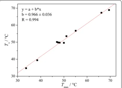
However, we found a very good correlation between
Tmax and Ts values of historical leathers (Fig. 4), as in
case of the new and artificially aged leathers (Fig. 2a). This behaviour is not so unexpected since both Tmax and Ts define the same process at various organisation levels
i.e. denaturation of collagen fibrils and shrinking of col-lagen fibres of the major (most abundant) colcol-lagen popu-lation (Table 4). We can therefore confirm the suitability of using Ts to classify the collagen major population into
one of the three groups defined earlier: “leather-like”, “parchment-like” and “gelatine-like”. It should be how-ever considered that the general behaviour of a his-torical leather is not always determined by its majority population. For example, BU-1 shows a “parchment-like” type majority collagen population (Tmax= 47.5 °C and Ts= 50 °C) while its behaviour is classified as “mixed”
since none of the collagen population contribute more than 50% of the total enthalpy. The fact that not always the majority population identified by MHT analysis is responsible for the general behaviour of the historical leather represents one of the limitations of this method.
From the shrinking activity illustrated in Fig. 5 it appears clearly that the investigated historical leathers can be grouped in three categories. The first group (BU-1, BU-2, BU-3 and BU-10) is characterised by lower Tf, Ts and Tl values and larger ∆T intervals compared to the
new leathers (Table 3), while ∆C intervals are slightly nar-rower. This behaviour can be attributed to a high hetero-geneity in the structural domains with distinct thermal
stabilities and clearly indicates the simultaneous presence of “leather-like” and “parchment-like” populations. When
Tf, Ts show very low values, the “gelatine-like” population
is also present. In the absence of micro DSC bulk analy-sis, such leather should be classified as having an “mixed” behaviour and its stability be evaluated according to its
Ts value.
The second group (BU-5, BU-6, BU-8 and BU-9) also display low Tf, Ts and Tl values but ∆T and ∆C
inter-vals are similar to those of new leathers. From Table 4 it comes out that these samples are mainly composed of a homogeneous collagen population and we can call them “homogeneous” leathers. For example, BU-5, BU-6 and BU-9 have “homogeneous parchment-like” behaviour, whereas BU-8 has a “homogeneous leather-like” behav-iour. Low Tf, Ts and Tl values accompanied by ΔT and ΔC
intervals like those of new leathers can therefore be used to identify the “homogeneous” historical leathers, while their behaviour (stability) is indicated by the Ts value.
The third group (BU-4 and BU-7) display the lowest values for Tf, Ts and Tl and ∆C. This latter group is
repre-sented by the two samples showing a prevalent “gelatine-like” behaviour (Table 4), with an enthalpy contribution of the gelatinised collagen population of 78% and 66%, respectively. We can thus state that detecting very low values for Tf, Ts, Tl and ΔC is a strong indication of
“gel-atine-like” behaviour and hence of an advanced state of deterioration.
By comparing the results obtained using micro DSC and MHT method clearly result that the most sta-ble “leather-like” populations (i.e. those displaying
Tmax > 80 °C) generally showed no shrinkage motion. This
can be partly ascribed to the low or very low content of such populations (Table 4) (that enhances the limitations
of surface sampling) and partly to the structural fea-tures of chemically modified (tanned) collagen fibres very tightly packed in compact and hard fibre bundles that prevents a good separation before measuring their shrinking activity. These drawbacks are overcome by the micro DSC technique which provides a bulk assessment and, due to its high sensitivity, ensures the detection of all collagen populations.
In conclusion, the capability of Ts to grade
deteriora-tion [13] in historical leather appears rather limited. When either micro DSC analysis is not available or sam-pling of 1–2 mg of sample is not allowed, it is necessary to also consider Ts, Tf, Tl, ∆C and ∆T variation for a more
reliable assessment of deterioration.
Conclusions
The micro DSC analysis provided quantitative results on the bulk material and unambiguously pictured the deterioration state of historical leather through several calorimetric indices of macromolecular change identi-fied for fibrous collagen, including (1) the temperature at the endothermic peak maximum (Tmax), (2) the
spe-cific enthalpy of denaturation (∆H), (3) heterogeneity in the structural domains with distinct thermal stabili-ties (∆T1/2). Separation through peak deconvolution of
the endothermic peaks associated with these structural domains confirms that historical leathers are complex blends of chemically modified, chemically unmodified and gelatinised collagen having distinct hydrothermal stabilities. Variation of the calorimetric indices and deconvolution of the overall DSC denaturation peak facilitated the interpretation of the changes induced by the natural ageing and identification of the key-steps of deterioration: thermal destabilisation of chemically modified collagen, de-tanning, thermal destabilisation of chemically unmodified collagen, gelatinisation and irre-versible denaturation.
The classification of the various collagen popula-tions depending on their Tmax in three main structural
domains (i.e. “leather-like”, “parchment-like” and “gela-tine-like”) and their quantification based on the enthalpy percent contribution to the overall denaturation enthalpy allowed for a deeper understanding of deterioration steps (dynamics) in ten historical leathers.
All this proves the potential of micro DSC technique to provide a greater practical understanding of deteriora-tion in collagenous materials and characterise the ability of historical leather to respond to environment and with-stand further deterioration. Studies of established and experimental treatments and preventive conservation practices would benefit from this method of quantifying deterioration based on the collagen population’s mass percentage distribution.
Besides, by correlating the MHT and micro DSC parameters, the suitability of using Ts to classify the
col-lagen major population into one of the three structural domains was confirmed. In addition, a more compre-hensive set of criteria, including Tf and Tl values, as well
∆C and ∆T intervals’ lengths’, was introduced to charac-terize the heterogeneity in the structural domains with distinct thermal stabilities. This is the first time that an in-depth correlation of micro DSC and MHT results was performed allowing for better interpreting the shrink-ing behaviour of collagen in historical vegetable tanned leathers. Our results substantiate MHT as a reliable method for in situ testing and evaluation of deterioration of leather artefacts.
Abbreviations
DSC: differential scanning calorimetry; MHT: micro hot table; INCDTP-ICPI: National Research and Development Institute for Textile and Leather, Division Leather and Footwear Research Institute.
Acknowledgements
We thank Prof. Giuseppe Della Gatta for encouragement, discussions, and allowing us to obtain early calorimetric data in his laboratory. Lucretia Miu (INCDTP-ICPI) generously provided the new leathers and parchments. The authors would also like to thank the National Military Museum “Ferdinand I”, the National Museum “Cotroceni”, the National Museum of Romanian History, and the History and Art Museum of Bucharest City for providing the historical samples.
Authors’ contributions
Both authors read and approved the final manuscript.
Funding
This work was supported by Grant of the Romanian National Authority for Scientific Research and Innovation, CCCDI-UEFISCDI, project number 56/2017, within PNCDI III.
Availability of data and materials
Not applicable.
Competing interests
The authors declare that they have no competing interests.
Author details
1 National Museum of Romanian History, Calea Victoriei 12, 030026 Bucharest, Romania. 2 Faculty of Sciences, University of Craiova, Calea Bucuresti 107 I, 200512 Craiova, Romania. 3 Advanced Research for Cultural Heritage Labora-tory (ARCH Lab), National Research and Development Institute for Textile and Leather, Division Leather and Footwear (INCDTP-ICPI), str. Ion Minulescu 93, 031215 Bucharest, Romania.
Received: 29 April 2019 Accepted: 3 July 2019
References
1. Forbes RJ. Leather in antiquity. Studies in ancient technology, vol. 5. 1st ed. Leiden: Brill Publishers; 1957. p. 1–77.
2. Haslam E. Vegetable tannage: where do the tannins go? J Soc Leather Technol Chem. 1997;81:45–51.
3. Hassan RRA. A preliminary study on using line seed oil emulsion in dress-ing archaeological leather. J Cult Herit. 2016;21:786–95.
4. Hassan RRA, Ali MF, Fahmy AA. Use of SEM, FTIR and amino acid analysis methods to assess the damage of some historical leather bindings from
the XIXth century, stored in National Archive, Cairo. Int J Conserv Sci. 2018;9(1):127–36.
5. Leather Thomson R. In: May E, Jones M, editors. Conservation science: heritage materials. Cambridge: The Royal Society of Chemistry; 2006. p. 67.
6. Falcão L, Araújo MEM. Tannins characterisation in new and historic veg-etable tanned leathers fibres by spot tests. J Cult Herit. 2011;12(2):149–56. 7. Bate-Smith EC, Swain T. Flavonoid compounds. In: Mason HS, Florkin AM,
editors. Comparative biochemistry. New York: Academic Press; 1962. p. 755–809.
8. Khanbabaee K, van Ree T. Tannins: classification and definition. Nat Prod Rep. 2001;18:641–9.
9. Covington AD. Modern tanning chemistry. Chem Soc Rev. 1997;26:111–26.
10. Weir CE. Rate of shrinkage of tendon collagen—heat, entropy, and free energy of activation of the shrinkage of untreated tendon. Effect of acid, salt, pickle, and tannage on the activation of tendon collagen. J Am Leather Chem Assoc. 1949;44:108–40.
11. Pankhurst K. “Incipient shrinkage” of collagen and gelatin. Nature. 1947;159:538.
12. Larsen R. Experiments and observations in the study of environmental impact on historical vegetable tanned leathers. Thermochim Acta. 2000;365:85–99.
13. Larsen R, Poulsen Sommer DV, Mühlen Axelsson K. Scientific approach in conservation and restoration of leather and parchments objects in archives and libraries. In: Engel P, editor. New approaches to book and paper conservation-restoration. Wien: Verlag Berger; 2011. p. 239–58. 14. Larsen R, Vest M, Nielsen K. Determination of hydrothermal stability
(shrinkage temperature) of historical leathers by Micro Hot Table tech-nique. J Soc Leather Technol Chem. 1993;77:151–6.
15. Carsote C, Kövari L, Albu C, Hadîmbu E, Badea E, Miu L, Dumitrescu G. Bindings of rare books from the collections of the Romanian Academy Library—a multidisciplinary study. Leather Footwear J. 2018;18(4):307–20. 16. Budrugeac P, Carşote C, Miu L. Application of thermal analysis methods
for damage assessment of leather in an old military coat belonging to the History Museum of Braşov-Romania. J Therm Anal Calorim. 2017;127(1):765–72.
17. Mühlen Axelsson K, Larsen R, Poulsen Sommer DV, Melin R. Degradation of collagen in parchment under the influence of heat-induced oxidation: preliminary study of changes at macroscopic, microscopic, and molecular levels. Stud Conserv. 2016;61(1):46–57.
18. Chahine C. Changes in hydrothermal stability of leather and parchment with deterioration: a DSC study. Thermochim Acta. 2000;365:101–10. 19. Budrugeac P, Miu L. The suitability of DSC method for damage
assess-ment and certification of historical leathers and parchassess-ments. J Cult Herit. 2008;9:146–53.
20. Miu OA, Badea E, Carsote C, Ciobanu S. Automatic detection of collagen fibres shrinkage activity using Σ-Δ filtering. In: Proceedings Book of the 5th international conference on advanced materials and systems (ICAMS 2014). Bucharest: Certex; 2014. p. 539–42.
21. Badea E, Poulsen Sommer DV, Mühlen Axelsson K, Larsen R, Kurysheva A, Miu L, Della Gatta G. Damage ranking in historical parchments: from microscopic study of fibres structure to collagen denaturation assess-ment by micro DSC. e-Preserv Sci. 2012;9:97–109.
22. Badea E, Şendrea C, Carşote C, Adams A, Blümich B, Iovu H. Unilateral NMR and thermal microscopy studies of vegetable tanned leather exposed to dehydrothermal treatment and light irradiation. Microchem J. 2016;129:158–65.
23. Sendrea C, Carșote C, Radu M, Badea E, Miu L. The effect of gamma irradiation on shrinkage activity of collagen in vegetable tanned leather. Rev Chim. 2017;68(7):1535–8.
24. Sebestyén Z, Jakab E, Badea E, Barta-Rajnai E, Şendrea C, Czégény ZS. Thermal degradation study of vegetable tannins and vegetable tanned leathers. J Anal Appl Pyrol. 2019;138:178–87.
25. Gill P, Moghadam TT, Ranjbar B. Differential scanning calorimetry techniques: applications in biology and nanoscience. J Biomol Tech. 2010;21(4):167–93.
27. Badea E, Usacheva T, Della Gatta G. The use of differential scanning calorimetry to characterise collagen deterioration in parchment. Rossiiskii Khimicheskii Zhurnal - Zhurnal Rossiiskogo Khimicheskogo Obshchestva im. D.I. Mendeleeva (Russian Chem J). 2015;59(1):28–41.
28. Miles CA. Kinetics of collagen denaturation in mammalian lens capsules studied by differential scanning calorimetry. Int J Biol Macromol. 1993;15:265–71.
29. Samouillan V, Delaunay F, Dandurand J, Merbah N, Gardou JP, Yousfi M, Gandaglia A, Spina M, Lacabanne C. Use of thermal techniques for the characterization and selection of natural biomaterials. J Funct Biomater. 2011;2:230–48.
30. Della Gatta G, Badea E, Ceccarelli R, Usacheva T, Mašić A. Assessment of damage in old parchment by DSC and SEM. J Therm Anal Calorim. 2005;82:637–49.
31. Badea E, Della Gatta G, Budrugeac P. Characterisation and evaluation of the environmental impact on historical parchments by DSC. J Therm Anal Calorim. 2011;104(2):495–506.
32. Carşote C, Badea E, Miu L, Della Gatta G. Study of the effect of tannins and animal species on the thermal stability of vegetable leather by differential scanning calorimetry. J Therm Anal Calorim. 2016;124(3):1255–66. 33. Badea E, Miu L, Budrugeac P, Giurginca M, Mašić A, Badea N, Della Gatta
G. Study of deterioration of historical parchments by various thermal analysis techniques, complemented by SEM, FTIR, UV-Vis-NIR and unilat-eral NMR investigations. J Therm Anal Calorim. 2008;91:17–27. 34. Budrugeac P, Badea E, Della Gatta G, Miu L, Comănescu A. DSC study of
deterioration caused by environmental chemical pollutants to parch-ment, a collagen based material. Thermochim Acta. 2010;500:51–62. 35. Carşote C, Budrugeac P, Decheva R, Haralampiev NS, Miu L, Badea E.
Characterization of a byzantine manuscript by infrared spectroscopy and thermal analysis. Rev Rou Chim. 2014;59:429–36.
36. Falcão L, Araújo MEM. Vegetable tannins used in the manufacture of historic leathers. Molecules. 2018;23(5):1081.
37. Tang HR, Hancock RA, Covington AD. Study on the composition and structure of commercial chestnut tanning agent. Basic Life Sci. 1992;59:221–43.
38. Pasch H, Pizzi A, Rode K. MALDI-TOF mass spectrometry of polyflavonoid tannins. Polymer. 2001;42(18):7531–9.
39. Miles CA, Burjanadze TV, Bailey AJ. The kinetics of the thermal denatura-tion of collagen in unrestrained rat tail tendon determined by differential scanning calorimetry. J Mol Biol. 1995;245(4):437–46.
40. Luescher M, Ruegg M, Schindler P. Effect of hydration upon the thermal stability of tropocollagen and its dependence on the presence of neutral salts. Biopolymer. 1974;13:2489–503.
41. Miu OA, Ciobanu S, Badea E. Automatic method for damage assessment of historical and archeological leather and parchment. Patent application A/001253-05.07.2014.
42. Hulmes DJ. Building collagen molecules, fibrils and suprafibrillar struc-tures. J Struct Biol. 2002;137(2):2–10.
43. Buehler MJ. Nature designs tough collagen: explaining the nanostruc-tures of collagen fibrils. Proc Natl Acad Sci USA. 2006;3(33):12285–90. 44. Wess TJ. Collagen fibrillas structures and hierarchies. In: Fratzl P, editor.
Collagen: structure and mechanics. New York: Springer; 2008. p. 49. 45. Covington AD. The mechanism of chrome tanning. Glob J Inorg Chem.
2010;1(2):119–31.
46. Covington A, Song L, Suparno O, Koon H, Collins MJ. Link-lock: an expla-nation of the chemical stabilisation of collagen. J Soc Leather Technol Chem. 2008;92:1–7.
47. Bella J, Brodsky B, Berman HM. Hydration structure of collagen. Structure. 1995;9:893–906.
48. Leikina E, Mertts MV, Kuznetsova N, Leikin S. Type I collagen is thermally unstable at body temperature. Proc Natl Acad Sci USA. 2002;99:1314–8. 49. George C Na. Monomer and oligomer of type I collagen: molecular
properties and fibril assembly. Biochemistry. 1989;28:7161–7. 50. Tiktopulo EI, Kajava AV. Denaturation of type I collagen fibrils is an
endo-thermic process accompanied by a noticeable change in the partial heat capacity. Biochemistry. 1998;37:8147–52.
51. Miles CA, Ghelashvili M. Polymer-in-a-box mechanism for the thermal stabilization of collagen molecules in fibers. Biophys J. 1999;76:3243–52. 52. Miles CA, Bailey AJ. Thermal denaturation of collagen revisited. Proc
Indian Acad Sci (Chem. ScL). 1999;111(1):71–80.
53. Lin SJ, Lo W, Dong CY. Prediction of heat-induced collagen shrinkage by use of second harmonic generation microscopy. J Biomed Opt. 2006;11(3):34020.
54. Bozec L, Odlyha M. Thermal denaturation studies of collagen by microthermal analysis and atomic force microscopy. Biophys J. 2011;101(1):228–36.
55. Cucos A, Gaidau C, Badea E, Miu L. Influence of glycerin on denaturation temperature of chrome- and vegetable-tanned leather. Rev Roum Chim. 2015;60(11–12):1093–6.
56. Yao Q, Wang Y, Chen H, Huang H, Liu B. Mechanism of high chrome uptake of tanning pickled pelt by carboxyl-terminated hyper-branched polymer combination chrome tanning. Chem Select. 2019;4(2):670–80. 57. Sebestyén Z, Czégény Z, Badea E, Carsote C, Sendrea C, Barta-Rajnai E,
János B, Miu L, Jakab E. Thermal characterization of new, artificially aged and historical leather and parchment. J Anal Appl Pyrol. 2015;115:419–27.
Publisher’s Note
![Fig. 2 Plots showing a good linear correlation between tanned leathers and 48 artificially aged samples [coefficient R a Tmax and Ts (correlation coefficient R = 0.961); b Tonset and Tf (correlation = 0.908) for a group of 12 samples from 6 new vegetable 32]](https://thumb-us.123doks.com/thumbv2/123dok_us/837901.2078528/6.595.56.292.335.681/correlation-leathers-artificially-coefficient-correlation-coefficient-correlation-vegetable.webp)