R E S E A R C H
Open Access
Mesenchymal stem cell repression of Th17
cells is triggered by mitochondrial transfer
Patricia Luz-Crawford
1, Javier Hernandez
2, Farida Djouad
2, Noymar Luque-Campos
1, Andres Caicedo
4,
Séverine Carrère-Kremer
2, Jean-Marc Brondello
2, Marie-Luce Vignais
2, Jérôme Pène
2and Christian Jorgensen
2,3*Abstract
Background:Mesenchymal stem cells (MSCs) are multipotent cells with broad immunosuppressive capacities.
Recently, it has been reported that MSCs can transfer mitochondria to various cell types, including fibroblast, cancer, and endothelial cells. It has been suggested that mitochondrial transfer is associated with a physiological response to cues released by damaged cells to restore and regenerate damaged tissue. However, the role of mitochondrial transfer to immune competent cells has been poorly investigated.
Methods and results:Here, we analyzed the capacity of MSCs from the bone marrow (BM) of healthy donors
(BM-MSCs) to transfer mitochondria to primary CD4+CCR6+CD45RO+T helper 17 (Th17) cells by confocal microscopy and fluorescent-activated cell sorting (FACS). We then evaluated the Th17 cell inflammatory phenotype and bioenergetics at 4 h and 24 h of co-culture with BM-MSCs. We found that Th17 cells can take up mitochondria from BM-MSCs already after 4 h of co-culture. Moreover, IL-17 production by Th17 cells co-cultured with BM-MSCs was significantly impaired in a contact-dependent manner. This inhibition was associated with oxygen consumption increase by Th17 cells and interconversion into T regulatory cells. Finally, by co-culturing human synovial MSCs (sMSCs) from patients with rheumatoid arthritis (RA) with Th17 cells, we found that compared with healthy BM-MSCs, mitochondrial transfer to Th17 cells was impaired in RA-sMSCs. Moreover, artificial mitochondrial transfer also significantly reduced IL-17 production by Th17 cells.
Conclusions:The present study brings some insights into a novel mechanism of T cell function regulation through mitochondrial transfer from stromal stem cells. The reduced mitochondrial transfer by RA-sMSCs might contribute to the persistence of chronic inflammation in RA synovitis.
Keywords:Mitochondria transfer, Mesenchymal stem cells, T cell, Th17, Immunomodulation
Background
Mesenchymal stem cells (MSCs) are multipotent and immunoregulatory stem cells that limit the progression
and severity of autoimmune diseases [1–3]. MSC
immunosuppressive functions are mediated through cell–
cell contacts and the secretion of soluble factors, particu-larly inducible nitric oxide synthase by murine MSCs and indoleamine-2,3-dioxygenase by human MSCs [4], as well
as IL-6, TGF-β1 and PGE2, IL-1RA, and HLAG5s [5–8].
We and others demonstrated that MSCs prevent T cell differentiation into T helper (Th)17 and Th1 cells and
induce T regulatory (Treg) cells in vitro [9–11]. This
MSC inhibitory effect requires cell–cell contacts
through ICAM-1 and the release of soluble factors,
such as PGE2 [9,11]. MSC immunomodulatory
proper-ties are not intrinsic, but are induced by inflammatory cytokines [9,12–14]. Thus, MSCs establish direct inter-actions with T cells in physiological conditions where MSCs play a key role in T cell homeostasis and also in pathological conditions where infused MSCs have immunoregulatory properties.
It has been shown that MSCs make tunneling nanotube (TNT) connections with various cell types, leading to intercellular exchange of mitochondria [15, 16]. We pre-viously demonstrated that the dynamic transfer of mitochondria to carcinoma cells induces changes in malignant cell proliferation and glucose uptake [17].
© The Author(s). 2019Open AccessThis article is distributed under the terms of the Creative Commons Attribution 4.0 International License (http://creativecommons.org/licenses/by/4.0/), which permits unrestricted use, distribution, and reproduction in any medium, provided you give appropriate credit to the original author(s) and the source, provide a link to the Creative Commons license, and indicate if changes were made. The Creative Commons Public Domain Dedication waiver (http://creativecommons.org/publicdomain/zero/1.0/) applies to the data made available in this article, unless otherwise stated. * Correspondence:christian.jorgensen@inserm.fr
2IRMB, Univ Montpellier, INSERM, Hôpital Saint-Eloi, 80 avenue Augustin Fliche, 34295 Montpellier CEDEX 5, France
3CHU Montpellier, F-34295 Montpellier, France
Moreover, mitochondrial transfer via the TNT connec-tions between MSCs and non-malignant cells with mitochondrial damage allows rescuing the mitochondrial
function in the receiving cells [16, 18]. TNT-mediated
transfer of MSC mitochondria to cancer cells increases their metabolic activity and resistance to therapy. It has been proposed that such transfer is regulated by the mito-chondrial Rho-GTPase Miro that favors mitomito-chondrial trafficking along cytoskeleton fibers [15, 16, 18]. In vivo, the transfer of MSC mitochondria to damaged bronchial epithelial cells modifies the metabolism and restores the functionality of the receiving cells, thus limiting cell damage and apoptosis [19, 20]. Thus, intercellular mito-chondrial transport modifies the recipient cell bioenerge-tics and stimulates their proliferation and functions. Concerning immune cells, it has been shown that MSCs extensively transfer mitochondria to macro-phages, partially through TNT, thus increasing their phagocytic activity. However, it is not known whether MSCs can transfer mitochondria to pathogenic Th17 cells and the consequence of this event, although it is widely acknowledged that MSCs regulate the balance between pathogenic Th17 cells and Treg cells to improve autoimmune disease outcomes.
In this study, we investigated the role of mitochondrial transfer in the MSC-mediated modulation of Th17 cell function. To this aim, we co-cultured memory Th17 cells and bone marrow (BM)-MSCs to investigate MSC capacity to transfer mitochondria to Th17 cells and the effect of this transfer on Th17 cell phenotype, function, and metabolism. Then, using an artificial method to transfer mitochondria (i.e., MitoCeption), we assessed the direct effect of BM-MSC mitochondrial transfer on the Th17 cell phenotype. Finally, to determine the in-fluence of the inflammatory environment in the joints of patients with rheumatoid arthritis (RA) on mitochon-drial transfer from MSCs to T cells, we compared mito-chondrial transfer from RA synoviocytes and healthy
BM-MSCs to CD4+ T cells. Altogether, our results
highlight that mitochondrial transfer from MSCs is a novel immunoregulatory mechanism for inhibiting Th17 cells that could be impaired in RA synoviocytes.
Methods
Isolation and characterization of human MSCs
Human MSCs, isolated from the BM of three healthy donors after written consent, were obtained from the Établissement Français du Sang (EFS; Grenoble, France) and used at the third or fourth passage. MSCs were
cultured in α-MEM medium (Lonza, Switzerland)
supplemented with 10% fetal calf serum (FCS) (Sigma, USA), 2 mM glutamine, 100 U/ml penicillin, 100 mg/ ml streptomycin (without antibiotics for PCR assays with mitochondrial DNA and for T cell bioenergetics
analysis), and 1 ng/ml basic FGF (Bio-Techne, UK). To simulate an inflammatory environment, MSCs were
pre-incubated with TNF-α (10 ng/ml) and IFN-γ (20 ng/ml)
(R&D, France) for 48 h.
Isolation and culture of human T cells
Polyclonal CD4+ T cell populations were prepared from
peripheral blood mononuclear cells (PBMCs) from five healthy donors (EFS, Montpellier, France) after written consent. CD4+ T cell isolation, stimulation, and culture protocols were as previously described [9]. Briefly, in
vivo polarized CD4+Th17 cells were sorted by flow
cy-tometry based on the fact that the chemokine receptor CCR6 is expressed almost exclusively by the Th17 cell
subset and not by CD4+Th1 and Th2 cells. CD4+CCR6+
and CD4+CCR6− T cells were propagated by weekly
stimulation with an irradiated feeder cell mixture (PBMCs and JY cell line) with PHA (Lonza, France) and recombinant IL-2 (2 ng/ml; Bio-Techne, UK). To prevent any possible derivation of the T cell lines due to culture conditions, cells were stimulated at most thrice, and their production of IL-17 and expression of RoRγt were regularly tested. T cells were used 10 to 15 days after the latest propagation phase, a time window that corresponds to T cells in a resting phase. All culture procedures were
carried out in Yssel’s medium, supplemented with 2%
human AB+serum (EFS, Lyon, France).
Isolation and characterization of human RA synovial stromal stem cells (RA-sMSCs)
The use of clinical biopsies and blood samples from patients with RA was approved by the ethics committee
of Montpellier University Hospitals (DC-2008-417—
co-ordinator C Jorgensen). Culture conditions were as for BM-MSCs, and isolation was performed as previously described [21].
Co-culture experiments
MSCs were labeled with the MitoTracker Green FM (M7514), MitoTracker Red CMXRos, or MitoTracker Deep Red FM (far red) fluorescent mitochondrial dyes (Molecular Probes, France) according to the manufac-turer’s instructions, 24 h before co-culture. In some ex-periments, T cells were labeled with the CellTracker Green fluorescent dye (Molecular Probes, France) accor-ding to the manufacturer’s instructions. After labeling, MSCs were harvested, extensively washed in PBS, and
co-cultured with Th17 cells (106 cells, MSC to T cell
T cells were harvested, and MitoTracker dye uptake (as a sign of mitochondrial transfer from MSCs) was analyzed by flow cytometry.
Flow cytometry
Flow cytometry analysis of T cells and MSCs was carried out as previously described [9] using PE-conjugated anti-CCR6 (Bio-Techne, UK), Fluo-conjugated anti-CD3 and anti-CD4 mAbs, and isotype control (BD, Germany) and a FACSCanto or FACSFortesa flow cytometer. Data (10,000 events/sample) were collected using the DIVA software (BD, Germany). Cell sorting was performed on a FACSAria® flow cytometer (BD, Germany).
Intracellular staining of T cells was performed after 4 h of activation with PMA (20 ng/ml; Merck, Germany) and ionomycin (1 mg/ml; Merck, Germany), in the presence of brefeldin A (10 mg/ml; Sigma, USA). When necessary, cells were stained with PE-conjugated anti-CCR6 (Bio-Techne, UK), Fluo-conjugated anti-CD3 and anti-CD4 mAbs, and isotype control (BD, Germany). Then, cells
were fixed with Cytofix/Cytoperm buffer
(Ther-moFisher, UK) at 4 °C overnight and stained with fluorochrome-conjugated mAbs diluted in Perm/Wash buffer (ThermoFisher, UK), according to the manufac-turer’s specifications. Cytokine production was assessed
by flow cytometry using anti-IL-17–Percp5.5,
anti-IL-CD25–APC, anti-IL-10–PECY7, and anti-IFN-γ–APCCY7 (ThermoFisher, UK) mAbs. FOXP3 expression was assessed with an anti-FOXP3 PE-conjugated mAb (ThermoFisher, UK). All mAbs were used at the optimal concentrations previously determined in independent experiments on the basis of the manufacturer’s recommendations.
T cell proliferation was assessed with the CellTrace Violet Cell Proliferation Kit for flow cytometry
(Mole-cular Probes, France) according to the manufacturer’s
instructions.
PCR assays with mitochondrial DNA
MSC mitochondrial DNA (mtDNA) in T cells was quanti-fied by PCR, based on the distinct haplotypes of the MSC and T cell donors. To increase the assay specificity, a sup-plementary modification was introduced at position n-2
relative to the SNP site and at the 3′ end of the PCR
primers. MSC mtDNA from one donor was amplified
with the primers 5′-TTAACTCCACCATTAGCACC-3′
[22] and 5′-AGTATTTATGGTACCGTCCG-3′
(posi-tions 15971–16143) and MSC mtDNA from the two
other donors with the primers 5′
-TAACAGTACATAGCA-CATACAA-3′ and 5′-GAGGATGGTGGTCAAGGGA-3′
(universal sequencing primer [22]) (positions 16298–16410).
Confocal microscopy
Cells were seeded on culture dishes with glass bottom, and images of live cells were taken with a Carl Zeiss
LSM 5 LIVE (LSM 510 META and LSM 5 LIVE) DuoS-can Laser SDuoS-canning microscope immediately after 4 h of MSC-T cell co-culture. 3D reconstruction was done using the Imaris Bitplane software.
T cell bioenergetics
Oxygen consumption rate (OCR) was measured using the XF24 analyzer (Seahorse Biosciences, North Billerica, MA, USA). Briefly, XF24 24-well plates were coated with Cell-Tak (BD Biosciences, France), as described in the Seahorse protocol. T cells (106cells/well) were plated in
unbuffered XF assay medium–modified DMEM
(Sea-horse Biosciences) at pH 7.4, supplemented with 10 mM glucose, 2 mM glutamate, and 1 mM sodium pyruvate, and incubated at 37 °C without CO2. OCR was measured (4 wells per condition) in basal conditions and after sequential addition of oligomycin (1μM),
carbonylcyanide-4-trifluoromethoxyphenylhydrazone (FCCP) (1μM),
rote-none (100 nM), and antimycin A (1μM). Three
readings were taken after each addition. Instrument background was measured in separate control wells using the same conditions but without cells. T cells from three independent co-cultures were used for these experiments.
Isolation of MSC mitochondria and transfer to T cells (MitoCeption)
Mitochondria were isolated using the Mitochondria Iso-lation Kit for Cultured Cells (ThermoScientific) follow-ing the manufacturer’s instructions. At least 1 × 10⁶ MitoTracker-stained MSCs were used to obtain between
50 and 100μg of mitochondria. To reduce
contamin-ation from cytosolic elements, a final centrifugcontamin-ation was
performed at 3000g for 15 min. Isolated mitochondria
were resuspended in Yssel’s medium, supplemented with
2% human AB+ serum (EFS, Lyon, France), and
main-tained on ice for immediate transfer into
CD4+CD45+CCR6+ T cells. To this aim, 10μg of mito-chondria was used for 2.5 × 10⁵T cells, as previously de-scribed [17]. Then, culture plates were centrifuged at 1500gat 4 °C for 15 min and incubated at 37 °C, 5%CO2, for 24 h. The following day MitoCeption efficiency was verified by flow cytometry analysis of the MitoTracker signal in T cells (Additional file1: Figure S1).
Statistical analyses
Data were analyzed using GraphPad Prism 6 (GraphPad Software Inc., San Diego, CA). The non-parametric Friedman test was used to evaluate statistical differences among paired multiple samples, and the non-parametric
Mann–Whitney test to compare two variables.
Dif-ferences were considered statistically significant when
Results
Uptake of mitochondria by primary human
CD4+CCR6+CD45R0+T cells co-cultured with BM-MSCs To determine whether mitochondria are transferred from BM-MSCs into primary lymphocytes, BM-MSCs were labeled with the MitoTracker Green probe and co-cultured with human PBMCs from healthy donors. After 4 h of co-culture, PBMCs were harvested and labeled
with fluorochrome-conjugated mAbs to identify CD4+,
CD8+, and CD19+ T cells. FACS analysis showed that
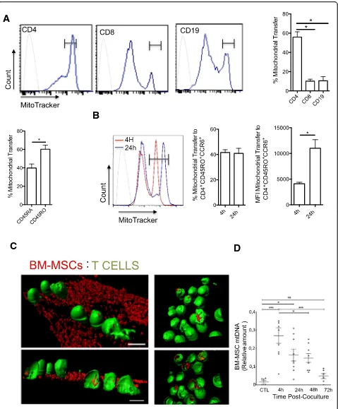
mitochondria were transferred from BM-MSCs into all studied lymphocyte subsets, particularly in CD4+ T cells (56% in CD4+vs 17% in CD8+T cells and 24% in B cells)
(Fig. 1a). Moreover, mitochondria uptake was higher in
CD4+ memory T cells (CD45RO+) than that in naive
CD4+ T cells (CD45RA+), and 60% of memory T cells
were CCR6+ cells (Th17 memory phenotype) (Fig. 1a).
As memory CD4+ T cells, and particularly those
expres-sing CCR6, were the best recipients of BM-MSC mitochondria, the subsequent analyses focused on the im-pact of mitochondrial transfer to pro-inflammatory Th17
cells. To this aim, FACS-sorted CD4+CCR6+CD45RO+ T
cells from PBMCs of five healthy donors were stimulated and amplified in vitro to obtain an enriched Th17 popula-tion. Then, day 10 resting Th17 cells were co-cultured with MitoTracker-labeled BM-MSCs isolated from three healthy donors for 4 h and 24 h. FACS analysis of non-ad-herent T cells showed that BM-MSCs could transfer mito-chondria to Th17 cells after 4 and 24 h of co-culture. Of note, although the percentage of mitochondrial transfer did not change between 4 and 24 h, the mean fluorescence intensity (MFI) was higher after 24 h of co-culture, sug-gesting that only a fraction of Th17 cells could receive
mitochondria from BM-MSCs (Fig. 1b). Mitochondrial
transfer was completely abolished by mechanical shaking of cells (data not shown), indicating that it occurred through a contact-dependent mechanism. Moreover, incu-bation of T cells with culture medium conditioned by MitoTracker-labeled BM-MSCs, but without BM-MSCs, did not lead to a fluorescence signal increase in T cells (data not shown), ruling out the possibility of passive staining of T cell mitochondria due to MitoTracker probe leak. To determine whether BM-MSC mitochondria enter Th17 cells, CellTracker-labeled T cells (green) were analyzed by confocal microscopy after co-culture (4 h)
with MitoTracker Red CMXRos-labeled BM-MSCs
(Fig.1c). MSC mitochondria (red) were observed in some
T cells attached to the BM-MSCs (Fig. 1c) and also in
some T cells in suspension (Fig.1c, right panel).
To confirm the transfer of MSC mitochondria in T cells during co-culture, MSC mtDNA was quantified in
T cells (Fig. 1d) by taking advantage of the different
mtDNA haplotypes of BM-MSCs and T cells (different donors for each cell type). First, mtDNA was isolated
from MSCs (three donors) and T cells (one donor), and their variable D-loop regions were sequenced. This allowed the detection of specific SNPs, and the design of PCR primers to specifically target and amplify MSC (and not T cell) mtDNA. After 4 h of co-culture (MSC to T cell ratio 1:25), non-adherent T cells were harvested and cultured with IL-2 and without BM-MSCs for up to 72 h. PCR analysis of MSC mtDNA in T cells after the 4 h of co-culture and then at 24, 48, and 72 h of incubation with IL-2 showed that MSC mtDNA could be detected in T cells after 4 h of co-culture and up to 48 h with IL-2, but not at the 72-h time point.
Altogether, these results indicate that mitochondria are efficiently transferred from human BM-MSCs into Th17 cells via cell-to-cell interactions already after 4 h of co-culture.
Co-culture with BM-MSCs leads to a reduction of Th17 cell responses and Treg cell generation
Our previous study showed that after long-term co-cul-ture, MSCs interfere with the functional activity of Th17 cells, a CD4+ T cell population that plays an important role in the development of autoimmune diseases, such as RA [9,11]. To investigate whether short-term (4 or 24 h) co-culture also could alter Th17 cell function and induce the expression of Treg markers, particularly FOXP3,
CD4+CCR6+CD45RO+ T cells sorted from PBMCs of five
healthy donors were stimulated and amplified in vitro to obtain a T cell population enriched in Th17 cells. Then, resting Th17 cells were co-cultured with BM-MSCs isolated from three healthy donors for 4 h, followed by harvesting of non-adherent Th17 cells and stimulation with anti-CD3 and anti-CD28 antibody-coated beads for 3 more days. Finally, FACS-based quantification of IL-17 and FOXP3 expression showed that compared with not co-cultured Th17 cells, production of IL-17 was reduced
only after co-culture with BM-MSCs for 24 h (Fig. 2a).
Conversely, the FOXP3 expression level was not
signifi-cantly affected by co-culture with BM-MSCs (Fig. 2a).
Then, to mimic a pro-inflammatory environment,
BM-MSCs were pre-incubated with TNF-α (10 ng/ml) and
INF-γ(20 ng/ml) for 48 h before co-culture. This pre-in-cubation step did not affect (1) mitochondrial transfer
from BM-MSCs to Th17 cells (Fig. 2b), (2) BM-MSC
suppressive effect on Th17 cell phenotype and function (Fig.2c), and (3) BM-MSC inhibitory effect on Th17 cell proliferation (Fig.2d).
Then, to monitor the fate of Th17 cells that acquired or not mitochondria from BM-MSCs, Th17 cells were co-cultured with BM-MSCs for 4 h, and Th17 cells that
received mitochondria (Mito+) or not (Mito−) were
separated by cell sorting (Fig. 3a). FACS analysis
A
B
C
D
Moreover, Mito+T cells produced less IL-17 than Mito−T cells, although the difference was not significant (Fig3c). Finally, the percentage of CD4+CCR6+CD25HighFOXP3+ cells (i.e., Th17 cells with a regulatory phenotype) was higher among Mito+ than Mito− cells. Altogether, these data suggest that mitochondrial transfer from BM-MSCs to Th17 cells results in a reduced production of pro-in-flammatory cytokines and induces a Treg phenotype in ef-fector memory Th17 cells.
Effect of BM-MSC mitochondrial transfer on the energetic metabolism of recipient Th17 cells
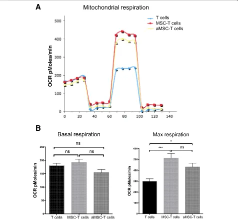
Mitochondria are crucial for the cell energetic meta-bolism and oxidative phosphorylation (OXPHOS). To determine the effect of BM-MSC-T cell co-culture on T cell metabolism, non-adherent T cells were collected after 4 h of co-culture with BM-MSCs and cultured in the presence of IL-2 for 24 or 48 h. OXPHOS quantification using the Seahorse extracellular flux technology showed that co-culture had no effect on the T cell basal respiration levels (OCR) compared with control (T cells alone) (Fig. 4a). Conversely, the maximal T cell respiration was increased (as observed upon FCCP addition) in co-cul-tured T cells. Similar results were obtained using BM-MSCs from three different donors (Fig.4b for basal respir-ation-level data). The maximal respiration was increased by 1.7-fold in T cells co-cultured for 4 h (Fig.4c).
Artificial transfer of BM-MSC mitochondria to T cells alters Th17 cell function
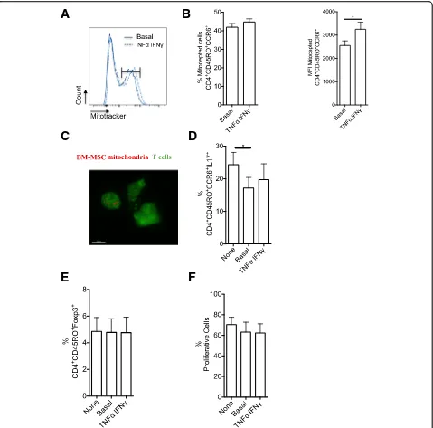
To assess whether BM-MSC mitochondria have a role in the regulation of CD4+ Th17 cell effector functions, the previously described MitoCeption technique [17] was used to transfer mitochondria isolated from BM-MSCs to the target cells without the need of co-culture. Before mitochondria isolation, BM-MSCs were labeled with MitoTracker Deep Red and T cells with CellTracker (green). Flow cytometry analysis of T cells after MitoCep-tion revealed that almost 40% of T cells had incorporated the MitoTracker dye (Fig. 5a, b), a percentage similar to the one obtained after co-culture of BM-MSCs and T cells
(Fig. 1b). 3D reconstruction of confocal microscopy
images after MitoCeption highlighted the presence of
BM-MSC mitochondria in T cells (Fig. 5c). Next,
un-labeled BM-MSCs were used as a source of mitochondria for MitoCeption of Th17 cells. After MitoCeption, Th17 cells were activated with CD3 and CD28 anti-body-coated beads for 3 days, and then their ability to pro-duce IL-17 was assessed. The percentage of IL-17-producing cells (Fig.5d) was significantly reduced among Th17 cells that underwent MitoCeption compared with controls (no MitoCeption). This indicates that BM-MSC mitochondria are sufficient to alter Th17 effector func-tions and suggests that mitochondrial transfer plays a role in MSC immunomodulation of Th17 cells. Finally, MitoCeption with mitochondria isolated from BM-MSC
activated with pro-inflammatory cytokines (TNFα and
IFNγ) did not affect the percentage of Th17 cells that incorporated mitochondria (Fig. 5a), but slightly increase the MFI (Fig.5b). Moreover, mitochondria from activated BM-MSCs did not change the percentage of IL-17-produ-cing Th17 cells after MitoCeption compared with
mito-chondria from non-activated BM-MSCs (Fig. 5d). The
immunoregulatory effect mediated by mitochondrial transfer to Th17 cells was not associated with increased FOXP3 expression level (Fig.5e). Then, to assess the effect of BM-MSC mitochondria transfer on T cell proliferation, MitoCepted T cells and control T cells were labeled with CellTrace violet. After 3 days of culture, the proliferation rate was comparable in control T cells and in T cells that underwent MitoCeption of mitochondria from
non-activated and activated BM-MSCs (Fig. 5f ).
Transfer of mitochondria derived from healthy BM-MSCs and from RA-sMSCs
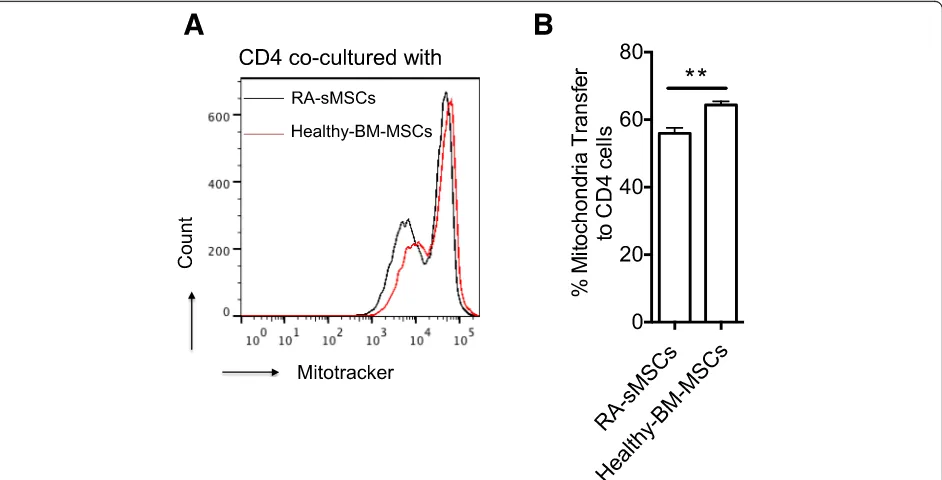
MSCs are the main stromal cells present in synovitis during active RA. Moreover, our previous findings showed that BM-derived and synovium-derived MSCs (sMSCs) share similar phenotypic properties and diffe-rentiation potential [21]. To determine whether these two cell types exhibit the same mitochondrial transfer poten-tial and whether RA affects this process, phenotypically
characterized RA-sMSCs (CD44+, CD29+, CD105+) and
BM-MSCs from healthy donors were labeled with Mito-Tracker before co-culture with CD4+T cells for 4 h. FACS analysis indicated that the mitochondrial transfer capacity
(See figure on previous page.)
A
B
D
C
Fig. 2Mitochondrial transfer from BM-MSCs significantly reduces IL-17 production independently of TNF-α/IFN-γstimulation.aRepresentative dot plots of FOXP3 and IL-17 expression (left panels) in Th17 cells after 3 days of activation by incubation with beads coupled to anti-CD3/CD28 antibodies following (+) or not (−) co-culture with BM-MSCs for 4 or 24 h. Graphs (right panels) show the quantification of the results.bBM-MSC mitochondrial transfer to Th17 cells after 4 and 24 h of co-culture. BM-MSCs were pre-incubated or not (Basal) with TNF-αand IFN-γ. Mitochondrial transfer is expressed as the percentage of MitoTracker-positive T cells relative to all T cells and as the mean fluorescence intensity (MFI).cIL-17 and FOXP3 expression quantification in Th17 cells co-cultured or not (None) with BM-MSCs pre-incubated or not (Basal) with TNF-αand IFN-γ.
of RA-sMSCs was significantly reduced compared
with BM-MSCs (Fig. 6a, b). Indeed, 56% and 66% of
CD4+CD45RO+ T cells were Mito+ after 4 h of
co-culture with RA-sMSCs and BM-MSCs, respectively (p< 0.05) (Fig.6b).
Discussion
The present study shows that BM-MSC immunoregula-tory effect on Th17 cells relies also on mitochondrial transfer. Mitochondrial transfer was observed already after 4 h of co-culture of BM-MSCs and T cells and did not require MSC activation by pre-incubation with pro-inflammatory cytokines. Moreover, after co-culture for
24 h, the MFI level was further increased, but not the percentage of T cells that acquired mitochondria. These findings suggest that a Th17 cell sub-population is resist-ant to mitochondrial transfer. Moreover, mitochondrial transfer was associated with increased oxygen consump-tion by Th17 cells that significantly impaired their capacity to produce IL-17. Additionally, the analysis of the Th17 cell phenotype after cell sorting according to the uptake or not of mitochondria showed a significant increase of Treg markers in Th17 cells that acquired mitochondria, suggesting that mitochondrial transfer from MSCs can promote the acquisition of an anti-in-flammatory phenotype by pro-inanti-in-flammatory Th17 cells.
A
B
C
D
Finally, our study showed that mitochondrial transfer is impaired in sMSCs from patients with RA compared with BM-MSCs from healthy donors.
Intercellular communication allows the transfer of small molecules and also of intracellular structures, including components of the plasma membrane, lysosomes, endoso-mal vesicles, and mitochondria, unidirectionally and/or bidirectionally [15]. MSCs can transfer their mitochondria to target cells [23] to restore mitochondrial function in recipient cells [19,20].
T cell activation and Th17 cell differentiation are asso-ciated with glycolysis increase, whereas mitochondrial alteration is associated with lipid oxidation [24]. T cell antigen receptor stimulation in the presence of inflam-matory co-stimulation leads to activation of the PI3K/ AKT/mTORC1 signaling pathway and promotes aerobic glycolysis and glutamine metabolism. This leads to increased lymphocyte proliferation and activation [25]. This metabolic phenotype is also associated with reduced expression levels of the Treg cell transcription factor
B
A
FOXP3 and a decrease of Treg suppressive functions [26]. In response to T cell activation, a metabolic switch from OXPHOS to glycolysis occurs concomitantly with an
increase in T cell size, indicating that activated T cells rely predominantly on glycolysis [27]. Mitochondrial lipid oxidation alters the electron flow through the succinate
A
B
C
E
D
F
Fig. 5Artificial transfer of BM-MSC mitochondria in Th17 cells significantly reduces IL-17 production independently of TNF-α/IFN-γactivation.
dehydrogenase complex, promoting the generation of re-active oxygen species and inflammation [28]. Further-more, it has been shown that memory T cell development depends on mitochondrial fatty acid oxidation [29]. It has been proposed that high glycolytic activity favors Th17 cell differentiation, whereas mitochondrial lipid oxidation promotes Treg cell differentiation, suggesting that meta-bolic programming is required for T cell differentiation. Accordingly, the local nutrient conditions are critical for the regulation of the ratio between pathogenic Th17 and Treg cells. Remarkably, various cellular factors that are implicated in the activation of the glycolysis pathway in T cells (e.g., mTOR and HIF-1) directly target and activate the key molecules of the Th17 cell signature (the master transcription factor RORγt, the cytokines 17A and IL-17F, and IL-23 receptor) and induce the expression of various genes that support Th17 cell survival [30]. On the other hand, HIF1 promotes the proteosomal degradation of FOXP3, a Treg cell marker [31]. However, it is not well understood how these metabolic switches operate and how they are related to T cell plasticity.
MSCs can transfer mitochondria to T cells to compen-sate for mitochondrial energetic metabolic alterations. We thus hypothesized that mitochondrial transfer from MSCs to Th17 cells regulates their function and also chronic inflammation. Our data show that the rapid uptake of BM-MSC mitochondria by Th17 cells after co-culture has a significant impact on Th17 cell metabolism
by increasing the maximal T cell respiration. In addition, IL-17 production by Th17 cells was decreased after artificial mitochondrial transfer, demonstrating that the suppressive effect associated with mitochondrial transfer in co-culture was driven only by mitochondria from MSCs. Importantly, this inhibitory activity is
indepen-dent of BM-MSC pre-activation by incubation with IFN-γ
and TNF-α. Altogether, these results suggest that MSCs exert an immunosuppressive activity on the Th17 cell sub-set via mitochondrial transfer that increases oxygen con-sumption and consequently induces OXPHOS. Additional studies are required to demonstrate the mechanism by which mitochondrial transfer modulates the phenotype and function of pathogenic Th17 cells, and the therapeutic relevance of this phenomenon in the context of auto-immune diseases.
Conclusions
In RA, chronic synovitis is characterized by proliferation of synovial stromal cells and accumulation of monocytes and Th17 cells. Synovial cell ability to adapt their meta-bolism in response to the inflammatory microenviron-ment is involved in RA pathogenesis. In the RA joint, mitochondrial dysfunction is associated with oxidative stress, angiogenesis, pro-inflammatory cytokines, and activation of the NLRP3 inflammasome [32]. Furthermore, metabolic studies have shown that in the RA synovium, the nicotinamide adenine dinucleotide phosphate oxidase
Fig. 6Mitochondrial transfer from RA-sMSCs to CD4+T cells is less efficient than that from BM-MSCs.aRepresentative histogram of mitochondrial transfer from synovium-derived MSCs (patients with rheumatoid arthritis; RA-sMSCs) and from BM-MSCs (healthy donors) to CD4+T cells.b
NOX2 is increased and cytochrome C oxidase is deficient [33]. In addition, alterations in mitochondrial function in response to hypoxia lead to a shift in synovial bioenerge-tics to glycolysis and induce pro-inflammatory me-chanisms. In autoimmune diseases, such as systemic lupus erythematosus, MSCs influence T cell response by repressing autophagy following mitochondrial transfer [34]. Here, we found that mitochondrial transfer from RA-sMSCs to Th17 cells is significantly reduced com-pared with BM-MSCs from healthy donors. This defect could be associated with reduced MSC immunoregulatory effects on Th17 cells in RA. This suggests a putative new mechanism responsible for the persistence of active pathogenic Th17 cells in chronic arthritis. Additional studies are needed to demonstrate that the impaired mitochondrial transfer from sMSCs alters Th17 cell function in RA.
Additional file
Additional file 1:Figure S1.Artificial transfer of mitochondria from BM-MSCs to Th17 cells using the“MitoCeption”protocol. Schematic representation of the different steps used for artificial mitochondrial transfer from BM-MSCs to Th17 cells. (PDF 583 kb)
Abbreviations
BM:Bone marrow; CTV: CellTrace Violet; FOXP3: Forkhead box protein P3; IDO: Indoleamine-2,3-dioxygenase; IFN: Interferon; IL: Interleukin; iNOS: Nitric oxide synthase; MSC: Mesenchymal stem cells; mtDNA: Mitochondrial DNA; OCR: Oxygen consumption rate; OXPHOS: Oxidative phosphorylation; PBMC: Peripheral blood mononuclear cells; PGE2: Prostaglandin 2; PHA: Phytohemagglutinin; RA: Rheumatoid arthritis; RORγ: Retinoid-related orphan receptor-γ; TNF: Tumor necrosis factor; TNT: Tunneling nanotubes; Treg: Regulatory T cell
Acknowledgements
We thank the“Montpellier RIO Imaging”facility and the IGF for access to the Seahorse equipment for the bioenergetics analysis.
Authors’contributions
PL-C, F-JH, FD, J-MB, M-LV, JP, and CJ designed all project and the experiments. PL-C, NL-C, AC, LV, and JP performed the experiments. PL-C, SC-C, J-MB, M-LV, JP, and CJ analyzed the results. PL-C, FD, and CJ wrote the manuscript with the input of F-JH, SC-C, J-MB, M-LV, and JP. All authors read and approved the final manuscript.
Funding
Work in the laboratory Inserm U1183 was supported by the Inserm Institute and the University of Montpellier. We acknowledge the Agence Nationale pour la Recherche (ANR) for the financial support with the project
“MITOSTEM”(ANR-14-CE12-0002) and the national infrastructure:
“ECELLFRANCE: Development of a national adult mesenchymal stem cell based therapy platform”(ANR-11-INSB-005).
Availability of data and materials
The datasets used and analyzed in this study are available from the corresponding author on reasonable request.
Ethics approval and consent to participate
Human MSCs, isolated from the bone marrow of three healthy donors, were obtained from the Établissement Français du Sang (EFS; Grenoble, France) after signature of the informed consent. Polyclonal CD4+T cell populations were prepared from peripheral blood mononuclear cells of five healthy donors (EFS, Montpellier, France) after signature of the written informed consent.
Consent for publication
All patients signed a consent form for their data to be used for research or publication.
Competing interests
The authors declare that they have no competing interests.
Author details
1Laboratorio de Inmunología Celular y Molecular, Centro de Investigación Biomédica, Facultad de Medicina, Universidad de los Andes, Santiago, Chile. 2IRMB, Univ Montpellier, INSERM, Hôpital Saint-Eloi, 80 avenue Augustin Fliche, 34295 Montpellier CEDEX 5, France.3CHU Montpellier, F-34295 Montpellier, France.4Universidad San Francisco de Quito, Hospital de los Valles, Quito, Ecuador.
Received: 12 January 2019 Revised: 13 June 2019 Accepted: 18 June 2019
References
1. Djouad F, Bouffi C, Ghannam S, Noel D, Jorgensen C. Mesenchymal stem cells: innovative therapeutic tools for rheumatic diseases. Nat Rev Rheumatol. 2009;5(7):392–9.
2. Yan L, Zheng D, Xu RH. Critical role of tumor necrosis factor signaling in mesenchymal stem cell-based therapy for autoimmune and inflammatory diseases. Front Immunol. 2018;9:1658.
3. Luz-Crawford P, Jorgensen C, Djouad F. Mesenchymal stem cells direct the immunological fate of macrophages. Results Probl Cell Differ. 2017;62:61–72. 4. Su J, Chen X, Huang Y, Li W, Li J, Cao K, Cao G, Zhang L, Li F, Roberts AI, et
al. Phylogenetic distinction of iNOS and IDO function in mesenchymal stem cell-mediated immunosuppression in mammalian species. Cell Death Differ. 2014;21(3):388–96.
5. Augello A, Tasso R, Negrini SM, Amateis A, Indiveri F, Cancedda R, Pennesi G. Bone marrow mesenchymal progenitor cells inhibit lymphocyte proliferation by activation of the programmed death 1 pathway. Eur J Immunol. 2005;35(5):1482–90.
6. Bouffi C, Bony C, Courties G, Jorgensen C, Noel D. IL-6-dependent PGE2 secretion by mesenchymal stem cells inhibits local inflammation in experimental arthritis. PLoS One. 2010;5(12):e14247.
7. English K, Ryan JM, Tobin L, Murphy MJ, Barry FP, Mahon BP. Cell contact, prostaglandin E(2) and transforming growth factor beta 1 play non-redundant roles in human mesenchymal stem cell induction of CD4+CD25(high) forkhead box P3+ regulatory T cells. Clin Exp Immunol. 2009;156(1):149–60.
8. Selmani Z, Naji A, Zidi I, Favier B, Gaiffe E, Obert L, Borg C, Saas P, Tiberghien P, Rouas-Freiss N, et al. Human leukocyte antigen-G5 secretion by human mesenchymal stem cells is required to suppress T lymphocyte and natural killer function and to induce CD4+CD25highFOXP3+ regulatory T cells. Stem Cells. 2008;26(1):212–22.
9. Ghannam S, Pene J, Torcy-Moquet G, Jorgensen C, Yssel H. Mesenchymal stem cells inhibit human Th17 cell differentiation and function and induce a T regulatory cell phenotype. J Immunol. 2010;185(1):302–12.
10. Rozenberg A, Rezk A, Boivin MN, Darlington PJ, Nyirenda M, Li R, Jalili F, Winer R, Artsy EA, Uccelli A, et al. Human mesenchymal stem cells impact Th17 and Th1 responses through a prostaglandin E2 and myeloid-dependent mechanism. Stem Cells Transl Med. 2016;5(11):1506–14. 11. Luz-Crawford P, Noel D, Fernandez X, Khoury M, Figueroa F, Carrion F,
Jorgensen C, Djouad F. Mesenchymal stem cells repress Th17 molecular program through the PD-1 pathway. PLoS One. 2012;7(9):e45272. 12. Djouad F, Plence P, Bony C, Tropel P, Apparailly F, Sany J, Noel D, Jorgensen
C. Immunosuppressive effect of mesenchymal stem cells favors tumor growth in allogeneic animals. Blood. 2003;102(10):3837–44.
13. Ren G, Zhao X, Zhang L, Zhang J, L'Huillier A, Ling W, Roberts AI, Le AD, Shi S, Shao C, et al. Inflammatory cytokine-induced intercellular adhesion molecule-1 and vascular cell adhesion molecule-1 in mesenchymal stem cells are critical for immunosuppression. J Immunol. 2010;184(5):2321–8. 14. English K, Barry FP, Field-Corbett CP, Mahon BP. IFN-gamma and TNF-alpha
differentially regulate immunomodulation by murine mesenchymal stem cells. Immunol Lett. 2007;110(2):91–100.
human MSC mitochondria to glioblastoma stem cells. J vis Exp. 2017;120: e55245.
16. Ahmad T, Mukherjee S, Pattnaik B, Kumar M, Singh S, Kumar M, Rehman R, Tiwari BK, Jha KA, Barhanpurkar AP, et al. Miro1 regulates intercellular mitochondrial transport & enhances mesenchymal stem cell rescue efficacy. EMBO J. 2014;33(9):994–1010.
17. Caicedo A, Fritz V, Brondello JM, Ayala M, Dennemont I, Abdellaoui N, de Fraipont F, Moisan A, Prouteau CA, Boukhaddaoui H, et al. MitoCeption as a new tool to assess the effects of mesenchymal stem/stromal cell mitochondria on cancer cell metabolism and function. Sci Rep. 2015;5:9073. 18. Babenko VA, Silachev DN, Popkov VA, Zorova LD, Pevzner IB, Plotnikov EY,
Sukhikh GT, Zorov DB. Miro1 enhances mitochondria transfer from multipotent mesenchymal stem cells (MMSC) to neural cells and improves the efficacy of cell recovery. Molecules. 2018;23(3).
19. Paliwal S, Chaudhuri R, Agrawal A, Mohanty S. Regenerative abilities of mesenchymal stem cells through mitochondrial transfer. J Biomed Sci. 2018;25(1):31.
20. Paliwal S, Chaudhuri R, Agrawal A, Mohanty S. Human tissue-specific MSCs demonstrate differential mitochondria transfer abilities that may determine their regenerative abilities. Stem Cell Res Ther. 2018;9(1):298.
21. Djouad F, Bony C, Haupl T, Uze G, Lahlou N, Louis-Plence P, Apparailly F, Canovas F, Reme T, Sany J, et al. Transcriptional profiles discriminate bone marrow-derived and synovium-derived mesenchymal stem cells. Arthritis Res Ther. 2005;7(6):R1304–15.
22. Lyons EA, Scheible MK, Sturk-Andreaggi K, Irwin JA, Just RS. A high-throughput Sanger strategy for human mitochondrial genome sequencing. BMC Genomics. 2013;14:881.
23. Wang J, Liu X, Qiu Y, Shi Y, Cai J, Wang B, Wei X, Ke Q, Sui X, Wang Y, et al. Cell adhesion-mediated mitochondria transfer contributes to mesenchymal stem cell-induced chemoresistance on T cell acute lymphoblastic leukemia cells. J Hematol Oncol. 2018;11(1):11.
24. Frauwirth KA, Riley JL, Harris MH, Parry RV, Rathmell JC, Plas DR, Elstrom RL, June CH, Thompson CB. The CD28 signaling pathway regulates glucose metabolism. Immunity. 2002;16(6):769–77.
25. Jones RG, Pearce EJ. MenTORing immunity: mTOR signaling in the development and function of tissue-resident immune cells. Immunity. 2017;46(5):730–42.
26. Gerriets VA, Kishton RJ, Johnson MO, Cohen S, Siska PJ, Nichols AG, Warmoes MO, de Cubas AA, MacIver NJ, Locasale JW, et al. Foxp3 and Toll-like receptor signaling balance Treg cell anabolic metabolism for suppression. Nat Immunol. 2016;17(12):1459–66.
27. van der Windt GJ, Pearce EL. Metabolic switching and fuel choice during T-cell differentiation and memory development. Immunol Rev. 2012;249(1):27–42. 28. Mills EL, Kelly B, Logan A, Costa ASH, Varma M, Bryant CE, Tourlomousis P,
Dabritz JHM, Gottlieb E, Latorre I, et al. Succinate dehydrogenase supports metabolic repurposing of mitochondria to drive inflammatory
macrophages. Cell. 2016;167(2):457–70 e413.
29. Wang R, Green DR. Metabolic checkpoints in activated T cells. Nat Immunol. 2012;13(10):907–15.
30. Park MJ, Lee SH, Lee SH, Lee EJ, Kim EK, Choi JY, Cho ML. IL-1 receptor blockade alleviates graft-versus-host disease through downregulation of an interleukin-1beta-dependent glycolytic pathway in Th17 cells. Mediat Inflamm. 2015;2015:631384.
31. Shi LZ, Wang R, Huang G, Vogel P, Neale G, Green DR, Chi H. HIF1alpha-dependent glycolytic pathway orchestrates a metabolic checkpoint for the differentiation of TH17 and Treg cells. J Exp Med. 2011;208(7):1367–76. 32. Biniecka M, Canavan M, McGarry T, Gao W, McCormick J, Cregan S,
Gallagher L, Smith T, Phelan JJ, Ryan J, et al. Dysregulated bioenergetics: a key regulator of joint inflammation. Ann Rheum Dis. 2016;75(12):2192–200. 33. Cillero-Pastor B, Martin MA, Arenas J, Lopez-Armada MJ, Blanco FJ. Effect of
nitric oxide on mitochondrial activity of human synovial cells. BMC Musculoskelet Disord. 2011;12:42.
34. Chen J, Wang Q, Feng X, Zhang Z, Geng L, Xu T, Wang D, Sun L. Umbilical cord-derived mesenchymal stem cells suppress autophagy of T cells in patients with systemic lupus erythematosus via transfer of mitochondria. Stem Cells Int. 2016;2016:4062789.
Publisher’s Note


