ISSN: 2321-7871 Impact Factor : 2.4210(UIF) [Yr.2014] Volume - 3 | Issue - 46 | (26 May 2016)
ISCHEMIC COLITIS : A CASE REPORT
Weekly Science
International Research Journal
1* 2 3
Thamilselvam P , Reddy S C and Khairuzi S
1
Department of Surgery , Hospital Sultanah Nora Ismail BatuPahat ,Malaysia. *Department of Surgery, Faculty of Medicine and Defence Health, National Defence
University of Malaysia, Kuala Lumpur, Malaysia.
2
Faculty of Medicine and Defence Health, National Defence University of Malaysia, Kuala Lumpur, Malaysia.
3
Department of Surgery , Hospital Sultanah Nora Ismail, BatuPahat , Malaysia.
ABSTRACT:
perfusion without vascular lesions. Abnormalities of veins rarely cause ischemia of the bowel. Phlebosclerosis of the mesenteric vein is a rarecause of ischemic colitis[4].
Ischemia, Colitis , Colon, Mesenteric thrombosis, Surgery.
The rapid onset of abdominal pain, tenderness over the affected bowel area (typically the left side of the colon) and mild to moderate hematochezia are the classic signs, although confusion can appear in cases that are considered to be inflammatory bowel disease or various forms of common bacterial colitis[5]. There is a lack of data regarding the natural history and outcomes of IC. A recent studyfound that IC is clinically presumed in only 24.2% of cases, and unfavorable outcomes (as defined by mortality and/or the need for surgery) occur at a rate of 12.9%, including an overall mortality rate of 7.7%[6].Vasculitis occurred withoutinvolvement of the mesenteric arteries and in the absence of systemic vasculitis or primary intestinal disease. The incidence and aetiology of Mesenteric inflammatory veno-occlusive disease remain unclear because only a few cases have been reported so far [7].
A 52 year old-female presented with severe abdominal pain, vomiting, bloody stools,abdominal distension and diarrhoea for 2 days duration, followed by absent bowel motions for three days. The pain was more in umbellical region. She was known case of hypertensive and no other comorbidities were noted.
On examination she was pale and toxic. Her pulse rate was120/minute, blood pressure 90/60 mm of Hg,respiratory rate 32/minute and the temperature 37.9 C. Abdominal examination showed mild distension and signs of peritonitis. Rectal examination revealed blood stained stool.
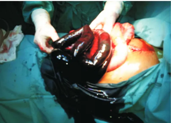
Laboratory investigations showed leucocytosis (WCC 15000cells/mcL), and Hb. (10 grams /dL). The patient was resuscitated well in ICU with IV fluids and fresh blood transfusion. The blood was sent for culture and sensitivity. Then cephalosporin antibiotic was started.We proceeded with emergency exploratory laparotomy under general anaesthesia.There was foul smelling peritoneal fluid with minimal amount of blood and gangrenous part of jejunum of 4 feet length (Figure 1). The rest of bowel was noted as healthy bowel with good vascularity and active peristalsis (Figure 2). The full length of the colon was found to be healthy. Thorough peritoneal wash was given with normal saline.The gangrenous portion of jejunum was removed ant healthy vascular borders of residual part of healthy jejunum and ileum were anastomosed as primary anastomosis. Peritoneal drainage tube was kept after complete wash with normal saline.
Blood culture showed no growth of microorganism. Mild sepsis during the postoperative period was controlled with I.V. Imipenem 1Gm 8 hourly and I.V. Metronidazole 500 mgm 8 hrly for 5 days. Clinically, there were no signs and symptoms of extension of gangrene in anastomotic site (jejuno ileostomy). The histopathology findings were consistent with ischemic colitis.
KEY WORDS:
INTRODUCTION:
CASE PRESENTATION
Figure 1 showing gangrenous jejunum.
Figure 2 showing healthy ileum.
DISCUSSION
Our case presented as acute ishemic colitis with involvement of major portion of jejunum. Ischemic colitis is the most common ischemic injury to the lower gastrointestinal tract [8].Although it is commonly seen in the elderly between 70 and79 years, it can occur in almost any age group. Atypical cases have been reported in young healthy endurance runners who would otherwise have no classical risk factors of hypertension, cerebrocardio¬vascular disease, or history of abdominal surgery[9,10,11].Colon ischemia is a clinicopathological condition that comprises a spectrum of disorders, from reversible colopathy and transient colitis to gangrene and fulminant colitis [12].
small vessels. The majority of patients are elderly. In this group, although many major predisposing factors arerecognised, such as aortic surgeryand obstructing coloniclesions, most cases are idiopathic and arise in the absence ofa proven precipitating event [14].Phlebosclerosis of the right side colon mesenteric vein is a rare cause of chronic ischemic colitis. The clinicopathologic findings of chronic phlebosclerotic ischemic colitis are different from those of conventional ischemic colitis. The pathogenesis remains unclear. The pathognomonic hallmark for radiological diagnosis of chronic phlebosclerotic ischemic colitis is the vascular calcifications in the colonic wall and mesenteric veins seen on plain abdominal radiography and abdominal CT scan [2].
Ischemic colitis is the most common form of gastro intestinal ischemia; the majority of cases resolve with nonsurgical management. Prompt recognition of full-thickness necrosis and gangrene is critical to good patient outcomes. The clinical scenario, and CT scan and endoscopic findings are essential in helping the clinician to identify patients with ischemic colitis who are at risk for full-thickness colonic necrosis. [15].CT angiogram is frequently obtained after diagnosis of ischemic colitis.CT angiogram detected more stenosis than the contrast-enhanced CT scan as the primary goal of performing the latter is not for detecting this finding. However, CT angiogram did not change the management or the prognosis of ischemic colitis. They recommend against the routine use of CT angiogram after the diagnosis of ischemic colitis. CT angiogram might be considered in more severe cases of ischemic colitis especially with right colon involvement or in instances of the need for excluding acute mesenteric ischemia based on clinical scenarios[16]. In our case, we could not proceed CT angiogram since this was already a established case of ischemic colitis. It would be better to do therapeutic angiogram for injection thrombolytic drugs into the involved mesenteric vessel.
Endovascular treatment includes the possibility of angiographically-directed catheter-aspiration embolectomy and/or catheter lysis with recombinant tissue plasminogen activator, urokinase, or pharmacotherapy with prostaglandin E1[17].
It has been shown that mesenteric atherosclerosis in surgical specimens is associated with worse survival and is an independent factor for poor long-term survival.Mesenteric atherosclerosis probably reflects systemic arterial atherosclerosis as most patients with ischemic colitis who expire in long-term followup die from cardiovascular diseases and it is judicious to treat those who survive from ischemic colitis with aggressive secondary prevention measures and should be considered as high-risk cardiovascular patients [18].
In such cases, fractionation of the thrombus using a guide wire increases the surface area that comes into contact with the fibrinolytic agent and so speeds up dissolution of the thrombus. The aim is to reopen the main arterial branches of the superior mesenteric artery, as this will allow even remaining occlusion to be well compensated for as a result of good collateral growth[19].When a definitive operation is performed for left-sided ischemic colitis, published opinion advocates for an end colostomy and rectal stump (Hartmann procedure) [15].In our case, we did not perform Hartmann procedure which is more useful procedure for distal part of gastro intestinal tract preferably colon gets involved since our case got gangrene in jejunum.
1.Boley SJ, Brandt LJ, Veith FJ. Ischemic disorders of the intestines. CurrProblSurg. 1978; 15:1–85.
2. Theodoropoulou A, Koutroubakis IE. Ischemic colitis: clinical practice in diagnosis and treatment. World J Gastroenterol. 2008;14:7302–7308.
3. Taourel P, Aufort S, Merigeaud S, Doyon FC, Hoquet MD, Delabrousse E. Imaging of ischemic colitis.RadiolClinNorth Am. 2008;46:909–924.
REFERENCES
4.Ying KS, HuangJC, Chan LP,Wu SH,Chang TY, Lee CH. Chronic Phlebosclerotic Ischemic Colitis. Chin J Radiol 2002; 27: 129-134
5. Elder K, Lashner BA, Al Solaiman F. Clinical approach to colonic ischemia. Cleve Clin J Med.2009;76:401–409.
6. Montoro MA, Brandt LJ, Santolaria S, GomollonF, Sánchez Puértolas B, Vera J et al. Clinical patterns and outcomes of ischaemic colitis: results of the Working Group for the Study of Ischaemic Colitis in Spain (CIE study). Scand J Gastroenterol. 2011;46:236–246.
7. EryigitE, HoentjenF, Barbe E, Meyel JJMV. Intestinal ischaemia caused by mesenteric inflammatory veno-occlusive disease. Netherlands J Medicine.2008;66:486-488.
8. Feuerstadt P, Brandt LJ. Colon ischemia: recent insights and advances. CurrGastroenterol Rep. 2010;12:383-90.
9. Grames C, Berry-Cabán CS. Ischemic colitis in an endurance runner. Case Rep Gastrointest Med. 2012;2012:356895.
10. CubiellaFernández J, NúñezCalvo L, González Vázquez E, GarcíaGar¬cía MJ, Alves Pérez MT, Martínez Silva I, et al. Risk factors associated with the development of ischemic colitis. World J. Gastroenterol. 2010 Sep 28;16(36):4564-9.
11. Moses FM. Exercise-associated intestinal ischemia. Curr Sports Med Rep. 2005;4:91-5.
12. Brandt LJ, Boley SJ. AGA technical review on intestinal ischemia. American Gastro-intestinal Association. Gastroenterology 2000;118:954–68.
13. Reinus JF, Brandt LJ, Boley SJ. Ischemic disease of the bowel. GastroenterolClin North Am 1990; 19: 319-343.
14.Radaelli F, Feltri M, MeucciG ,Spinz G, Terruzzi V, Minoli G. Ischemic colitis associated with rofecoxib. Digestive and Liver Disease .2005;37:372–376.
15.Washington C, Carmichael JC.Management of Ischemic Colitis.Clinics in Colon and Rectal Surgery.2012; 25(4): 228–235.
16. Sherid M, Samo S, Sulaiman S, Husein H,SethuramanSN,VainderJA. Is CT Angiogram of the Abdominal Vessels Needed following the Diagnosis of Ischemic Colitis?
A Multicenter Community Study.ISRN Gastroenterology;2014: Article ID 756926, 8pageshttp://dx.doi.org/10.1155/2014/756926 (accessed on 11th August 2016).
17. Schoots IG, Levi MM, Reekers JA, Lameris JS, van Gulik TM. Thrombolytic therapy for acute superior mesenteric artery occlusion. J VascIntervRadiol. 2005;16:317–329.
18. Reissfelder C, Sweiti H, Antolovic D, et al. Ischemic colitis: who will survive? Surgery. 2011;149:585–592.