161
Author for correspondence: Dr. Saiqa Ishtiaq
Assistant Professor, Department of Pharmacognosy, Punjab University College of Pharmacy, University of the Punjab, Allama Iqbal Campus, Lahore Pakistan
E-mail: saiqa.pharmacy@pu.edu.pk Tel.: +92-3004399827
Pharmacognostic studies of aerial parts of Colebrookea oppositifolia Sm.
Saiqa Ishtiaq, M ehvesh Bas hir Meo, Muhammad Shahar yar Khan Afridi, Shehla Akbar* and Shahid Raso ol** Punjab University College of Pharmacy, University of the Punjab, Allama Iabal Campus, Lahore Pakistan
*Lahore College of Pharmaceutical Sciences, Lahore Pakistan **Department of Pharmacy, University of Sargodha, Sargodha Pakistan
Received November 10, 2016: Revised November 25, 2016: Accepted November 29, 2016: Published online December 30, 2016
Abstract
Colebrookea oppositifolia Sm., a well-known traditional medicinal plant, belongs to the family, Lamiaceae. The research work is about the pharmacognostic characterization of C. oppositifolia which includes; macro and microscopic evaluation, phytochemical and physicochemical properties of leaf, stem and inflorescence. Transverse section (T.S.) of leaf and stem showed the arrangement of the different cells, i.e. vascular bundle, collenchyma, parenchyma, cortex, cambium, pith, etc. respectively. Histochemistry of T.S. of leaf and stem gave positive results with conc. HCI, phloroglucinol, ferric chloride and Sudan III which indicated the presence of Ca+2 oxalate crystals, lignin, tannins and volatile oils, respectively. Powder study along with histochemical analysis of leaf showed the presence of glandular trichomes, lignified fibres, spiral vessels and lignified tracheids while, stem and inflorescence powder showed collenchyma, parenchyma, covering trichomes, papillose cells, uniseriate tapering trichomes, round pollen grains and corolla. In fluorescence analysis, different colors were observed under different lights. Phytochemical analysis of MeOH extract indicated the presence of alkaloids, glycosides, flavonoids, sterols, triterpenoids and tannins. Physicochemical analysis, i.e. ash values and extractive values were performed. These results will help in identification and quality control of C. oppositifolia medicinal material.
Key words: Colebrookea oppositifolia, histochemical analysis, pharmacognostic, phytochemical
PHYTOMEDICINE An International Journal
1. Introduction
Plants are widely used as raw ingredients for many preparations in conventional medicine system. To confirm the genuineness of the raw ingredients and to detect the presence of adulterant stains. Comprehensive and detailed pharmacognostic evaluations are required for each raw drug. Usually these raw drugs were collected by the traditional workers who were engaged in herbal, ayurvedic or any other system of complementary system of medicine. Their identification is commonly based on macroscopic structural features or other unique visible characteristics. Therefore, in such manual practices there is a chance of accidental collection of improper or wron g plant material. Therefore, an extensive an atomical, physicochemical and phytochemical screening was required that is helpful to avoid any ambiguity (Vaibhav and Kamlesh, 2007). Anatomical findings were supportive in elucidation of a unique drug with a major focus on quantitative microscopy, such as starch grains, Ca+2oxalate crystals, stomatal index and trichomes and
qualitative microscopy, such as arrangement of vascular bundles,
i.e. xylem and phloem tissues and other tissues (Brinda et al., 2000).
Colebrookea is a genus of plants in the Lamiaceae, first described in 1806. It contains only one known species, C. oppositifolia Sm. The plant possesses antimicrobial, antifungal and antioxidant
Annals of Phytomedicine 5(2): 161-167, 2016 Journal homepage: www.ukaazpublications.com
Print ISSN : 2278-9839 and Online ISSN : 2393-9885
attributes due to high content of flavonoids and polyphenols. It is used for treating sore eyes, corneal opacity or conjunctivitis due to its anti-inflammatory effects (Torri, 2012; Sandhu et al., 2011). The plant material is generally used to cure the diseases like epilepsy, fever, headache, and urinary problems. It possess hepatoprotective, cardioprotective and anti-inflammatory attributes. The essential oil of Colebrookea possesses fungitoxic property (Holley and Cherla, 1998; Sharma et al., 2013). Colebrookea has anthelmintic properties which is used in the management of ringworms and it is also employed in the treatment of dermatitis, nose bleeds, bleeding, bloody coughs and dysentery (Venkateshappa and Sreenath, 2013). Despite, a lot of therapeutic uses were ascribed to this plant material, there is not any comprehensive pharmacognostical information available on structural morphology and other physicochemical standards, generally needed for the quality control for the plant. Therefore, the present research comprises of anatomical and structural evaluations, assessment of physicochemical parameters and phytochemical nature of the C. oppositifolia.
2. Materials and Methods
2.1 Plant material
The plant was collected from Botanical garden of Government College University, Lahore and was authenticated by Miss Uzma Hanif, Lecturer, Department of Botany, Government College University, Lahore Pakistan, based on authenticity established by Gamble, Benthum and Hooker. A specimen of plant was deposited in herbarium of Government College University, Lahore under Voucher Specimen No: GC. Herb. Bot. 2941. Aerial parts of the plants, i.e. leaves, stem and inflorescences were separated. All plant parts were dried under shade, were powdered and preserved in brown containers in dry place.
Copyright @ 2016 Ukaaz Publications. All rights reserved. Email: ukaaz@yahoo.com; Website: www.ukaazpublications.com
Original article
2.2 Chemicals and instruments
Analytical grade alcohol, methanol, chloroform, n-hexane, ethyl acetate, glacial acetic acid, sulphuric acid, hydrochloric acid, ferric chloride, lead acetate, phloroglucinol, safranin, fast green, Sudan
III, glycerin were used in the present study. All the chemicals were purchased from Sigma Aldrich, Germany. Distilled water and chemical reagents: (distilled water and all the reagents were prepared in th e laboratory durin g histochemical an d ph ytochemical evaluation): Wagner’s reagent, Dragendroff’s reagent, Millon’s reagent, Molisch’s reagent. Microscope (Labomed, U.K.) and Canon Powershot SX 220HS Japan.
2.3 Macroscopic evaluation
All macroscopic evaluations of leaf, stem and inflorescence were carried out on 5 samples of each part. The taxonomical description was made according to the related articles and the data given in different books (Kokate et al., 2006).
2.4 Microscopic evaluation
2.4.1 Transverse section cutting
Fresh leaf and stem were immediately fixed in formalin: acetic acid:70% alcohol (5:5:90) for 24 h. T.S. of leaf and stem were made by commonly used blade and razor method. Sections were stained with safranin and fast green dye (Sylevester and Ruzin, 1994).
2.4.2 Histochemistry of T.S.
The sections of leaf and stem were treated with various chemicals to note the chemical reaction and color change. Inferences were made to evaluate the particular reaction for histochemical analysis in the plant tissues accordingly. Phloroglucinol, Conc. HCl, iodine solution, ferric chloride and Sudan III solution were used to locate the presence of Ca+2 oxalate crystals, lignin, starch, tannins and oil
globules, respectively. After treatment with particular reagents, the sections were observed under light microscope and their photographs were taken using digital camera. (Christodoulakis et al., 2015).
2.4.3 Powder study
Chloral hydrate (75%) solution was used as clearing reagent prior to observation of powder drug using microscope. Slides of powdered leaf, stem and inflorescence were prepared according to the prescribed procedures. Photographs were taken, using microscope by means of digital camera (Upton et al., 2016).
2.4.4 Histochemistry of powder
The prepared slides of powdered leaf, stem and inflorescence were treated with chloral hydrate, phloroglucinol, HCl and safranin reagents to view and stain the elements. The slides were seen under microscope and were photographed (Biggs, 1985).
2.5 Fluorescence analysis
U.V. fluorescence analysis of powdered leaf, stem and inflorescence was carried out by treating them with different reagents and was observed in ordinary light and U.V. light (short wavelength of 254 nm and long wavelength of 366 nm) (Schoor et al., 2015).
2.6 Phytochemical analysis
The phytochemical analysis was carried out according to the standard procedures (Kumar et al., 2016).
2.7 Physicochemical analysis
Powdered samples were subjected to physicochemical analysis for their extractive values along with total ash, water soluble ash, and acid insoluble ash (Evans, 1989; Harborne, 1998).
2.8 Statistical analysis
The simple statistical analysis was carried out for calculating the mean and the standard error of mean.
3. Results
3.1 Macroscopic characters
Plants is 1-3 m tall, with pale hairy stout square branches (Figure 1). Petiole (0.82.5 cm), leaf blade (1020 × 37 cm), base broadly cuneate to rounded, margin crenulateserrulate, apex -long acuminate, adaxially rugulose and puberulent, abaxially densely-tomentose to lanate-densely-tomentose. Numerous tiny white flowers occur in panicles of upright spikes - (10-15 cm long) branches - (4-7 cm); verticillasters - 10-18 flowered, globose; bracteoles - (1 mm), densely tomentose outside, glabrous inside. Flowers - (2 mm), pistillate: calyx campanulate - (1.5 mm-6 mm) in fruit, tube very short, visibly ribbed; teeth subulate, later spinescent, ± purple. Corolla tube puberulent, lower lip slightly longer than upper lip, with middle lobe ovate. Stamens inserted on apical part of tube, included. Style 2 × as long as corolla. In bisexual flowers: calyx minute - (0.6 mm), corolla (3 mm); upper lip ovate-oblong - (0.5 mm), straight, emarginate; lower lip elongated, spreading - (1.5 mm), middle lobe ovate-oblong, 2 × as long as ovate lateral lobes. Style erect, slightly longer than corolla. Nutlets are obovoid - (1 mm), yellow-brown in color, with a small basal white scar (Table 1). Flowering period ranges from January-March, through March-April (Madhavan et at., 2011).
Table 1: Macroscopic characters of C. oppositifolia Plant Part Fe at ur e s O bse r v at io ns Leaf Leaf blade (10-20 × 3-7 cm)
Base Broadly cuneate to rounded Margin Crenulate-serrulate Apex Long acuminate, adaxially
rugulose and puberulent, abaxially densely-tomentose to lanate-tomen tose
Color Dirty dark green
Textur e Hairy
Fracture Crisp
St e m Height 1-3 m
Color Dust brown
Textur e Glabrous
Fracture Woody and fibrous I nf lo re s c enc e Appea rance Panicles of upright spikes
Color White
Length of 4-7 cm flowering
br anch
Size Tiny (2 mm)
Bracteoles Present (1 mm) Pistillate Yes
Calyx Campanulate
Corolla Puberulent
Stamens Inserted on apical part Style Erect and long Flowering January-March, through
period March-April.
3.2 Microscopic characters
3.2.1 Transverse section
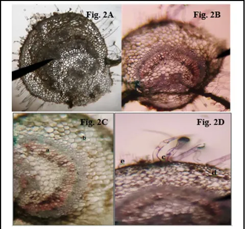
T.S. of leaf showing; vascular bundles with xylem and phloem in a lunar shape, ground tissue and collenchymas, trichomes (uniseriate, multicellular), epidermis, cuticle (Figure 2).
Figure 2 ( A,B,C,D): Micr oscopy of the leaf of C. op positifo lia. (A) T.S. of leaf before staining, (B) T.S. of leaf after staining, (C) T.S. of leaf showing: a) vascular bundles with xylem and phloem in a lunar shape, b) ground tissue and collenchyma, (D) c) Trichomes (uniseriate, multicellular), d) epidermis, e) cuticle.
T.S. of stem shows; raphides, druses and Ca+2oxalate crystals along
with cellular content and oil globules in collenchyma of hypodermis, sclerenchyma. Vascular bundles were visible showing xylem and phloem tissue. Cambium and sclerenchyma was also observed, pith, ground tissue and parenchymatous tissue with medullary rays of primary parenchymatous tissue were also seen (Figure 3).
Figure 3 (A, B, C, D, E): Microscopy of the stem of C. oppositifolia. (A) T.S. of stem before staining, (B) T.S. of stem after staining, (C) T.S. of stem where: a) raphides and druses and Ca+2oxalate crystals along with cellular content and oil globules in collenchyma of hypodermis, b) sclerenchyma, c) vascular bundles showing xylem and phloem, (D) T.S. of stem where: a) cambium and sclerenchyma between cortex and vascular bundles, b) vascular bundles showing primary and secondary xylem, c) pith and parenchymatous tissue, (E) T.S. of stem where: a) pith and ground tissue, b) oil globules in collenchyma: c) vascular bundles and medullary rays of primary parenchymatous tissue.
3.2.2 Histochemical analysis of T.S.
Histochemistry of leaf shows the presence of Ca+2 oxalate crystals,
tannins, volatile oils and lignins (Figure 4).
Figure 4 (A, B, C, D, E) : Histochemical analysis of powder of leaf of C. oppositifolia. (A) Conc. HCl gave effervescence with Ca+2-oxalate
crystals, (B) A magenta coloration in the vascular bundle indicated positive result, (C) Iodine solution did not give blue color to the starch granules, (D) Ferric chloride turned the tannins black giving a +ve result, (E) Sudan III gave +ve result and turned the oil globules red.
Histochemistry of stem shows the presence of Ca+2 oxalate crystals,
Figure 5 (A, B, C, D, E): Histochemical analysis of powder of stem of C. oppositifolia. (A) Conc. HCl gave effervescence with Ca+2-oxalate
crystals, (B) A magenta coloration in the vascular bundle indicated positive result for lignins, (C) Iodine solution did not give positive result to the starch granules, (D) Ferric chloride turned the tannins black giving a +ve result, (E) Sudan III gave +ve result and turned the oil globules red.
3.2.3 Powder microscopy
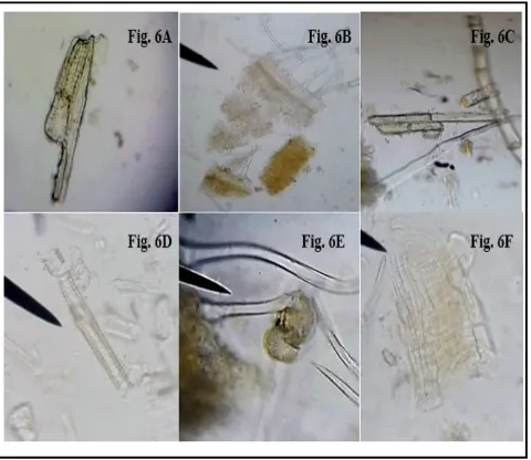
The powder microscopy of leaf shows; glandular trichomes with spiral vessels. The lignified spongy mesophyll tissue and along with tracheid vessels and parenchyma tissues of upper epidermis was also observed (Figure 6).
Figure 6 (A, B, C, D, E, F): Powder microscopy of the leaf of C. oppositifolia. (A) Lignified fibres with moderately thickened and pitted walls, (B) Capitate glandular trichomes in surface view and part of underlying palisade, (C) Lignified fibro-vascular structures in spongy mesophyll, (D) Fragment of lignified vessel with spiral thickening, (E) Glandular trichome with unicellular stalk and radiate bladder-like head with common cuticle, (F) Tracheid vessel and parecnchyma of upper epidermis.
The powder microscopy of stem shows; trichomes, lignified vessels, paranchyma and collenchyma tissues (Figure 7).
Figur e 7 (A, B, C, D): Powder micr oscopy of the stem of C. op pos itifolia. (A) Large cells with slightly thickened walls of parenchyma from pith, (B) Thin-walled, longitudinally elongated cells with occasional and scattered covering trichome base in epidermis (surface view), (C) Surface view of collenchyma in hypodermis, (D) Group of lignified vessels along with underlying parenchyma.
The powder microscopy of inflorescence revealed the presence of tapering and glandular trichomes and tracheids. There was sinous walled cells in outter epidermis along with papillose straight walled cells (Figure 8).
3.2.4 Histochemical analysis of powder
Histochemistry of powdered leaf shows venation in palisade mesophylls in hypodermis, lignified fibres, parenchyma and collenchyma tissues (Figure 9).
Figure 9 (A, B, C, D): Histochemical analysis of powder of leaf of C.oppositifolia. (A) Vein terminations in palisade with underlying hypodermis, (B) Large-celled, lignified and pitted par enchyma, (C) Group of lignified fibres with pitted walls, (D) Collenchyma in upper epidermis with underlying hypodermis showing lignification.
The histochemistry of powdered stem shows lignified vessels, glandular and conical covering trichomes and tracheids (Figure 10).
Figure 10 (A, B, C): Histochemical analysis of powder of stem of C.oppositifolia. (A) Glandular trichome with multiple thin-walled cells radiating with a common cuticle to form spherical head, (B) Lignified vessels and tracheids form pith, (C) Short conical covering trichomes with swollen base.
The histochemistry of inflorescence shows papillose cells and glandular trichomes (Figure 11).
Figure 11 (A, B): Histochemical analysis of powder of inflorescence of C. oppositifolia. (A) Inner epidermis of corolla with papillose cells, (B) Multicellular base of glandular trichome.
3.3 Fluorescence analysis
The fluorescence analysis revealed various colors of the extracts under ordinary light, short wavelength (254 nm) U.V. light, and Long wavelength (366 nm) U.V. light (Table 2).
Table 2: Fluorescence analysis of C. oppositifolia
Black Yellowish Orange Neon Yellow Black Orange Sharp Yellow Black Orange Amber Yellow Picric acid 12 Neon Blue White Light Green Neon Blue White Light Green Neon Red Brown Emerald Green MeOH 11 White Orange Dirty White White Light Yellow Hazy Brown Peach Yellow Amber Yellow Petroleum ether 10 Black Yellowish Brown Ruby Red Yellowish Brown Orange Maroon Orange Dark Brown Blackish Brown Aniline 9 Peach Orange Brown Greenish Yellow Peach Brown Brown Neon Red Brown Emerald Green CHCl3 8 Light Green Orange Brown Greenish Brown Light Green Orange Brown Dirty Yellow Light Green Dark Brown Translucent Green 50%HCl 7 Light Green Yellowish Orange Brownish Yellow Light Green Orange Mustard Yellow Pistachio Green Orange Light Brown 50% HNO3 6 Light Green Orange Brown Greenish Brown Light Green Orange Light Brown Pistachio Green Brown Light Green 50%
H2SO4 5 Black Yellowish Brown Dark Brown Black Brown Dark Brown Black Yellowish Brown Dark Brown
5% FeCl3 4 Green Orange Dark Brown Green Orange Brown Amber Yellow Orange Brown Brown Yellowish Green 5% NaOH 3 Light Green Light Brown Dirty Brownish Green Light Green Light Brown Light Brown Light Green Light Brown Dirty Green Water 2 Light Green Brownish Green Brownish Green Light Green Light Brown Saw dust Brown Light Green Dark Green Dirty Green Powder 1 Long waveleng th (λ=366
nm) Short
wavelength (λ=254nm) Ordinary
light Long
wavelength (λ=366 nm) Short
wavelength (λ=254 nm) Ordinary
light Long
wavelength (λ=366 nm) Short
wavelength (λ=254 nm) Ordinary light Inflorescence Stem Leaf Reagent S. No. Black Yellowish Orange Neon Yellow Black Orange Sharp Yellow Black Orange Amber Yellow Picric acid 12 Neon Blue White Light Green Neon Blue White Light Green Neon Red Brown Emerald Green MeOH 11 White Orange Dirty White White Light Yellow Hazy Brown Peach Yellow Amber Yellow Petroleum ether 10 Black Yellowish Brown Ruby Red Yellowish Brown Orange Maroon Orange Dark Brown Blackish Brown Aniline 9 Peach Orange Brown Greenish Yellow Peach Brown Brown Neon Red Brown Emerald Green CHCl3 8 Light Green Orange Brown Greenish Brown Light Green Orange Brown Dirty Yellow Light Green Dark Brown Translucent Green 50%HCl 7 Light Green Yellowish Orange Brownish Yellow Light Green Orange Mustard Yellow Pistachio Green Orange Light Brown 50% HNO3 6 Light Green Orange Brown Greenish Brown Light Green Orange Light Brown Pistachio Green Brown Light Green 50%
H2SO4 5 Black Yellowish Brown Dark Brown Black Brown Dark Brown Black Yellowish Brown Dark Brown
5% FeCl3 4 Green Orange Dark Brown Green Orange Brown Amber Yellow Orange Brown Brown Yellowish Green 5% NaOH 3 Light Green Light Brown Dirty Brownish Green Light Green Light Brown Light Brown Light Green Light Brown Dirty Green Water 2 Light Green Brownish Green Brownish Green Light Green Light Brown Saw dust Brown Light Green Dark Green Dirty Green Powder 1 Long waveleng th (λ=366
nm) Short
wavelength (λ=254nm) Ordinary
light Long
wavelength (λ=366 nm) Short
wavelength (λ=254 nm) Ordinary
light Long
wavelength (λ=366 nm) Short
3.4 Phytochemical screening
The phytochemical screening of plant material mainly revealed the presence of terpenoids, sterols, glycosides, flavonoids, alkaloids, carbohydrates, tannins, phenols and lignins (Table 3).
Table 3: Phytochemical analysis of C. oppositifolia
Group Name of tests Leaf Stem Inflorescence
Terpenoids Liebermann’s test + + +
Sterols Salkowaski test + +
-Glycoside Keller-Killiani test + - +
Flavonoids NaOH test + + +
Alkaloids Dragendroff’s test + + +
Proteins Millon’s test - -
-Carbohydrates Molisch’s test + + +
Saponin Foam test - -
-Lipids Soap formation test - -
-Tannins Braymer’s test - + +
Phenols Ferric chloride test + - +
Lignins Lignin test - +
-Fixed oils
and fats Spot test - -
-3.5 Physicochemical constants
The extractive values of leaf in distilled H2O and MeOH are high
while, for inflorescence and stem, ethyl acetate showed more extractive value as compared to the rest of the solvents (Table 4).The ash values of leaf and stem showed high content of water-soluble ash followed by acid inwater-soluble ash but inflorescence showed the highest acid insoluble ash (Table 5).
Table 4: Extractive values of C. oppositifolia
Parameters Percentage yield ± (SEM)
Leaf Stem Inflorescence
n-Hexane 1.367 ± 0.001 0.359 ± 0.004 1.287 ± 0.006 Chloroform 1.867 ± 0.009 0.532 ± 0.005 1.352 ± 0.005 Methanol 2.629 ± 0.001 1.759 ± 0.020 2.584 ± 0.006 Ethyl acetate 1.296 ± 0.002 4.392 ± 0.015 3.967 ± 0.012 Distilled H2O 2.416 ± 0.001 3.265 ± 0.008 2.470 ± 0.015
Table 5: Ash values of C. oppositifolia
Parameters Values % (w/w)
Leaf Stem Inflorescence
Total ash 20.8233 ± 0.0052 5.8473 ± 0.0327 20.933 ± 0.6187 Acid insoluble
ash 27.7908 ± 2.1744 7.9894 ± 0.08694 28.0743 ± 4.2496 Water soluble
ash 72.0166 ± 1.4668 157.4138 ± 2.6247 16.480 ± 5.5786
4. Discussion
Natural remedies recorded from medicinal plants are favorable choices over synthetically formulated drugs. It is of importance to individualize the consumption of herbal products not merely based on the knowledge of folklore use but through systematic studies (Dewick, 2 00 2). P harmacogn ostic, ph ysicochemiscal an d
phytochemical evaluations are of robust entitlement of entire crude drug profile in context of its pharmaceutical and pharmacological importance (Panda, 2004).
C. oppositifolia is a local plant, commonly used by natives for folklore use. The transverse section of leaf and stem showed the basic profile of botanical anatomy with a little contrast in having lunar shaped vascular bundles in the leaf while a continuous vascular bundle with primary and secondary xylem and phloem with presence of sclerenchyma all around; fibres and trachieds with annular and spiral thickening present in the medullary region and parenchyma along with collenchyma in papillose form with oil globules were indicated in prominence are all represented in Figures 1 and 2.
Histochemical evaluation of transverse sections were performed to get a clear picture on preliminary scale at cellular level using conc.
HCl, phloroglucinol which showed presence of lignins in the leaf and stem while iodine solution gave negative result for starch, Ferric chloride solution specified the presence of tannins and Sudan III
dye coloured the oil globules in the ground tissue of leaf and pith area of stem (Figures 4 and 5).
The photomicrograghic evaluations revealed microscopic features particular to each part of C. oppositifolia used for powder microscopy in general and basic characters; they were prominent especially the covering trichomes shown in Figures 6, 7 and 8 (Beck, 2010). While, the HCl and phloroglucinol treated powder macerates of C. oppositifolia leaf, stem, and inflorescence were stained with safranin and observed the results were given in Figures 9, 10 and 11 (Jensen, 1962)
The fluorescence analysis was a valuable and modest method for the identification of fluorescent compounds. Different compounds give fluorescence when exposed to U.V. light. The powder of leaf, stem and inflorescence gave various fluorescence in short and long wavelength of U.V. light given in Table 2 (Joshi, 2012).
The methanolic extracts of C. op positifo lia leaf, stem and inflorescence established the presence of various important active constituents like terpenoids, sterols, glycosides, flavonoids, alkaloids, carbohydrates, etc. (Table 3), which may be responsible for the folklore uses of this plant.
Quantitative analysis using the physicochemical parameters of extractive values of C. oppositifolia leaf, stem and inflorescence were determined for further standardization of the powder and evaluation was performed with n-hexane, chloroform, MeOH, ethyl acetate and distilled water; the results were tabulated in Table 4 (Mukherjee, 2002). Ash values for the powdered plant parts was helpful in the determination of extraneous material content adhered to the plant as well as the amount of siliceous matter in the left over residue, respectively; the results were presented in Table 5 (Khandelwal, 2008).
5. Conclusion
values of leaf and ethyl acetate for stem and inflorescence were significan t. Ash valu es added more stren gth to crude drug standardization with prominent results indicating the involvement of extraneous matter. Such study on the macro and microscopic anatomy, prelimin ary ph ytoconstitu ent screen in g an d physicochemical parameters are important informations which may be useful in verification and contamination for quality control of this therapeutic plant afterwards.
Acknowledgements
The present research work was funded by the Punjab University College of Pharmacy, University of the Punjab, Lahore Pakistan.
Conflict of interest
We declare that we have no conflict of interest.
References
Beck, C.B. (2010). An introduction to plant structure and development: Plant anatomy for the Twenty-First Century. 2ndRev. Cambridge University Press, U.K., pp:260,334.
Biggs, A.R. (1985). Detection of impervious tissue in tree bark with selective histochemistr y a nd fluorescence microscopy. Sta in Technology, 60(5):299-304.
Brinda, P.; Saraswathy, A. and Jayaraman, P. (2000). Micromorphological identification of the medicinal bark of Ventilago madraspatana. J. Med. Arom. Plant Sci., 23:619-622.
Christodoulakis, N.S.; Mamoucha, S.; Termentzi, A. and Fokialakis, N. (2015). Leaf str ucture and histochemistry of the hardy ever green Euphorbia characias L. (Mediterranean spurge). Floral morpho-logy, Distribution, Functional Ecology of Plants, 210:13-18. Dewick, P.M. (2002). Medicinal Natural Products: A biosynthetic approach.
Ill Ed. John Wiley and Sons, New York, U.S.A., pp:2-5.
Evans, W.C. (2003). In: Trease and Evans Pharmacognosy, 15th edn., Saunders, London, pp:545-547.
Harborne, J.B. (1998). Phytochemical Methods, (3rd Ed), Chapman and Hall, London, U.K., pp:6-7.
Holley, J., and Cherla, K. (1998). Medicinal plants sector in India. A review of medicinal and aromatic plants program in Asia (MAPPA). SARO/ IDRC, New Delhi, India, pp:109.
Jensen, W.A. (1962). Botanical History: Principles and Practice. Vol. 12. W. H. Freeman and Company, New York, U.S.A., pp:72, 74, 93, 197, 205, and 260.
Joshi, D.D. (2012). Herbal drugs and fingerprints: Evidence based herbal drugs. 1st Ed. Springer, New Delhi, India, pp:116.
Khandelwal, K.R. (2008). Practical Pharmacognosy. 9th Ed. Pragati Books Pvt. Ltd. Pune, India, pp:158-160.
K okate , C.K .; Pu rohit, A.P. and Gokhal e, S.B . (2 006). Test book of Pharmacognosy, 42nd Ed. Nirali Prakashan. Pune, India, pp:42. K umar, P.; Kumar, J.; K umar, R. and Dubey, R.C. (2016). Studies on
phytochemical constituents and antimicrobial activities of leaves, fruits and stems of Solanum nigrum L. Asian Journal of Plant Science and Research, 6(4):57-68.
Madhavan, V.; Yadav, D. K.; Gurudeva, M. and Yoganarasimhan, S. (2011). Pharmacognostical studies on the lea ves of Co lebr ook ea oppositifolia Smith. A.J. Trad. Med., 6(4):134-144.
Mukharjee, P.K. (2002). Quality control of herbal drugs: An approach to evaluation of botanicals. 1st Ed. New Delhi, India, pp:187-189. Panda, H. (2004). Handbook on herbal drugs and its plant sources.
National Institute of Industrial Research, Delhi, India, pp:4. Schoor, S.; Lung, S.C.; Sigurdson, D. and Chuong, S. D. (2015). Fluorescent
staining of living plant cells. In: Plant microtechniques a nd protocols, Springer International Publishing, pp:153-165. Sylevester, A.W. and Ruzin, S.E. (1994). Light microscopy I: Dissection and
Microtechnique. The Maize Handbook. Springer, New York, USA, pp:8 3 .
Torri, M.C. (2012). Mainstreaming local health through herbal gardens in India: A tool to enhance women active agency a nd primary healthcare. Environ. Dev. Sustain., 14:389-406.
Upton, R.; Graff, A.; Jolliffe, G.; Länger, R. and Williamson, E. (2016). American Herba l Phar macopoeia : Bota nical Pha rma cognosy-Microscopic Characterization of Botanical Medicines. CRC Press.
U.K., pp:197.
Vaibhav, S. and K amlesh, D. (2007). Pharmacognosy: The Changing Scenario, Pharmacog. Rev., 1(1):1-6.