World Journal of Advance Healthcare Research Volume 1, Issue 1. 2017
REVIEW OF ANIMAL LISTERIOSIS, DIAGNOSIS, ETIOLOGY AND ITS PUBLIC
HEALTH
Dr. Haftom Abay Hagos*
Ministery of Livestock and Fisheries, Export Abattoir Inspection and Certefication Directorate Adissabeba Ethiopia.
Received date: 20 April 2017 Revised date: 08 May 2017 Accepted date: 25 May 2017
LIST OF ABBREVIATIONS
AFL: Pamplified fragment length polymorphism HSIM: Hydrogen Sulphide, Indol and Motility LLO: listeriolysin–O
LPM: Lithium Chloride Phenylethanol Moxacalatam medium
MHA: Mueller-Hinton agar MOX: Modified Oxford Agar OXA: Oxford Agar
PALCAM: Chrisite Atkins Muench Peterson PCR: polymerization chain reaction
PFGE: pulsed-field gel electrophoresis
PI– PLC: Phosphatidylinositol specific phospholipase RAPD: Random amplified polymorphic DNA SPP: Species
TNF: Tissue Necrotizing Factor
TSYEA: Tryptic Soya Yeast Extract Agar
INTRODUCTION
Listeriosis is an infectious disease affecting animals, birds and man. It is caused by an intracellular bacterium
L.monocytogenes. There has been an increasing concern in recent years about the role of foods of animal origin in the transmission of listeriosis to man. The disease is characterized by septicaemia and nervous symptoms in animals as well as man. Listeriosis is one of the important emerging food-borne bacterial zoonotic diseases of human being acquired through consumption of contaminated food of animal origin.[31] Food safety has appeared as a significant worldwide issue with international trade and public health implication.[29] The available current literature shows that caused by Listeria monocytogenes, have been reported from a wide variety of food types and clinical samples in various countries of the world. Human listeriosis is a public health problem of low incidence but high mortality.[39]
Review Article www.wjahr.com
Issue: 1. Page N. 9-24Year: 2017
HEALTHCARE RESEARCH
ABSTRACT
Listeriosis is a sporadic bacterial infection that affects a wide range of animals, including people and birds. It is seen worldwide, more frequently in temperate and colder climates. There is a high incidence of intestinal carriers. Encephalitis or meningoencephalitis in adult ruminants is the most frequently recognized form. They are 7 types of listeria species but L.monocytogenes is the only pathogenic for both humans and animals. The aim of this manuscript was to review and compile the available literature of listeriosis in animals, diagnosis, etiology and its public health. The major clinical forms of listeriosis in cattle are encephalitis, abortion, Mastitis, Ophthalmitis, iritis and keratoconjunctivitis. There are two main forms of illness in human associated with L.monocytogenes infection such as invasive (fatal form) and non-invasive forms (mild form). People at risk of invasive listeriosis include pregnant women and their fetuses, newborn babies, the elderly and immune compromised individuals (such as cancer, transplant and HIV/AIDS patients). Every effort should be made to produce silage of good quality, with early cutting of grass, minimal contamination with soil or feces and ensuring optimal anaerobic fermentation, which will insure that the pH falls below 5.0; at that level, growth of Listeria species is inhibited. People susceptible for acquiring listeriosis should not consume unpasteurized milk and milk products. In the world, this disease is becoming an emerging bacterial disease, with low incidence but high case fatality rate. Animal food products serve as important source of many food pathogens including Listeria monocytogenes. The bacteriumis ubiquitous in the nature and which is rod shaped, Gram-positive, motile and psychotropic and has been isolated from a variety of foods (Milk and meat products) and environmental sources like soil, dust, water, sewage, decaying vegetation, etc.
KEYWORDS: Food of animal origin, Listeria specie, L.monocytogene.
Corresponding author: Dr. Haftom Abay Hagos
World Journal of Advance Healthcare Research Volume 1, Issue 1. 2017 Bovine Listeriosis a sporadic bacterial infection most
commonly manifested encephalitis and meningo encephalitis in adult animals associated with feeding poorly fermented silage during the winter months.[58] The genus Listeria consisted of seven species Listeria monocytogenes, L. ivanovii, L. innocua, L. welshimeri, L.murray, L. seeligeri and L. grayi.[32] Of these, L grayi L. monocytogenes are pathogenic[25] While, L. monocytogenes has been known to cause listeriosis in humans and animals.[42] L. ivanovii, which causes abortion in animals.[26] Sporadic human infections due to
L. seeligeri and L. innocua have also been reported,[36] the rest species reported are not identified properly.[42] Many researchers consider an infective dose of 102-105
Listeria monocytogenes of food stuff to be enough to cause listeriosis in humans.[33] Among the 13 serotypes of Listeria monocytogenes (1/2a, 1/2b, 1/2c, 3a, 3b, 3c, 4a, 4b, 4c, 4d, 4e, 4ab, and 7), the most common causes of listeriosis are: 1/2a, 1/2b, and 4b.[23]
Listeriosis is ubiquitous in the environment and they are often found in animal products such as raw milk and raw meat due to unsanitary practices during milking and slaughtering.[41] Listeria species with its unique physiological characteristic can survive in temperatures from -7°C to 45°C.[12] Its optimum growing temperature is around 37°C and it is able to grow, although slowly, at temperature as low as 0°C. As a result, it can multiply in properly refrigerated food after contamination. The organism can also tolerate a pH between 5.4 and 9.6.[37] Listeriosis is a serious disease, primarily transmitted from man to man or from animal to man mostly through ingestion of the organism with contaminated through various foods: raw milk products, raw meat products, raw vegetables and sea foods as sources of food borne listeriosis. Of particular concern are ready-to-eat foods that are refrigerated before consumption and those that do not undergo any substantial heat treatment.[26] Thus, farm animals and their environment may present an important source of food contamination and infections for humans[21] Listeria monocytogenes, is a pathogen of food safety concern as it can induce disease in humans and can be transferred to food products derived from animals.[44] listeriosis is transmitted via three main routes: direct contact with animals, cross infection of newborn babies in hospital and food borne infection.[11] Listeriosis is a relatively rare but serious disease with high case fatality rates 20–30%.[31] Listeriosis is a high-risk disease for pregnant women and alcoholics, drug abusers, patients with corticosteroid therapy, AIDS patients and the elderly.[55] Infection acquired in early pregnancy may lead to abortion, still birth or premature delivery. When listeriosis is acquired late in pregnancy it can be transmitted transplacetally and lead to neonatal listeriosis. The first human case of listeriosis has been reported by Nyfeldt in 1929.[56] Today listeriosis is regarded as food born disease of serious public health concern due to the great mortality rate (20-30%).[57]
Listeriosis has so far been reported, which were known to cause serial deaths in a number of individuals and in different regions, especially in Europe and the USA.[105] However, in most African countries, there are a few reports on Listeria species and listeriosis, when compared to the Europe and USA. This is because; the organism has not been given much attention, might be due to lack of adequate facility and life style differences. Published information on the status of food borne Listeriosis and Listeria species is limited in Ethiopia. A study conducted in Addis Ababa city showed that the prevalence of 32.6% and 5.1% and Gonder town 18.75% and 6.25% for other Listeria species and L. monocytogenes to in foods of animal origin.[59]
Listeriosis has been treated by various drugs which were effective but reports showed that the trend goes towards resistance.[104]
LITERATURE REVIEW
Etiology
The Genus Listeria contains seven species but one is most pathogenic for both animal and human being. The most important species is L. monocytogenes a gram-positive, facultatively anaerobic bacillus 0.5 to 2 microns long and 0.5 microns in diameter that is motile at temperatures between 20°C and 25°C. It is beta-hemolytic in blood agar and forms a narrow band of hemolysis around the colonies (unlike L. ivanovii, which forms a wide band). A noteworthy characteristic of L. monocytogenes is its ability to grow at low temperatures; at a pH between 6 and 9, it can reproduce at temperatures from 3°C to 45°C. It is a facultative, intra-cellular parasite of the reticuloendothelial system. For purposes of epidemiological LISTERIOSIS 169 research, L. monocytogenes is subdivided into 11 serovars. Most human (92%) and animal cases are caused by serovars 4b, 1/2b, and 1/2a. Listeria can be divided into 16 serovars on the basis of somatic and flagellar antigens and there is considerable genetic diversity between serovars.[56] Listeria monocytogenes has thirteen serotypes, but only three serotypes; serotype 4b, 1/2a and 1/2b are responsible for the majority of veterinary and human listeriosis cases. Serotype 4b has been identified as the cause of most human listeriosis cases where as serotypes 4a and 4c are most of the time limited to animals.[68] However, Bundrant et al.[69] that the outbreak was caused by an unusual L.monocytogenes serotype 4b strain, which was classified into lineage III with clinical signs of listeriosis in cattle.
World Journal of Advance Healthcare Research Volume 1, Issue 1. 2017 Lineage III isolates is very rare, but significantly more
common in animal than human isolates.[70]
Morphology and growth characteristics of listeriosis Listeria species are small, gram-positive, non acid fast, non spore forming and non capsulate coccobacilli measuring 0.5 to 2mm x 0.4 to 0.5 mm. listeria has typical Gram positive cell wall. They are facultative anaerobes that grow best under reduced oxygen and increased carbon dioxide concentration. Growth occurs at 4 to 45 co, with an optimum temperature of 30 to 37 co. simple laboratory media support growth preferably at an alkaline or neutral PH. Listeria tolerates 0.04% potassium tellurite, 0.025% thallium acetate , 3.75% potassium thiocyanate, 10% NaCl and 40% bile in media. Most strains grow over a PH range of 5.5 to 9.6. It has greater heat tolerance than other non spore forming bacteria; however, short time high temperature pasteurization is effective for killing listeria.[72]
Epidemilogy
Occurrence and distribution
Listeriosis was belived to be a sporadic disease, predominantly of ruminants and found worldwide. Although listeriosis is worldwide distribution it has been an important problem in North America, Europe, Britain, New Zealand and Australia.[73,74,75] In the northern hemisphere, listeriosis has a distinct seasonal occurrence with the highest prevalence in the winter months (between December and may).[56]
Abortion is obviously associated with pregnancy, the majority of encephalitis cases occur in February and March.[76,77] The reason for this season distribution is obscure but the peak of infection is coincidental with late pregnancy when they are fundamental changes in animal immune status.
Cattle show a similar age incidence to sheep with the majority of cases two to three –olds although cattle appear much less susceptible to listeriosis than sheep.[78] The organism was found to survive for 13 years in milk, 16 years in brain sample, 12 years in feces and 12 years in silage. It is reported to persist two years in dry soil, 11.5 months in damp soil, and two years in dry feces, three months in sheep feces, 16.5 months in cattle feces and up to 7 months in dry straw.[74] In human’s age and pregnancy related defects in cell mediated immunity have been described[79] which may explain the prevalence of listeric infections in neonates and during pregnancy. Similar mechanisms may be involved in listeric infections of ruminants.
Source of infection: the organism is commonly isolated from animal feces, human fece, farm slurry, sewerage sludge, soil, farm water trough, surface water, plants, animal feed and the walls, floors, drains etc of farms and other environments since listeria has ubiquitous nature in the environment.[74,80] L.monocytogenes have the
potential to be found in most feeds like hay, grains and formulated feeds but its multiplication is restricted due to low level of available water in food. In ruminants L.monocytogenes can be isolated from the feces and nasal secretion of healthy animals known as asymptomatic carriers of germs and therefore constitute an important reservoir of germs. The main source of L. monocytogenes in raw milk is the gastrointestinal tract of animals and the environment, skin of the teats, in particular shedding of listeria species in to milk due to mastitis.[72]
L.monocytogenes is commonly available in the silage but it does not multiply to any significant extent in effectively preserved silage which is characterized by anaerobic storage, high density, high concentration of organic acid and a PH below 4.5.it may be present in silage which is poorly fermented. When silage contains soil its risk to contaminate by listeria is higher than non contaminated silage. Moist preserved feeds other than grass silage are at risk for listeria growth. Infected animals can be serve as a source of infection from their feces, milk, urine,aborted fetus and uterine discharges.[72] Transmission
The primary mode of transmission for listeria is through soil contamination and ingestion of contaminated feed. Calves which develop septicemic disease may acquire infection from contamination of the cow teat, from the ingestion of milk containing the organism or from cow with sub clinical bacteremia, through the navel from the environment and also as congenital infection.[72] The encephalitic form of the disease results from infection of the terminals of the trigerminal nerve consequent abrasion of the buccal mucosa form feed and or browse or infection from tooth cavities. Spinal myelities is believed to result from growth up to spinal nerves subsequent to body area infections.[73,74]
Risk factors: a number of predisposing factors have been observed or proposed as risk factors for the disease. These include factors that cause a lowering of the host animal’s resistance and factors that increase the infection pressure of the organism. In farms the latter appears the most important as risk factor for the occurrence of outbreak listeriosis. They are three risk factors for listeriosis.[72,74]
Host risk factors: regarding sensitivity, all wild and domestic animals are susceptible for to infection caused by listeria.[75] Listeriosis in cattle may develop sporadically or enzootic, having a stationary character, without tendency to disseminate in the outbreak.[72] Morbidity may vary between 5-15% sometimes 20-25% and mortality varies according to clinical form, being very high in nervous form (over 90%) and much lower in the abortive form.[76]
Management risk factors: the most common
World Journal of Advance Healthcare Research Volume 1, Issue 1. 2017 response includes poor nutritional status, transport,
sudden changes of weather to very cold and wet, late pregnancy and parturition stresses, long period of flooding with resulting poor access to pasture and overcrowding and insanitary conditions with poor access to feed supplies.[72,73]
Pathogen risk factor: the pathogen risk factor of infection increases due to a massive multiplication of L.monocytogenes in the feed or environment. The most important aspect in food hygiene is the ability of the bacteria to survive in a wide range of temperatures and to make biofilms on solid surfaces, including food processing facilities, which are more resistant to disinfectant and sanitizing agents.[77]
The organism is a facultative intracellular pathogen that can infect cells, including intestinal cell, by directed endocytosis. Bacterial superoxide dismutase protects against the bactericidal activity of the respiratory burst of the phagocyte and listerolysin O disrupts lysosomal membranes’ allowing the organism to grow in the cytoplasm.[74]
Host range: Listeria monocytogenes is a widespread zoonotic pathogen that infects variety of species including mammals (cattle, cats, rabbits, sheep, deer, Guinea pigs, goat, pig, horse, dogs, foxes, humans etc.), birds, fish and crustaceans. The most clinical listeriosis cases occur in ruminants; pigs rarely develop disease and birds are generally subclinical carriers of the organism.[81]
Listeria species are found worldwide and everywhere, in animals, in foods, in humans, in soil, in the food processing environment, in contact surfaces and also in food containers (Peeler and Bunning, 1994; FDA/ CFSAN, 2003). Until about 1960 it was associated almost exclusively with infections in animals and less frequently in humans. However, in the last 30 years it has been known that it was widely distributed in nature.[52]
L. monocytogenes has been reported several times from raw milk, in different countries; in USA, in 1987 (4.2%), in 1992 (4.1%), in 1997 (4.6%); in Canada, in 1988 (1.3%, and 5.4%), and in 1998 (2.7%); in South Africa, in 1990 (5.2%); in Ireland, in 1992 (4.9%) and in England and Wales (5.1%).[45] The source of L. monocytogenes in raw milk is mostly the gastrointestinal tract of animals and the environment, skin of the teats, in particular shedding of Listeria into milk due to mastitis.[30] In cows with mastitis, L. monocytogenes may be shed at 10,000-20,000 cells per ml of milk, with the appearance of the milk being normal and there being no inflammation of the affected quarter. So that raw milk collection has the potential of delivering
L.monocytogenes to the cheese making facility.[2] However, in most African countries, there are a few reports on listeriosis, as compared to the Europe and
USA. This could be associated with lack of awareness or lack of diagnostic facilities and limited resources together with the presence of other disease epidemics that claim more priority than listeriosis in developing countries including Ethiopia. However, nowadays there are some reports on prevalence of L.monocytogenes in different samples. For example, in Addis Ababa, Ethiopia, study conducted in 2004 showed that over all prevalence of 32.6%.out of the total 316 examined samples.[27] Also, of the samples examined (391) in Addis Ababa in 2010, (26.1%) were found to be positive for Listeria. L. monocytogenes was detected in 5.4% of the samples analyzed. It was isolated mainly from raw milk (13%).[13] Another study, reported that the carriage rate of Listeria in animal farms showed that silage had the maximum prevalence rate of the organism (75%), followed by fodder (43.5%), feed (33.5%), fecal matter (20.5%), sewage (16.6%) and soil (4.8%). It was also isolated from 2-6% of fecal samples fromhealthy people [38], which shows, humans and animals can be asymptomatic execrators, thus introducing L. monocytogenes into the environment.[51]
The incidence of listeriosisin humans is very low, in the order of 3-8 cases per million inhabitants per year in the industrialized countries.[22] Most countries within the EU have an annual incidence between 2-10 reported cases per million populations per year. However, because of its high case fatality rate, listeriosis ranks among the most frequent causes of death due to food borne illnesses: it ranks second, after salmonellosis, in the USA and France; and fourth in England and Wales.[34] The number of new illness cases, in comparison with other food borne pathogens, per year per 100,000 population in the EU and the USA, respectively, was reported recently as, Salmonella (42 vs. 14.5), Campylobacter (48 vs. 20), Listeria (48 vs. 20), E. coli (1.3 vs. 0.9), and Yersinia (2.4 vs. 3.9).[16]
Pathogenesis
Once organism is ingested then penetrates to the intestinal mucosa and results in a clinically in-apparent infection with localization of bacteria in various organs, or a fatal septicemia.According to Coetzer and Tustin[84] listeria localize themselves in the uterus of pregnantanimals and usually cause abortion if infection takes place early in pregnancy. It is not knownprecisely how bacteria reach the brain in animals developing meningoecephalitis; however,they probably gain entrance through wounds in the mucosa of the oral cavity.
World Journal of Advance Healthcare Research Volume 1, Issue 1. 2017 development of unilateral micro abscesses and the
perivascular infiltration of lymphocytes largely restricted to the brain stems (pons and medulla oblongata) are typically seen which seems to support this assumption. Recent studies in different models indicated non-neuronal cells take up bacteria more avidly than neurons, with microglia being the most easily infected. A more reliable means for L. monocytogenes to infect neurons seems to be through cell to cell spread from an infected macrophage.[84,85] Several molecular virulence determinants have been indentified that play a role in the cellular infection by L.monocytogenes and the unraveling of their mechanism of action has made of L.monocytogenes one of the most exciting models of host-pathogen interaction at the cellular and molecular levels. These virulence determinants include, among others, the internalins listeriolysin O (LLO), ActA protein, two phospholipases, a metalloprotease, Vip protein, a bile exclusion system (BilE) and a bile salt hydrolase.[86-88] Although there is polymorphism among different strains of L.monocytogenes for some of these virulence determinants, it cannot be correlated with the ability or inability of the organism to produce disease.[89] Nevertheless, internalin A or B mediates entry of L.monocytogenes into some human-cultured cell lines and crossing of the intestinal in gerbil or transgenic mice expressing its receptor, human E-cadherin, in enterocytes.
Clinical sign: Clinical outcome depends on the number of organisms ingested, pathogenic properties of the strains of listeria and the immune status of the host. Although listeriosis is manifested by three major clinical signs; meningoencephalities, abortion and septicemia, only one clinical form of the disease usually occurs in a group of animals or an individual animal. However an overlap of clinical forms of disease has been reported. In addition to these three major signs of disease, mild, mastitis, uveitis, ophthalmitis, iritis and keratoconjuctivitis have also been associated with L.monocytogenes[56]
Symptoms of disease with listeria species in human: they are two main forms of illness associated with L.monocytogenes infection such as invasive and non-invasive forms. Non non-invasive form of listeriosis is the mild form of disease, while invasive listeriosis is the severe form of disease and can be fatal.[91] The likelihood that invasive listeriosis will develop depends upon a number of factors, including host susceptibility, the number of organisms consumed and the virulence of the particular strain.[92]
Symptoms of non invasive listeriosis can include fever, diarrhea, muscle aches, nausea, vomiting, drowsiness and fatigue. The incubation period is usually 1 day (range 6 hours to 10 days).[91,93] Non invasive listeriosis is also known as listerial gastroenteritis or febrile listeriosis. Invasive listeriosis is characterized by the presence of L.monocytogenes in the blood, in the fluid of
the central nervous system (leading to bacterial meningitis) or infection of the uterus of pregnant women. The latter may result in spontaneous abortion or still birth (20% of cases) or neonatal infection (63% of cases). Influenza like symptoms, fever and gastrointestinal symptoms often occur in pregnant women with invasive listeriosis. In non pregnant adults, invasive listeriosis presents in the form of bacterial meningitis with a fatality rate of 30%, symptoms of invasive listeriosis include fever, malaise, ataxia, seizures and altered mental status[93] and the incubation period occurs before onset of invasive listeriosis and ranges from 3 days to 3 months.[91]
The invasive form of listeriosis in humans include septicemia, meningitis (or meningoencephalitis) and encephalitis (rhombencephalitis). Gastro enteric manifestations with fever also occur. Although the morbidity of listeriosis is relatively low, the mortality can reach values around 30%. In pregnant women, infection may result in abortion, still birth or premature birth and may be influenza-like signs including fever.[93] Necropsy finding: aborted fetuses of ruminants show very few gross lesions, but autolysis may be present if the fetus was retained before being expelled.[94] In abortion the pathological picture depends on the stage of pregnancy. Metritis usually occurs and results in retention of the fetal membranes. Gross lesions are tiny pin point yellow foci in the liver. Similar foci but visible only microscopically are seen in the lung, myocardium, kidney, spleen and brain[56] aborted fetuses are usually edematous and autolysed, with very large numbers of bacteria visible microscopically in a variety of tissues. Aborted fetuses due to L.ivanovii have suppurative
bronchopneumonia and lack the multifocal
hepatocellular necrosis commonly seen in abortions associated with L.monocytogenes.[74]
There are usually no remarkable lesions in the brain of affected animals, but occasionally slight clouding or pin point grayish-white foci of the meninges may be observed. Microscopic lesions are always confined to the pons, medulla and anterior spinal cord. Both white and grey matter may be involved. In the brain substance and sometimes in the meninges a remarkable perivascular cuffing cuffing with varying degrees of focal necrosis develops which is typical of listeria encephalitis.[56] In the encephalatic form, the cerebrospinal fluid may be cloudy and the meningeal vessels congested. Gross lesions are generally subtle and characterized by vascular congestion and mild discoloration of the brain stem. On occasion, the medulla shows areas of softening (malacia) and abscession. The medulla and pons are most severely involved.[94]
Diagnosis Techniques
World Journal of Advance Healthcare Research Volume 1, Issue 1. 2017 Laboratory diagnosis: confirmatory diagnosis can be
achieved by isolating the pathogen from appropriate specimens. Appropriate specimens for laboratory examination depend on the form of the disease. Cerebrospinal fluid (CSF) and tissue from the medulla and pons of animals with neurological signs should be sampled. Fresh tissue is required for isolation of organisms and fixed tissue for histopathological examination. Specimens for cases of abortion should include cotyledons, fetal abomasal contents and uterine discharges. Suitable samples from septicemic cases include fresh liver, spleen or blood.[72]
Direct Microscopy: Smears from cotyledons or from liver lesions may reveal Gram positive coco bacillary bacteria. Histopathological examination of fixed (10% formalin) brain tissue can often give a presumptive diagnosis of neural listeriosis. Micro abscesses in the brain stem usually unilateral together with perivascular cuffing are very characteristics of listeriosis.[77]
Isolation of Listeria Species
Conventional cultural method
A number of methods are available for the detection of
Listeria species. However, culture isolation is best with a detection power of 102 CFU/ml. Colonies of Listeria species in general are small, smooth, translucent, and bluish gray when viewed in normal light, but blue green sheen is visible by oblique light. The most widely used selective media is OXA (Oxford agar) agar which was developed in 1989. Most literatures recommended to use OXA agar and either one of the following media; PALCAM (Polymixin Acriflavine, Lithium chloride, Ceftazidime Aesculine Mannitol), MOX (Modified
Oxford Agar), and LPM (Lithium Chloride
Phenylethanol Moxacalatam medium) with esculine and ferric iron. The principle of the media is the ability of esculine hydrolysis by Listeria species. Since all Listeria
species utilize B-D-Glucosidase, they cleaved esculine (esculinase), evidenced by blackening of the medium. In addition, different antimicrobial agents like acriflavin, nalidixic acid, cyclohexamide are supplemented with those media to suppress the growth of other non-Listeria
species of bacteria and other microbial agents.
The two-stage enrichment method for detection of L. monocytogenes with isolation on PALCAM agar and Oxford agar[18] are widely used. The CAMP reaction is useful for identifying Listeria species. This test uses horse blood agar and streaks of hemolytic
Staphylococcus aureus and Rodococcus equi in combination with Listeria isolates. L. monocytogenes
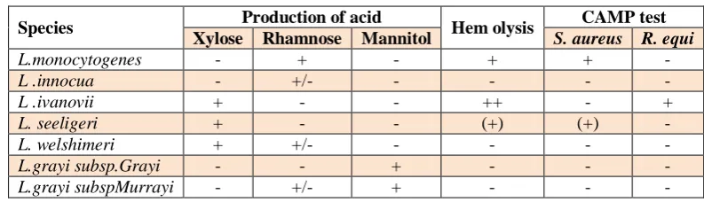
and L. seeligeri hemolytic reactions are enhanced in the zone influenced by the S. aureus streak, while the other species remain non-hemolytic in this zone. In contrast, the hemolytic reaction of L. ivanovii is enhanced in the zone influenced by R. equi.[17] For confirmed and specification, different standard biochemical tests can be used. The biochemical test used for identification and confirmation of Listeria species, tests can be done by picking pure colonies and transferring into the following biochemical media and broths. These are motility test medium (motility), blood agar (haemolysis), mannitol, rhamnose, and galactose, xylose, and Hippurate hydrolysis and xylose broths for carbohydrate fermentation testing.[20]
In general, L. monocytogenes is catalase positive, glucose fermenter with acid production but no gas formation, ferment lactose (after 3-5 days of incubation), urease negative, oxidase negative, methyl red and Voges Proskauer positive, indole test negative, express hemolytic which produces clear zone on blood agar (Beta hemolytic), do not utilize citrate, do not produce hydrogen sulfide and also do not hydrolyze urea, gelatin, and casein.[52]
Table 1: Biochemical chart for Listeriaspecies identification.
Species Production of acid Hem olysis CAMP test
Xylose Rhamnose Mannitol S. aureus R. equi
L.monocytogenes - + - + + -
L .innocua - +/- - - - -
L .ivanovii + - - ++ - +
L. seeligeri + - - (+) (+) -
L. welshimeri + +/- - - - -
L.grayi subsp.Grayi - - + - - -
L.grayi subspMurrayi - +/- + - - -
+/-: variable; (+): weak reaction; +: 90% positive reaction; -: no reaction (-). R. equi;
Rhodoccocus equi.Source:[9,21]
Hemolysis is an important characteristic which would seem to be directly related to the pathogenicity of
Listeria, since non–haemolytic Listeria species can be considered as non–pathogenic[31] and the pathogenicity of the pathogen is highly correlated to the haemolytic factor. Considering haemolysis, only L. monocytogenes
and L. ivanovii produced characteristic haemolysin like
World Journal of Advance Healthcare Research Volume 1, Issue 1. 2017 Figure 1: Haemolysin activity of L. monocytogenes
isolates.[9,21]
In CAMP test, haemolysin acts synergistically with the β– haemolysis of S. aureus on sheep erythrocytes and gives synergistic zone of hemolysis towards S. aureus
which is due to either phosphatidylinositol specific or phosphatidyl–choline specific C from L. monocytogenes
and sphingomyelinase from Staphylococcus aureus.[5]
Evaluation of the pathogenicity of Listeria species by phosphatidylinositol specific phospholipase C (PI– PLC) based assay has been reported to be reliable indicator to discriminate pathogenic and non–pathogenic Listeria species.[46] Copious workers have been used CAMP test to charecterised L. monocytogenes isolates[48,28,13] (Figure.4).
Figure 2: CAMP activity by L. monocytogenes
isolates.
Source:[9,21]
All species of Listeria produce catalase though some catalase-negative strains of L. monocytogenes have been isolated. L. monocytogenes, L. seeligeri and L. ivanovii
hemolyse red blood cells on blood agar; L. monocytogenes and L. seeligeri produce a narrow zone of hemolysis while a wider zone surround colonies of L. ivanovii. Sugar fermentation tests are also important in differentiating between them. L. monocytogenes is the main human pathogen even though L .ivanovii and L.
seeligeri has at least once been associated with listeriosis.[47]
Rapid Listeria test/Chromogenic Substrate
A more recent development is the production and commercial availability of chromogenic media. Rapid identification of bacterial enzymes is provided by the use of chromogenic substrates, which are incorporated into plating media to allow direct identification of colonies by their characteristic colour. Phosphatidylinositol-specific phospholipase C (PIPL-C) is an enzyme that is produced only by L. monocytogenes and L. ivanovii and activity of this enzyme is measured using chromogenic media. Many chromogenic media are commercially available and are gradually gaining acceptance by regulatory authorities.
In addition, alternative new chromogenic differential selective agars like BCM, ALOA, CHROM agar Listeria
and Rapid L.monocytogenes can be used in parallel with one of the selective agars. FDA recommended and validated such chromogenic rapid kits with threshold of detection greater than 104 CFU/ml of enrichment culture and incubation period of 4-6 hours. The advantage of such tests, are their principle lies on identifying of a specific virulence factors like phosphatidylinositlol specific phospolipase and in enumeration of the organism in the food samples.[9]
Serological methods
Listeria species possess group-specific surface proteins, such as somatic (O) and flagellar (H) antigens that are useful targets for serological detection with corresponding monoclonal and polyclonal antibodies. While there are 15 Listeriasomatic (O) antigen subtypes (I–XV), flagellar (H) antigens comprise four subtypes (A–D),[42,43] with the serotypes of individual Listeria strains being determined by their unique combinations of O and H antigens. Through examination of group-specific Listeria O and H antigens in slide agglutination, at least 12 serotypes (i.e. 1/2a, 1/2b, 1/2c, 3a, 3b, 3c, 4a, 4b, 4c, 4d, 4e and 7) have been recognized in L. monocytogenes, several (e.g. 1/2a, 1/2b, 3b, 4a, 4b, 4c and 6b) in L. seeligeri, one (i.e. 5) in L. ivanovii, and a few (e.g. 1/2b, 6a and 6b) in L. innocua, L. welshimeri
and L. grayi.[43,24] Since slide agglutination is not easily adapted for high-throughput testing, an ELISA has recently been developed to improve efficiency.[35] Molecular methods
Identification of Listeria spp. and L. monocytogenes
World Journal of Advance Healthcare Research Volume 1, Issue 1. 2017 more recent addition to the genetic detection methods for
pathogen identification and diagnosis. Among several elegant approaches to nucleic acid amplification, PCR was the first and remains the most widely applied technique in both research and clinical laboratories. PCR employs two primers (usually 20–30 nucleotides long) that flank the beginning and end of a specific DNA target, a thermostable DNA polymerase that is capable of synthesizing the specific DNA, and double stranded DNA to function as a template for DNA polymerase. The PCR process begins at a high temperature (e.g. 940C) to denature and open the double-stranded DNA template into single-stranded DNA, followed by a relatively low temperature (e.g. 540C) to enable annealing between the single-stranded primer and the singlestranded template, and then a temperature of 720C to allow DNA polymerase copying (extension) of the template. The whole process is repeated 25–30 times so that a single copy of DNA template can turn into billions of copies within 3–4 hrs. As PCR has the ability to selectively amplify specific targets present in low concentrations (theoretically down to a single copy of DNA template), it offers exquisite specificity, unsurpassed sensitivity, rapid turnover, and ease of automation for laboratory detection of L. monocytogenes from clinical specimens, in addition to its value for identifying both cultured and non-cultivable organisms. The amplified DNA products can be separated by agarose gel electrophoresis and detected with a DNA stain, or alternatively detected via labelled probes, DNA sequencing, microarray and other related techniques.[54]
Phenotypic methods such as serotyping and phage typing hold certain drawbacks owing to the existence of non-typable strains, and the low discriminative power of such techniques. Therefore, more discriminatory genotypic methods are needed. To this end, ribotyping , pulsed-field gel electrophoresis (PFGE),[24] amplified fragment length polymorphism (AFLP),[15] and random amplified polymorphic DNA (RAPD)[53] have been developed. PFGE and RAPD-PCR are the techniques most often used to type L. monocytogenes strains.[3] PFGE is one of the most discriminatory methods, but it is time consuming, the five to seven days are needed before results are available and it requires an expensive ap-paratus.[10] However, the RAPD technique is appropriate for monitoring strains on a wide scale and for determining whole genome diversity.[47] A previous report indicated that the genetic diversity of the inlA gene might be useful for discrimination among L. monocytogenes isolates from foods, animals and environmental samples by PCR-Restriction Fragment Length Polymorphism (PCR-RFLP) analysis.[40]
In RFLP analysis, bacterial genomic DNA is digested with restriction enzymes to yield hundreds of fragments, which are then separated by conventional agarose gel electrophoresis to form distinctive banding patterns for individual strains. Given its complex band patterns, however, the interpretation of RFLP results is notably
tedious and technically demanding. PFGE uses selected restriction enzymes to yield between 8 and 25 large DNA bands of 40–600 kb in size, and alternating currents to cause DNA fragments to move back and forth, resulting in a higher level of fragment resolution. For this method,
L. monocytogenes bacteria are first placed in agarose plugs, where they are lysed, and the DNA is then digested with selected restriction enzymes. The plugs containing the digested DNA are transferred into an agarose gel and electrophoresed for 30–50 h with alternating currents. On the basis of distinct DNA band patterns, PFGE classifies L. monocytogenes into subtypes (pulsotypes), providing sensitive subtype discrimination that is considered the reference standard.[1,14] Indeed, after a comparative examination of 35 L. monocytogenes strains by serotyping, esterase typing, ribotyping, RAPD and PFGE, PFGE along with ribotyping produced the most discriminatory outcomes for L. monocytogenes.[24] However, due to its time-consuming nature (taking 30 h or longer to perform) and its requirement for special equipment, PFGE is not widely used outside reference laboratories.
Public Health Significance of listeriosis
World Health Organization (WHO) defines zoonosis as those diseases and infections which Are naturally transmitted between vertebrate animal and man. There are approximately 1415 Pathogens known to affect humans of which about 61% of all human pathogens are zoonotic.[40] Nearly half of all human infectious diseases known today can be classified as Emerging and about 75% of emerging infectious diseases are caused by zoonotic pathogens [55] Human population encounters animal disease with varying frequency depending on their Occupation geographical location and the prevailing culture of the country. Whether living in Urban or rural environment animals constantly may have close contact with human on farm (Food producing animals) at area of residence (dogs, cats, cage birds) through leisure.
Activities (horse, wild life) or by virtue of the occupation of individual as veterinarians or Animal nurses. This close contact can result in the occurrence and transmission of zoonotic Disease which is naturally transmitted between vertebrate animal and man.[55] L. monocytogenes is the most important species in the genus Listeria creating human health hazard and having a worldwide distribution with an extensive host range includes mammal, poultry, fish, crustaceans and ticks. The name of intracellular organism emphasizes the relationship between infection and development of monocytosis in the host which is now considered to be inconsistent feature of listeriosis in all animal species.
World Journal of Advance Healthcare Research Volume 1, Issue 1. 2017 sporadic cases almost in all type of foods. The fist proof
that milk products could be responsible for Listeriosis out breaks was corroborated by by Fleming et al. (1985). This involved in 49 cases, seven of them in the infants and 42 in the immune compromised adults.[56]
Although L. monocytogenes is infective to all human population groups, it has a propensity to cause especially severe problems in pregnant women, neonates, the elderly and immune suppressed individuals.[96] Direct transmission is possible especially among veterinarians performing gynecological interventions with aborted animals. Animals may be diseased or asymptomatic carriers of L. Monocytogenes shedding the organism in their feces. Thus, earlier it was believed that L. monocytogenes was causing disease by direct transmission from animals to humans. Indirect transmission may occur simply by consumption of food products from diseased animals, for example, Danielsson-Tham et al.[97] reported that on-farm manufactured raw milk cheese made from cattle with subclinical infection caused an outbreak with febrile gastrointestinal listeriosis involving 120 people. Raw or contaminated milk, vegetables and ready-to-eat meat have been implicated in overseas outbreaks.
People at risk: the risk of listeriosis for different ages and conditions found different categories that increased the risk.[104]
Age: the most age at risk of listeriosis is new born babies and the elderly with age, starting around age 65 and 70 years. In elderly as age increase risk of infection increase.
Pregnancy: the risk of maternal listeriosis increases during pregnancy, particularly in the third trimester. As compared to the general population, pregnancy increases the risk of listeriosis by 2 to 17 fold. Most infected pregnant women have mild illness unless they have other under lying illness. However about 20 % cases have result in spontaneous abortion and or neonatal death. About 2/3 of surviving infants develop neonatal listeriosis (presenting as sepsis and meningitis).[104] Cell mediated immunity decrease during pregnancy, so pregnant women are at higher risk getting L.monocytogenes infection. Listeriosis is most common in third trimester stage of pregnancy but listeriosis case has been reported in all stages of pregnancy. Pregnant women may also prone to listeriosis tropism of internalin
for E cadherin molecules present on the
syncytiotrophoblasts.[105]
Immunocompromised and medication: usually
individuals having weakened cell mediated immunity are more susceptible to L.monocytogenes.[31] Medical conditions and medications that decrease T cell mediated immunity increase the risk of listeriosis. Transplants and blood related cancers confers the greatest risk HIV/AIDS seems to rarely lead to listeriosis since the advent of
highly active retroviral therapy (HAART) and trimethoprim sulfamethoxazole prophylaxis. Pre – HAART HIV/AIDS increased the risk of listeriosis by 865 –fold. Other cancers, dialysis, liver disease and diabetes all confer a moderate risk of infection greater than that of caused advanced age and pregnancy.[104] Treatment, Prevention and Control
Treatment: although the optimal antibiotic treatment regimens for the various forms of listeriosis have not been established in experimental and clinical trial, cases with non-nervous signs (abortion, septicemia) respond well to antibiotic treatments. The treatment is more effective in cattle because the course of the disease is longer and less severe in cattle. Animals that remain ambulatory are likely to recover but recumbent or comatose animals rarely survive and spontaneous recovery rarely occurs.[56]
The difficulty in treating encephalitic listeriosis has resulted in several in-vitro and in vivo experimental studies to determine the best possible treatment regimens. In-vitro studies have shown that the majority of antibiotic; pencillin, ampicillin, erythromycin, rifampicin, chloramphenichol, tetracycline and the aminoglycosides, with the exception of cephalosporin are effective against L.monocytogenes. However, in vivo use has proved controversial. Several drugs and their combinations were used in experimental Listeriosis in laboratory animals. A combination of trimethprim and tetracycline was more effective than a combination of trimethprim and pencillin and these combinations were better than the use of each antibiotic alone.[56]
Cases of spinal myelitis are poorly response to treatment. Treatment of listeria iritis is with systemic antibiotics in the early stages coupled with sub palpebral corticosteroid and atropine to dilate the pupil. In vitro resistance to the tetracycline group of antimicrobials is reported.[74] Treatment of uveitis includes the use of systemic antibiosis, but subconjunctival corticoids and topical atropine are essential for effective resolution.[78] The animals recovered after treatment with parentral ampicillin and topical ceprovin in case of ocular listeriosis.[106]
Prognosis: The animals that were recumbent, excited, with an absent or weak menace reflex, nystagmus, high numbers of leukocytes in the CSF. High serum concentrations of urea and calcium and high serum activities of aspartate aminotransferase and creatine kinase and an acid-base deficit, had a smaller chance of surviving. When a logistic regression model was constructed, only recumbency, excitement and a weak or absent menace reflex remained significant factors affecting the likelihood of survive.[107]
World Journal of Advance Healthcare Research Volume 1, Issue 1. 2017 rates and minimizing ingestion of soil contaminated
pastures. Spoiled silage should be avoided and corn ensiled before being too mature and grass silage containing additives are likely to have a more acid PH, which discourages multiplication of L.monocytogenes should be used.[108] Vaccines are available in some countries; however results are questionable, which leads to questions about the cost-benefit of vaccinations.
Epidemiology investigations have demonstrated that nearly all types of food can transmit listeria. Most sporadic cases and all large outbreaks have been associated with manufactured foods.[109] Pasteurization eliminates Listeria from items that are inadequately pasdteurized or contaminated after pasteurization. Women should wash all utensils and surfaces well after preparing meat dishes or cutting prepared foods. Patients should know to contact their provider if they have any of the common symptoms listed in. providers should then maintain enough suspicion for listeria infection to draw blood cultures for any women at risk.[110]
The control of listeria in foods relies largely on a HACCP approach and the establishment of effective critical control points in the process. The careful design and layout of processing equipment in conjunction with the implementation of regular, thorough cleaning regimens of the processing environment can significantly reduce the level of Listeria contamination in many processed foods. However, because of its ubiquitous nature it is virtually impossible to totally eliminate the pathogen from many food products. Vulnerable individuals, especially pregnant women, the elderly and the immunosuppressed are advised to avoid consuming unpasteurized dairy products to reduce the risk from listeriosis.[78] Early detection of a listeriosis outbreak and efficient intervention are important in preventing the epidemic from continuing. Typing of food isolates and comparison with clinical isolates may also lead authorities to contaminated food processing plants. However, in addition to typing results, epidemiological evidence is needed for the incrimination of a food or a food processing plant.[110] Standards/legislation for the pasteurization of ice cream/frozen desserts adapted in various countries has an importance in reducing listeriosis.[104]
CONCLUSION AND RECOMMENDATION
L. Monocytogenes has gained recognition as a global human pathogen because of the increasing incidence, diagnosis of infections and also it was a widespread in nature and lives naturally in plants and soil environment and has a potential to introduce food plant. It can grow in wide range of temperature and ph. milk and milk products are important vehicles of L.monocytogenes, regularly causing Listeriosis out breaks in different countries of the world. Good manufacturing and hygiene practices particularly maintaining hygiene of processing machines are the key in preventing L.monocytogenes contamination. It is also equally important to notice that
products which may be subjected to post processing contamination should be properly reheated before consumption by highly immune compromised persons in order to eliminate possible contamination. A food safety management system based on the principles of HACCP with regular reviews should be developed and implemented in diary plant.
Based on the fact and information mentioned in the review the following recommendations are forwarded:-
proper disposal of aborted fetus and feces of infected animal to avoid spread of the disease
Create awareness about the importance of the listeriosis through different mechanisms(mass media, radio television conference)
People susceptible for acquiring listeriosis should not consume unpasteurized milk and milk products
Efferent should be done to prevent the feed of animals from contamination of soil and dirty materials.
Meat products should be treated by heat before consumption which can kill listeria species and reduce to undetectable level.
Should be wear protective clothing during handling of fetus and specimen from aborted cow.
ACKNOWLEDGEMENTS
I would like to thank my Lord and also my special coworkers in the ministry of livestock and fisheries for their advice and support in materials.
REFERENCES
1. Brosch R., Chen J. and Luchansky J. B. Pulsed-field fingerprinting of listeriae: identification of genomic divisions for Listeria monocytogenes and their correlation with serovar. Appl Environ Microbiol, 1994; 60: 2584–2592.
2. Bunning V.K., Donnelly C.W., Peeler J.T., Briggs E.H., Bradshaw J.G., Crawford R.G., Beliveau C.M. and Tierney J.T. Thermal inactivation of Listeria monocytogenes within bovine milk phagocytes. Appl. Environ. Microbiol, 1988; 54: 364-370. 3. Cocolin L., Stella S., Nappi R., Bozzetta E., Cantoni
C. and Comi G. Analysis of PCR-based methods for characterization of Listeria monocytogenes strains isolated from different sources. International Journal of Food Microbiol, 2005; 103: 167–178.
4. Eyasu, T., Seyoum, Prevalence of Listeria monocytogenes in raw bovine milk and milk products from central highlands of Ethiopia. Aklilu Lemma Institute of Pathobiology, Addis Ababa University, Addis Ababa, Ethiopia, 2015.
5. Farber JM. And Peterkin PI. Listeria
monocytogenes, a foodborne pathogen. Microbiol, 1991; 55: 476–511.
World Journal of Advance Healthcare Research Volume 1, Issue 1. 2017
2012; 100–104.
http://www.fda.gov/Food/FoodborneIllnessContami nants/CausesOfIllnessBadBugBook/ucm2006773.ht m.
7. FDA/CDC, Reducing the Risk of Listeria monocytogenes. Update of the Listeria Action Plan, 2003b; www.foodsafety.gov.
8. FDA/CFSAN. Detection and Enumeration of L. monocytogenes in foods. Bacteriological Analytical Manual, 2003a; Online. http//www.cfsan.fda.gov. 9. FDA-CFSAN, Detection and enumeration of
Listeria monocytogenes in Foods. Bacteriological Analytical Manual online. Available at file:
WWW/A/FDA-CFSAN BAM-Listeria
monocytogenes. Tm. Access date, 2003; June 5: 2003.
10. Franciosa G., Pourshaban M., Gianfranceschi M. and Aureli P. Genetic typing of human and food isolates of Listeria monocytogenes from episodes of listeriosis. European Journal of Epidemio, 1998; 14: 205–210.
11. Frye, D.,M., Zweig, R., Sturgeon, J., Tormey, M., Le Cavalier, M., Lee, I., Lawani, L. and Mascola, L. An outbreak of febrile gastroen-teritis associated with delicatessen meat contaminated with Listeria monocytogenes. In: Clin Infe Dise., 2002; 35: 943-949.
12. Gandhi, M., Chikindas, M., L., (2006): Listeria A foodborne pathogen that knows how to survive. Int J Food Microbiol, 2007 Jan 1; 113(1): 1-15. Epub Sep 28.
13. Gebretsadik S., Kass T., Alemayehu w., Huruy K. and Kebede N. Isolation and characterization of Listeria monocytogenes and other Listeria species in foods of animal origin in Addis Ababa, Ethiopia. J. Infect. Pub. Health, 2010; 4: 22-29.
14. Graves L., Swaminathan B., Reeves M., Hunter S. B., Weaver R. E., Plikaytis B. D. and Schuchat A. Comparison of ribotyping and multilocus enzyme electrophoresis for subtyping of Listeria monocytogenes isolates. J Clin Microbiol, 1994; 32: 2936–2943.
15. Guerra MM., Bernardo F. and McLauchlin J. Amplified fragment length polymorphism (AFLP) analysis of Listeria monocytogenes. Systematic and Applied Microbiol, 2002; 25: 456–461.
16. Gurtler Joshua, Hot Topics in Food Safety. Pathogens and Antimicrobials– Listeria. In Session Summaries. Members of the IAFP Student Professional Development Group. Food Pro, 2006; 20: 824-845.
17. Hitchins A.D. Detection and enumeration of Listeria monocytogenes in foods. In: Bacteriological Analytical Manual online. 8th ed, 2002.
18. ISO (International Organization for
Standardization). Microbiology of food and animal feeding stuffs – Horizontal method for the detection and enumeration of Listeria monocytogenes –Part 1: Detection method International Standard ISO 11290-1, Geneva, Switzerland, 2004.
19. ISO, Microbiology of food and animal feeding stuffs - Horizontal method for the detection and enumeration of Listeria monocytogenes - Part 1: Detection method, Geneva, Switzerland, 2004; 1. 20. James M. Jay., Martin J.L. and David A.G. Modern
Food Microbiology. Springer Science Business Media, USA, Seventh edi, 2005; 9: 591-612.
21. Jemmi T. and Stephan R. Listeria monocytogenes: food-borne pathogen and hygiene indicator. Rev. sci. tech. Off. int. Epiz, 2006; 25(2): 571-580. 22. Johan V., Neveltlaan O. and Haag D. 2nd
Informatory Note on Refrigeration and Food Listeria Monocytogenes in Refrigerated Foods. International institute of refrigeration, 2004. iifiir@iifiir.org - Web: www.iifiir.org
23. Kasalica, A., Vuković, V., Vranješ, A. and Memiši. N. Listeria monocytogenes in Milk and Dairy Products: Biotechnol. in Animal Husbandry, 2011; 27(3): 1067-1082 № 40: 1327-1332.
24. Kerouanton A., Brisabois A., Denoyer E., Dilasser F., Grout J., Salvat G. and Picard B. Comparison of five typing methods for the epidemiological study of Listeria monocytogenes. International Journal of Food Microbiol, 1998; 43: 61–71.
25. Liu, D., Identification, sub typing and virulence determination of Listeria monocytogenes, an important foodborne pathogen: J Med Microbiol, 2006; 55: 645-659.
26. Low, J.,C and Donachie, W., A review of Listeria monocytogenes and listeriosis. Vet. J, 1997; 153: 9– 29.
27. Molla, B., Yilma, R., and Alemayehu, D., Listeria monocytogenes and other Listeria species in retail meat and milk products in Addis Ababa, Ethiopia: Ethiop.J.Health Dev., 2004; 18: 208-212.
28. Muhammed, W., Muleta, D., Deneke, Y., Gashaw, A. and Bitew, M., Studies on occurence of Listeria monocytogenes and otherspecies in milk and milk products in retail market of Jimma town, Ethiopia.: Asian J. Dairy and Food Res., 2013; 32: 35-39. 29. Mulu, S., Studies on the prevalence, risk factors,
public health implication and antibiogram of Listeria monocytogenes in sheep meat collected from municipal abattoir and butcher shops in Addis Ababa.O’Donnell E.T. (1995): The Incidence of Salmonella and Listeria in raw milk from farm bulk tanks in England and Wales. Int. J. Dairy Technol, 2014; 48: 125–129.
30. OIE Listeria monocytogenes: OIE terrestrial manual,
2008; 1239-1254.
www.oie.int/eng/normes/manual/A-index htm. 31. OIE Listeria monocytogenes. Version adopted by
the World Assembly of Delegates of the OIE in May 2014.Paris, 2014.
32. Ooi, S., T. and Lorber B., Gastroenteritis due to Listeria monocytogenes. Clin. Infect. Dis., Issue, 2005.
World Journal of Advance Healthcare Research Volume 1, Issue 1. 2017 monocytogenes. European commission .Scientific
health opinions, 1999. http://europa.eu.int.
34. Palumbo J. D., Borucki M. K., Mandrell R. E. and Gorski L. Serotyping of Listeria monocytogenes by
enzyme-linked immunosorbent assay and
identification of mixed-serotype cultures by colony immunoblotting. J Clin Microbiol, 2003; 41: 564– 571.
35. Perrin M., Bemer M., Delemare C., Fatal Case of Listeria innocua Bacteremia. J Clin Microbiol, 2003; 41: 5308–5309.
36. Ramaswamy, V., Cresence, V.,M., Rejitha, J.S., Lekshmi, M.U, Dharsana, K.S., Prasad, S.P., Vijila ,H.M Listeria--review of epidemiology and pathogenesis. JMicrobiol Immunol Infect, 2007 Feb; 40(1): 413.
37. Rocourt J. and Cossart P. Listeria monocytogenes. In: Doyle MP, Beuchat LR and Montville TJ (eds), Food microbiology fundamentals and frontiers. ASM Press, Washington D.C. U.S.A, 2001; 337-351.
38. Rodas-Sua´rez,O., R., Flores-Pedroche, J., F., Betancourt-Rule, J., M., Quin˜ones-Ramı´rezE. I. and Va´zquez-Salinas C. Occurrence and Antibiotic Sensitivity of Listeria, 2006.
39. Saito A., Sawada T., Ueda F. and Hondo R. Classification of Listeria monocytogenes by PCR-restriction enzyme analysis in the two genes of hlyA and iap. New Microbiol, 1998; 21: 87–92.
40. Schukken, Y., H., Grohn, Y.,T. and Wiedemann, M., Epidemiology of listeriosis. In: Torrence ME and Issacson RE (eds), Microbial food safety in animal agriculture – Current topics. Iowa State Press, Iowa, USA, 2003; 221-232.
41. Seeliger H. P. R. and Ho¨ hne K. Serotyping of Listeria monocytogenes and related species. Methods Microbiol, 1979; 13: 31–49.
42. Seeliger H. P. R. and Jones D. Listeria. In Bergey’s Manual of Systematic Bacteriology, 1986; 2: 1235– 1245.
43. Selamawit, M., studies on the prevalence, risk factors, public health implication and antibiogram of listeria monocytogenes in sheep meat collected from municipal abattoir and butcher shops in Addis Ababa. Msc. Thesis, Addis Ababa University Department of Microbiology, Immunology and public health Faculty of Medicine, Addis Ababa, Ethiopia, 2014.
44. Siegman-Igra Y., Levin R. and Weinberger M. Listeria monocytogenes: infection in Israel and review of cases worldwide. Emerg. Infect. Dis, 2002; 8: 305-310.
45. Soni D.K., Singh R.K., Singh D.V. and Dubey S.K. Characterization of Listeria monocytogenes isolated from Ganges water, human clinical and milk samples at Varanasi, India. Appl. Environ. Microbiol, 2013; 68: 6273-6282.
46. Stephan R., Cernela N., Ziegler D., Pflüger V., Tonolla M., Ravasi D., Fredriksson- Ahomaa M. and Hächler H. Rapid species specific identification and
subtyping of Yersinia enterocolitica by MALDI-TOF mass spectrometry. J. Microbiol, 2011; 87: 150–153.
47. Tasci F., Turutoglu H. and Ogutcu H. Investigation of Listeria species in milk and silage produced in Burdur province. Kafkas Univ Vet Fak Derg, 2010; 16: 93 – 97.
48. Teuber M. Spread of antibiotic resistance with foodborne pathogens. Cell Mol Life Sci, 1999; 56: 755–63.
49. Todar's online textbook of Bacteriology Listeria monocytogenes and Listriosis. Kenneth Todar University of Wisconsin-Madison Department of Bacteriology, 2003.
50. Uyttendael M., Neyts K., Lips R. and Debevre J. Incidence of Listeria monocytogenes in poultry and poultry products obtained from Belgian French abattoir. Int. J Food Microbiol, 1997; 14: 339-343.
51. Vázquez-Boland J.A., Kuhn M., Berche P., Chakraborty T., Domínguez-Bernal G., Goebel W., González-Zorn B., Jürgen Wehland J. and Kreft J. Listeria Pathogenesis and Molecular Virulence Determinants. Clin. Microbiol, 2001; 14: 584-640. 52. Vogel BF, Jorgensen LV., Ojeniyi B., Huss HH. and
Gram L. Diversity of Listeria monocytogenes isolates from cold-smoked salmon produced in different smokehouses as assessed by random amplified polymorphic DNA analyses. International Journal of Food Microbiol, 2001; 65: 283–292. 53. Wang R F., Cao W W., Wang H. and Johnson M. G.
A 16S rRNA-based DNA probe and PCR method specific for Listeria ivanovii. FEMS Microbiol, 1993; 106: 85–92.
54. WHO Foodborne listeriosis: Report of a WHO
informal working group. World Health
Organization. Geneva. Switzerland, 1988; 2-18. 55. Erdogan, H.M., 1998. An epidemiological study of
listeriosis in dairy cattle. Ph.D. thesis. Division of Animal Health and Husbadry, Department of Veterinary Clinical Science, University of Bristol. 56. Painste, J. and L. Slutsker, Listeriosis in humans,
listeriosis and Food Safety, 3rd ed. Eds,, Ryser,, E.. and E.H. Marth. CRC press, Tayler & Francis Group, Boca Raton, Florida, USA, 2007; 85-110. 57. Cahn, CM. and S. Line, The Merck Veterinary
Manual. 9th edition. Merck & Co; INC. White house Station, N.J.,USA, 2005; 531-533
58. Yan, H., S.B Neogi, Z. Mo, W. Guan, Z. Shen, S. Zhang, L.Li, S.Yamasaki, L.Shi and characterization of antimicrobial resistance of foodborne Listeria monocytogenes isolates in Hebei province of Northern China, 2005-2007. International Journal of Food and Microbiology, 144: 310-316.
59. Woon-Sam, Listeriosis in a Holstein cow. The Canadian Veterinary Journal, 1999; 40: 506.
World Journal of Advance Healthcare Research Volume 1, Issue 1. 2017 61. Roberta, J. and Wiedemannm, Pathogen, host and
environmental factors contributing to the pathogenesis of listeriosis. Cellular an Molecular Life Science, 2003; 60: 904-918.
62. Pal, M., Zoonoses. Second edition. Satyam publisheries, Jaipure, India, 2007; 118-119.
63. Gebretsadik, S., T. Kass, Alemayehu, K. Huruy and N. kebede, Isolation and characterization of listeria monocytogenes and other listeria species in foods of animal origin in Addis Ababa, Ethiopia. Journa of Infection and public health, 2010; 4: 22-29.
64. Esteban, J.I., B. Oporto, G. Aduriz, R.A. Juste and A.Hurtado, Faecal shedding and strain diversity of listeria monocytogenes in healthy ruminants and swine in Northern Spain. Biomedical Central Veterinary Research, 2009; 5(2).
65. Bibek, R and B. Arun, Fundamental Food Microbiology. CRC press, United States, 4th ed, USA, 2008; 288-294.
66. Chakraborty, T and T. Hain, Comparative genomics:The genus listeria. In the compendium of international symposium on problems of listeriosis (ISOPOL XVIII), 2013; 19-22. Goa India. Abstract No-BIO-22.
67. Molla, B., R. Yilma and D.Alemayehu, Listeria monocytogenes and other listeria species in Retail Meat and Milk Products In Addis Ababa, Ethiopia. Ethiopia Journal of health development, 2004; 18(3): 208-212.
68. Bundrant, N.B.,T. Hutchins, H.C. Bakker, E. Fortes and M.Wiedmann, listeriosis outbreak in dairy cattle caused by an unusual Listeria monocytogenes serotype 4b strain. Journal of Veterinary Diagnostic Investigation, 2006; 23: 155- 158.
69. Wiedmann, M., G. T. Jeffers, J. L. Bruce, p.L. McDonough, J. Scarlett and K.J. Boor, Comparative genetic characterization of Listeria monocytogenes isolates from human and animal listeriosis cases. Microbiology, 2001; 147(pt5): 1095-1104.
70. Scott, V.N., Gray, R.N. Zadoks, E.D, Fortes, B. Dogan, S Cai and Y. Chen, Listeria monocytogenes isolates from Foods and Humans Form Distinct but Overlapping Populations. Applied and Environment Microbiology, 2004; 70(10): 5833-5841.
71. Tewdros, F. and F. Atsedewoyne, Listeriosis in small Ruminants: Advance In Biological Reasearch, 2012; 6(6): 202-209.
72. Hirsh, C.D., J.N.Maclachlan and L.R. Walklers, Veterinary Microbiology. 2nd ed, Blackwell publishing, USA, 2004; 185-189.
73. Radostits, O.M., C.C. Gay, K.W. Hincheliff and P.D. Constable, Diseases associated with Listeria species: Veterinary Medicine, a Textbook of the disease of cattle, sheep, pigs, Goats and Horses. 10th edition Sauders Elsevier Published Ltd., London, 2007; 805-810.
74. Larpent, J.P., Listeria, 2nd Edition, Ed. Tec et DOnosis C, paris, 2000.
75. Perianu, T and Bolile, Infectious Animal or Domestic, Paris, 2004.
76. Anon., Veterinary Investigation Diagnosis Analysis III. Ministry of Agriculture, Fsheries and Food. Central Veterinary Laboratory, Weybridge, 1992. 77. Andrew, A.H., R.W. Blowely, H. Boyd and R.H
Eddy, Bovine Medicine Diseases and Husbandry of cattle. 2nd Ediion. Blackwell Science Ltd a Blackwell publishing Company, 2004; 156-251, 581-2582, 789 and 904-906.
78. Oortolussi, R., D.D McGregor, P.A.L. Kongshavn, S. Galsworthy, W. Albritton, J.W. Davies and H.P.R. Seeliger, Host defense mechanisms to perinatal and neonatal Listeria monocytogenes infection. Pathology and Immunopathology Res., 1984; 3(4): 311-332.
79. Quinn, P.J., B K. Markey, W.J.Donnelly, A.Carter and F.C Leonard, Veterinary Microbiology and Microbial Disease. 1st published Blackwell publishing; UK (GreaRCt Britain), 2002; 162-165. 80. Wesley, Listeriosis in animals. Listeria, Listeriosis
and Food Safety, Third Edition, Ryser E.T. & Marth E.H., eds. CRC Press, Taylor & Francis Group, Boca Raton, Florida, USA, 2007; 55-84.
81. Derra, F. A., S. Karlsmose, D.P. Mong, A. Mache, C. A.Svenden, B. Felix and R.S. Hendriksen, Occurrence of Listeria spp. In retail meat and dairy products in the area of Addis ababa, Ethiopia. Food born pathogens and Disease, 2013; 10(6): 577-579. 82. Selamawit, M., The prevalence, Risk Factors, public
health implication And Antibiogram of Listeria monocytogenes In Sheep Meat Collected From Municipal Abattoir And Butcher Shops In Addis ababa, 2014.
83. Coetzer. J.W. and R.C Tustin, Infectious Disease of livestock. Second edition. Oxford University prss Southern Africa, Cape Town, 2004; 46-56.
84. Drevets, D.A. and M.S Bronze, Listeria monocytogenes, epidemiology, human disease and mechanisms of brain invasion immunology and Medical Microbiology, 2008; 53: 151-165.
85. Dussurget, O., D. Cabanes, p. Dehou, M. Lecuit, C. Buchrieser, P Glassr and p. Cossart, Listeria monocytogenes bile salt hydrolase is a prfA-regulated virulence factor involved in the intestinal and hepaticphases of listeriosis Molecular Microbiology, 2002; 45: 1095-1106.
86. Cabanes, D., S.Sousa, A.Cebria, M.Lecuit, F. Garcia-Del portillo and p. Cossart, Gp96 is a receptor for a Novel listeria monocytogenes virulence factor, Vip, a surface protein. The EMBO Journal, 2005; 24(15): 2827-2838.
87. Cossart, p and A. Toledo- Arana, 2008. Listeria monocytogenes, a unique model in infection biology: an over view. Microbes and Infection, 2002; 10: 1041- 1050.
![Figure 1: Haemolysin activity of L. monocytogenes isolates.[9,21]](https://thumb-us.123doks.com/thumbv2/123dok_us/8701638.1738507/7.595.62.286.428.625/figure-haemolysin-activity-l-monocytogenes-isolates.webp)
