BMC Physiology (2001) 1:4 http://www.biomedcentral.com/1472-6793/1/4
BMC Physiology (2001) 1:4
Research article
Calcium-mediated transductive systems and functionally active gap
junctions in astrocyte-like GL15 cells
Maria A Mariggio*
1
, Giovanna Mazzoleni
2
, Tiziana Pietrangelo
1
,
Simone Guarnieri
1
, Caterina Morabito
1
, Nathalie Steimberg
2
and
Giorgio Fano
1
Address: 1Dipartimento di Scienze del Farmaco, Laboratorio di Fisiologia Cellulare, Università "G. D'Annunzio", I-66013 Chieti and and
2Sezione di Patologia Generale ed Immunologia, Dipartimento di Scienze Biomediche e Biotecnologie, Facoltà di Medicina e Chirurgia, Università di Brescia, I-25123 Brescia, Italy
E-mail: Maria A Mariggio* - [email protected]; Giovanna Mazzoleni - [email protected]; Tiziana Pietrangelo - [email protected]; Simone Guarnieri - [email protected]; Caterina Morabito - [email protected]; Nathalie Steimberg - [email protected];
Giorgio Fano - [email protected] *Corresponding author
Abstract
Background: It has been proposed that GL15, a human cell line derived from glioblastoma multiforme, is a possible astroglial-like cell model, based on the presence of cytoplasmic glial fibrillary acidic protein.
Results: The aim of this work was to delineate the functional characteristics of GL15 cells using various experimental approaches, including the study of morphology, mechanism of induction of intracellular Ca2+ increase by different physiological agonists, and the presence and permeability of the gap-junction system during cell differentiation.
Immunostaining experiments showed the presence and localization of specific glial markers, such as glial fibrillary acidic protein and S100B, and the lack of the neuronal marker S100A. Notably, all the Ca2+ pathways present in astrocytes were detected in GL15 cells. In particular, oscillations in intracellular Ca2+ levels were recorded either spontaneously, or in the presence of ATP or glutamate (but not KCl).
Immunolabelling assays and confocal microscopy, substantiated by Western blot analyses, revealed the presence of connexin43, a subunit of astrocyte gap-junction channels. The protein is organised in characteristic spots on the plasma membrane at cell-cell contact regions, and its presence and distribution depends on the differentiative status of the cell. Finally, a microinjection/dye-transfer assay, employed to determine gap-junction functionality, clearly demonstrated that the cells were functionally coupled, albeit to varying degrees, in differentiated and undifferentiated phenotypes.
Conclusions: In conclusion, results from this study support the use of the GL15 cell line as a suitable in vitro astrocyte model, which provides a valuable guide for studying glial physiological features at various differentiation phases.
Published: 17 May 2001
BMC Physiology 2001, 1:4
This article is available from: http://www.biomedcentral.com/1472-6793/1/4
(c) 2001 Mariggio et al, licensee BioMed Central Ltd.
Background
Astrocytes are the most abundant cell type of the central nervous system, where they are closely involved in the modulation of the activity of neuronal components. As-trocytes play a pivotal role in several physio-pathological brain events that involve the synthesis and secretion of neurotrophic growth factors [1]. In addition, it has been shown that neurotrophin-mediated signalling may not be the only mechanism involved in astrocyte-neuron in-teractions. In fact, the presence of specific intercellular connections (gap junctions) between these two cell pop-ulations, which allow direct and selective cell-to-cell ex-change of chemical signals (ions, small metabolites), may represent an additional, rapid and unique way for astrocytes to communicate with each other and to inter-act with adjacent neurons [2].
In mammalian astrocytes, extracellular physiological ag-onists are able to increase the concentration of intracel-lular Ca2+ ([Ca2+]i) via voltage-dependent channels or controlled release from internal stores (via inositol tri-phosphate receptors and/or ryanodine receptors) [3, 4, 5]. This is one of the most utilised mechanisms for
mod-ulating astrocyte functions. However, Ca2+ waves, which
are transmitted from cell to cell via gap junctions (gjs), are thought to be important for co-ordination of astro-glial function [6, 7, 8]. The genesis and propagation of
Ca2+ waves were originally observed in brain-derived
cell populations in culture and, more recently, this event has also been demonstrated in more integrated systems, such as brain slice preparations [4, 9] and living rat brain [10]. In spite of the large number of contributions pub-lished in the last decade, the mechanism(s) involved in
the genesis and propagation of Ca2+ waves are not yet
clear [11]. Moreover, there is insufficient data from in
vivo experiments, especially those on human astrocytes.
About ten years ago, the GL15 cell line was established from human glioblastoma multiforme [12]. GL15 cells were characterised as an astroglial-like cell line by the study of the cell karyotype and immunohistochemical and cytogenetic demonstration of glial fibrillary acidic protein (GFAP) expression [12]. Moreover, other bio-chemical properties peculiar to astroglials were found in the GL15 cellular population that confirmed their astro-glial origin; for example, expression of glutamine syn-thetase, taurine transport, transforming growth factor receptor expression and interleukin-induced apoptosis [13, 14, 15, 16]. Although the data derived from the previ-ous studies support the presence of an astroglial
pheno-type, as yet no determination has been made concerning the main physiological characteristics of the GL15 cells in relation to their differentiation.
Therefore, we decided to focus our attention on one of the most important aspects of astrocyte physiology: the mechanism(s) of cell communication. Considering that in vivo astrocytes are capable of cellular communication both via membrane surface receptor-operated systems and/or gjs between two neighbouring cells, the investi-gation of the presence and activity of these mechanisms
is fundamental in proposing GL15 cells to be an in vitro
model of astrocytes.
For these reasons, we define the characteristics of this model by analysing some morphological aspects, the
mechanism of [Ca2+]i increase induced by different
ex-tracellular physiological agonists and the expression and functional capacity of the gjs system in relation to the dif-ferentiative pathway.
Results
Morphological analysis
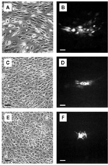
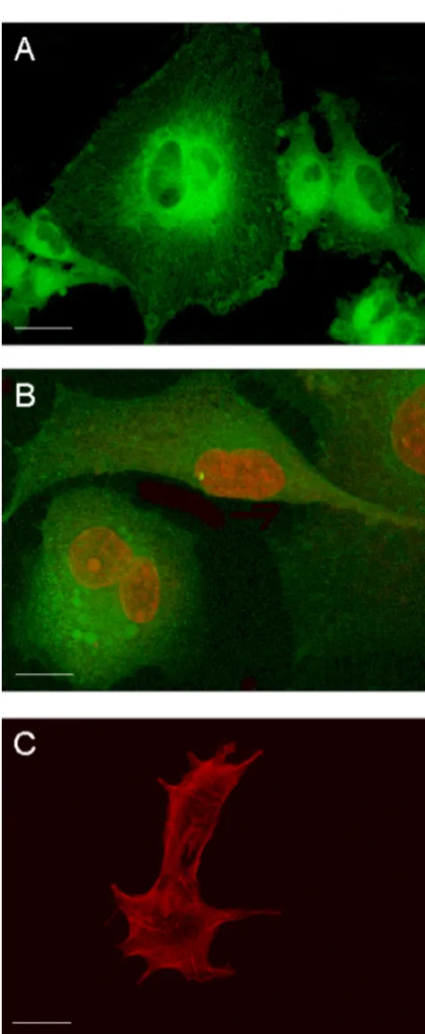
Undifferentiated GL15 cells have a heterogeneous mor-phology. Figure 1 shows confocal microscopy images of the different typologies of these cells. Fluorescent mark-ers were used to make evident the structural characteris-tics of these cells, namely, the microtubule array of the cytoskeleton (Paclitaxel-Bodipy, green fluorescence) (Fig. 1A), the nuclear structure and the mitochondria network (Propidium Iodide, red fluorescence, and Mi-totracker-Green488, green fluorescence, respectively) (Fig. 1B), and the F-actin organization (Phalloidin-Alexa594, red fluorescence) (Fig. 1C). These data made it clear that cytoskeleton organization and lobate nuclei were characteristic of each cellular element, confirming the population heterogeneity.
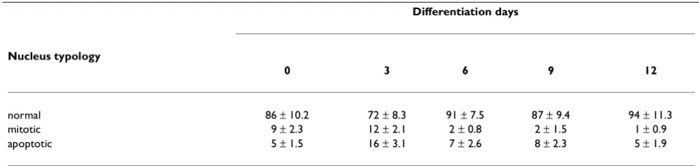
Replacement of the 10% FCS in the culture medium with 2% HS, in order to induce cell differentiation, caused the GL15 cell growth rate to slow and apoptosis to be trig-gered. Therefore, the differentiation phase was complet-ed within 9-12 days. The percentage of apoptotic and dividing nuclei (assayed by staining with the fluorescent nuclear probe DAPI) present during the differentiation period (0-12 days) are reported in Table 1. The percent-age of mitotic cells fell sharply from 9-12% (0-3 days) to 1-2% after 12 days, whereas the presence of apoptotic nu-clei increased during the first 3 days (from 5% to 16%) and regained its initial value at day 12 of differentiation. The characteristics of the proliferation rate and
apopto-sis process in GL15 cells can also be correlated with the morphological analysis carried out over the same time interval. In particular, in 10% FCS-supplemented DMEM or after only 1-2 days in 2% HS-supplemented
BMC Physiology (2001) 1:4 http://www.biomedcentral.com/1472-6793/1/4
Figure 1
(Fig. 2A). In contrast, the 10-day confluent cells cultured in the presence of 2% HS showed apparent homogeneous morphology. Most of the cells were of small size with a regular, round shape (Fig. 2B); the extremely rare mi-toses and numerous thin cell elongations, which form a sort of web within the culture, were strongly suggestive of resting or differentiated cells (Fig. 2B).
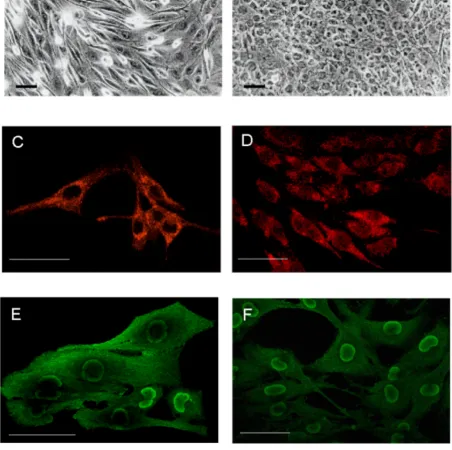
To better define the glial features of GL15 cells, we also tested for the presence of specific astrocytic markers (GFAP and S100B). Immunolabelling for GFAP and the use of confocal microscopy highlighted, in both undiffer-entiated and differundiffer-entiated phenotypes, a diffuse expres-sion of the protein in the cytoplasmic compartment (Figs 2C and 2D). Under the same conditions, we also tested
the expression of S100A and S100B, two Ca2+-binding
proteins characteristically distributed in the central nervous system, but with different modalities: S100A is predominantly expressed in neurons, whereas S100B is normally expressed in and secreted by astroglial cells [17]. Both undifferentiated and differentiated GL15 cells were S100B-positive (Figs 2E and 2F, respectively). A marked distribution of S100B was found in the perinu-clear area, above all in the differentiated phenotype (Fig. 2F), and the undifferentiated phenotype also showed a diffuse cytoplasmic localization (Fig. 2E). In contrast, S100A was not detected (data not shown). This result is particularly significant, because it indicates a specific as-troglial feature of the GL15 line.
[Ca2+]i levels
We also studied the membrane activating systems of GL15 cells by analysing, in single cells, the [Ca2+]i varia-tions triggered by extracellularly applied stimuli for which the concentration and effect on astroglial cells are well known. Figures 3, 4, 5 and 6 show the temporal
anal-ysis of single cell [Ca2+]i variation expressed as normal-ized fluorescence values (see Materials and Methods).
The main graph in Fig. 3 shows the repetitive Ca2+
vari-ations induced by 50 mM KCl that were found in 100% of the differentiated cells (∆ of [Ca2+]i increase = 7 ± 2; n = 60). In the undifferentiated GL15 cells, only 70% of the cell population responded to KCl, with [Ca2+]i variations showing a kinetic shape similar to that observed for the
differentiated ones, but with a lower amplitude (∆ of
[Ca2+]i increase = 1 ± 0.5; n = 47; trace in the box). We also tested the cell response to glutamic acid (L-glu) and ATP which, like other physiological extracellular signals, induce [Ca2+]i variation in astroglial cells via membrane-receptor systems. Starting from a concentration of 300
µM, L-glu induced in 83% of undifferentiated cells a
rap-id increase in [Ca2+]i that regained the basal value within 4 minutes (∆ of [Ca2+]i increase from 1 to 3.5; n = 60) (Fig. 4A). In contrast, 100% of differentiated cells were
responsive to 300 µM L-glu (n = 70), but with different
kinetics and time-course. In fact, as shown in Fig. 4B,
three different Ca2+ pathways, with the same probability
percentage, were recorded in this phenotype: i) a single transient [Ca2+]i spike, ii) a [Ca2+]i variation with a shape similar to that observed in the undifferentiated phenotype, and iii) [Ca2+]i oscillations. Another physio-logical stimulus able to trigger an intracellular Ca2+
re-sponse in GL15 cells was the presence of 200 µM ATP in
the medium. While this purinergic agonist did not in-duce [Ca2+]i variation in the undifferentiated population (0 responsive cells out of 53 tested cells; data not shown),
in the 10-day low serum-treated cells 200 µM ATP
caused single Ca2+ spikes or oscillations in 50% (n = 69)
of the stimulated cells, with a ratio of 2:1 between the two possibilities (Figs 5A and 5B). Another important obser-vation is that the different types of [Ca2+]i variation are derived from different involvements of intracellular Table 1: GL15 nucleus morphology analysis during differentiation phases
Differentiation days
Nucleus typology
0 3 6 9 12
normal 86 ± 10.2 72 ± 8.3 91 ± 7.5 87 ± 9.4 94 ± 11.3
mitotic 9 ± 2.3 12 ± 2.1 2 ± 0.8 2 ± 1.5 1 ± 0.9
apoptotic 5 ± 1.5 16 ± 3.1 7 ± 2.6 8 ± 2.3 5 ± 1.9
BMC Physiology (2001) 1:4 http://www.biomedcentral.com/1472-6793/1/4
Figure 2
Ca2+-stores. In fact, in 18% of the differentiated cells that
showed Ca2+ oscillations, 200 µM ATP almost
complete-ly emptied the intracellular Ca2+-stores. This was
con-firmed by the further addition of thapsigargin, a
well-known blocker of internal store Ca2+-pumps [18], which
caused a slight increase in [Ca2+]i (Fig. 5B). In contrast, if the same thapsigargin concentration was added to the
cell that responded to the presence of 200 µM ATP with
a single spike, the alkaloid induced a large and sustained increase in Ca2+ (Fig. 5A).
Another distinctive aspect of this cell line was the
pres-ence of spontaneous Ca2+ variations, a phenomenon
fre-quently observed in cells of astroglial origin. GL15 cells
showed at least three different kinetics of Ca2+
oscilla-tions: i) single transient variation (Fig. 6A) ii) low fre-quency oscillations (Fig. 6B) and iii) high frefre-quency oscillations (Fig. 6C) (see also additional data: Movie 1 for the original data used to perform this analysis).
Spon-taneous Ca2+ variations were observed in both GL15
phe-notypes. The phenomenon seemed to be independent of
the presence of external Ca2+, because spontaneous
[Ca2+]i variations were detectable both in the presence of
1.8 mM extracellular Ca2+ and in Ca2+-free external
me-dium containing 5 mM EGTA (data not shown). To quan-tify this phenomenon, we tested more than 200 undifferentiated and differentiated cells; among these, 50% of each cell population seemed to be able to prime
spontaneous intracellular Ca2+ movements. However,
considering the variability of the genesis of spontaneous [Ca2+]i oscillations, this cell percentage could be even higher.
Gap-junction analysis
The complex process of astrocytic Ca2+ signalling
in-volves not only the inner cellular pathway (information flow throughout the sub-cellular compartments) and the Figure 3
KCl-evoked Ca2+ spikes in GL15 cells. The graph shows the trace of intracellular Ca2+ variations in a single cell responding to extracellular 50 mM KCl pulses (arrows). The solid trace represents the [Ca2+]i variations found in 100% (n = 60) of the differentiated tested cells. The trace in the box represents the [Ca2+]i variations found in 70% (n = 47) of the undifferentiated cells. Time (s=seconds) is indicated on the abscissa; the ordinate gives the normalized fluorescence value (f/f0).
Figure 4
Temporal analysis of [Ca2+]i variations induced by L-glutamic acid in GL15 phenotypes. Of the undifferenti-ated cells (n = 60), 83% were responsive to addition of 300
BMC Physiology (2001) 1:4 http://www.biomedcentral.com/1472-6793/1/4
extracellular, receptor-mediated, chemical signal trans-duction (i.e. neurotransmitters - or stimuli triggered by growth factors), but also the gap junction intercellular communication (GJIC).
In order to assess the astrocytic properties that are shared by the GL15 cell line, it was important to investi-gate their capacity to establish functional GJIC. To achieve this aim, functional, immunocytochemical and Figure 5
ATP-induced [Ca2+]i variations in differentiated GL15 cells. Panel A shows a single Ca2+ spike induced by 200 µM ATP (arrow) in 32% of differentiated cells (n = 69); addition of 2 µM thapsigargin (Tg) (arrow) evoked a further increase in [Ca2+]i. In 18% (12 out of 69) of differentiated cells, 200 µM ATP (arrow) induced Ca2+ oscillations, partially inhibiting the thapsigargin-induced Ca2+ increase (B). Time (s=seconds) is indicated on the abscissa; the ordinate gives the normalized fluorescence value (f/f0).
Figure 6
Spontaneous [Ca2+]i variations in both undifferenti-ated and differentiundifferenti-ated GL15 cells. Panels A, B and C show, respectively, a single [Ca2+]i spike, low frequency
molecular analyses were performed on GL15 pheno-types.
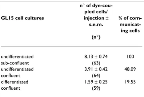
i) Functional analysis of GL15 junctional cou-pling. A microinjection/dye-transfer assay was em-ployed to determine GJIC strength in differentiated and undifferentiated GL15 monolayers, analysed at different culture densities. Cells from undifferentiated, proliferat-ing (sub-confluent) monolayers were clearly shown to be junctionally coupled. When the saturation density (con-fluence) of the cultures was reached, a significant reduc-tion of the cell coupling was observed (48% of the value found in the proliferating counterpart). In the case of dif-ferentiated, resting GL15 cells (confluent cultures), dye-transfer was almost completely restricted to the cells in-itially loaded with the dye (Table 2). The GJIC capacity of GL15 cultures is shown in Fig. 7.
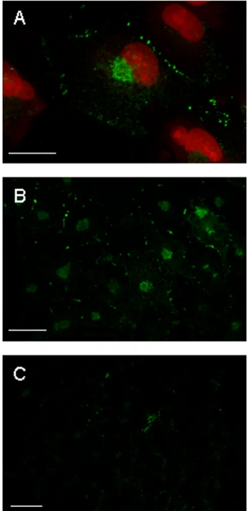
ii) Immunocytochemical analysis of connexin 43 (cx43) in GL15 cells. It is well known that cx43 is the main gap-junction protein expressed by astrocytes (both in vivo and in vitro); hence, its expression could have been responsible for the GJIC observed in GL15. The im-munocytochemical localization of cx43 protein was per-formed by confocal microscopy on GL15 cultures kept in differentiating and proliferating conditions identical to those described above. The results showed the presence of the cx43 antigen in all the GL15 populations tested; however, the antigen distribution differed between pop-ulations, and its quantity was strictly related to the GJIC extent specific to each culture condition. In the highly communicating, sub-confluent, undifferentiated cells the punctate immunopositive reaction, typical of cx43
gjs aggregates, was localized close to the plasma mem-brane at cell-cell contact areas (Figs 8A and 8B; see also additional data: Movie 2 for the original data used to per-form this analysis). The quantity gradually decreased when the cells reached the confluence status (Fig. 8C), and almost completely disappeared in the non-commu-nicating, confluent differentiated GL15 cells (Fig. 8D). In all cases, randomly distributed cx43-positive staining was also found in the cytoplasmic compartment of GL15 cells. This could correspond to the unphosphorylated cx43 isoform that is usually defined as non-functional. Also in this case, an inverse relation was found between the GJIC capacity of the culture and the quantity of cx43 antigen located in the cytoplasm.
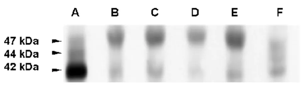
iii) Immunoblot analysis of cx43 in GL15 cells.
The results of the immunoblot analysis of cx43 expres-sion in the various GL15 cell populations (shown in Fig. 9), confirmed the results reported above. The anti-cx43 antiserum recognised, in control samples (rat heart and IAR 203 cell line), both the functional phosphorylated (44 and 47 kDa) and the unphosphorylated (42 kDa) iso-forms of the connexin, whereas in GL15 cells only a small quantity of the 42 kDa antigen was detected. This obser-vation may explain the very low communication capacity of this cell line. The quantity of the functional phosphor-ylated cx43 isoform could have been, in this case, under the limit of immunoblot sensitivity. Only in the samples of GL15 cells was an upper signal sized around 50 kDa detectable. This signal is probably due to a non-specific reaction of the anti-rabbit secondary antiserum with a protein only expressed by human cells. The possibility that it was caused by a slowly migrating, altered (and non-functional) isoform of cx43, typical of this cell line, is very unlikely. If this were the case, a very high total quantity of the cx43 antigen/single GL15 cell would be expected (much more than in the case of the IAR 203 cells, used as a positive control because of their extreme-ly high cx43 expression level). However, the data ob-tained by the immunolocalization of the protein showed the quantity of cx43 antigen/single cell to be very low.
Discussion
The name glia derived from the Greek word for glue -per se indicates why this cell compartment was consid-ered the "less interesting element" of the nervous system until only a few years ago. Today the conception that re-searchers have of this cell population has completely changed, and, in general, theories concerning the role of astrocytes have been radically modified [19]. A recent, novel hypothesis put forward the possibility that this cell type is directly involved in the activity of brain tissue, and not just as a growth-factor producer. This hypothesis is strongly supported by the observation that astrocytes display rapid electrical responses to neuronal activity Table 2: Quantitative analysis of dye-coupling in GL15 cell
cul-tures
n° of dye-cou-pled cells/ GL15 cell cultures injection ±
s.e.m.
% of com- municat-ing cells (n°)
undifferentiated 8.13 ± 0.74 100
sub-confluent (63)
undifferentiated 3.91 ± 0.42 48.09
confluent (64)
differentiated 1.59 ± 0.25 19.55
confluent (59)
BMC Physiology (2001) 1:4 http://www.biomedcentral.com/1472-6793/1/4
Figure 7
and/or modifications of transductive systems [2, 4], like those of extracellular purines, which are known to be ac-tive in primary cultures of astrocytes [3].
Our experiments demonstrate that GL15 cells possess characteristics typical of astrocytes. The glial nature of this cell line was demonstrated by a number of experi-mental approaches, which collectively show that they have several astrocyte-like physiological properties. Im-munostaining with astrocyte protein-specific anti-bodies against GFAP and S100 showed the presence of specific glial markers GFAP and S100B, and the lack of the neuronal marker S100A, a particular isoform of S100 [17]. S100B and GFAP proteins were present in GL15 cul-tures, and the distribution pattern of S100B was relative to the differentiation status of the examined cells.
The presence of voltage-gated channels, correlated di-rectly or indidi-rectly with the possible generation of differ-ent transmembrane potdiffer-entials, was indicated by data obtained from incubation of GL15 mature phenotype with 50 mM KCl, which induces membrane depolarisa-tion. Under these conditions, a transient increase of
[Ca2+]i was observed, similar to that obtained in other
excitable cells such as neurons, muscle cells or mature astrocytes [20]. By employing [Ca2+]i variation as an
in-dicator for the presence of Ca2+-related receptors in
these cells, it was further possible to demonstrate that the mature phenotype of GL15 cells is associated with different agonist-receptor systems. The presence of ap-propriate glutamate or ATP concentrations in the
exper-imental medium induced significant [Ca2+]i variation
with specifc and agonist-related characteristics. Howev-er, upon testing with undifferentiated GL15 cells, these effects were either not present (i.e. ATP-induced [Ca2+]i variation), or less marked (i.e. KCl-induced), or evident in a different way (i.e. L-glu-induced).
Our decision to examine [Ca2+]i as probe for definitive
astrocytic features of GL15 cells was based on the fact that the specific role played by astrocytes in many brain
functions is achieved by Ca2+ signalling mechanism(s).
Astrocytes express many different pathways, through which they react to external stimuli by variation of [Ca2+]i. In fact, these cells contain different forms of
in-ositol triphosphate-coupled receptors that increase Ca2+
signalling, for example via the glutamate pathway and purine-activated systems. Ionotropic receptors, which open Ca2+ channels, are also found in this cell type.
Fur-thermore, the presence of voltage-activated Ca2+
chan-nels that permit Ca2+ fluxes from outside was
demonstrated in both primary cultures and brain slices [4, 11].
Figure 8
BMC Physiology (2001) 1:4 http://www.biomedcentral.com/1472-6793/1/4
Therefore, it is well elucidated in the literature that the
mechanisms involved in intracellular Ca2+ regulation
(release from and uptake to Ca2+-stores, capacitative
and inductive Ca2+ currents, Ca2+ oscillations, and Ca2+ waves, etc) are present in astrocytes. If one considers that in these cells changes in [Ca2+]i underlie a reciprocal communication system between neurons and astrocytes,
assessing the presence and definition of the Ca2+-system
signalling in the GL15 cell line is fundamental in examin-ing the reliability of this cell line as an astrocyte model.
All the Ca2+ pathways previously described for astrocytes
are present in GL15 cells, although they are differentially regulated according to the differentiation status of the cell. It is possible to record oscillations in intracellular
Ca2+ levels, generated either spontaneously or in the
presence of ATP or Glutamate. On the other hand, depo-larising agents like 50 mM KCl are unable to induce this phenomenon, which may therefore be considered
inde-pendent from the voltage-operated Ca2+ channel status.
This interpretation is credible, especially considering os-cillating responses were also observed when
extracellu-lar Ca2+ was absent in the experimental medium. We
hypothesise from the data that in the GL15 population, Ca2+ oscillations originate during Ca2+-release from in-ternal stores. The presence of a complete fan of receptor-operated responses in GL15 cells is correlated with the mature phenotype, whereas intercellular communica-tion of astrocytes is associated with the immature status of GL15 cells. In vivo, astrocytes have the possibility to form 'functional syncytia' by establishing cytoplasmic connections through specific intercellular channels (gap
junctions), which in turn provide pathways for the direct exchange of ions, small metabolites and water. Cultured astrocytes, like their in vivo counter-parts, are extensive-ly coupled by gjs (cx43 being the predominant junctional protein expressed by these cells) [21].
Cx43 is expressed in specific brain regions by different glial populations like the other connexins (e.g. cx30), de-pending on the developmental stage of the tissue, differ-ent physio-pathological conditions and/or growth-factor influence [22, 23, 24].
Our results demonstrate that GL15 cells express the cx43 protein and form junctional channels in which permea-bility is directly related to the proliferation rate, and de-creases when the differentiative status is reached.
The reduction of cell coupling when confluence is achieved supports the proposed role of GJIC in regulat-ing astrocyte migration. In the differentiated phenotype, the very low levels of GJIC compared to that found in non-proliferating, undifferentiated confluent monolay-ers, demonstrated that even in a situation of 'mitotic ar-rest', factors other than those linked with cell-cycle progression (although probably dependent on the devel-opmental features of the cells) may be involved in the regulation of GL15 cell coupling. These data confirm that physiologically, GL15 behave like glial cells with respect to both cell-cycle-dependent and differentiation-related regulation, and astrocytic junctional coupling (cx43 ex-pression) [25]. A further point to note is that in the in vit-Figure 9
ro conditions cx43 expression is limited to type I astrocytes [26].
It was observed that even when subjected to culture con-ditions allowing maximal junctional communication (sub-confluent, undifferentiated monolayer), the extent of GL15 dye-coupling was low, especially when com-pared to results obtained from other cultured cells of as-trocytic origin [27]. This functional evidence closely reflects the data from immunocytochemical and immu-noblot analyses, which indicate that in GL15 cells the junctional protein cx43 is expressed at low levels and its localization is mainly cytoplasmic, probably in the non-functional, unphosphorylated isoform. The presence of low levels of cx43 and limited intercellular coupling of GL15 cells is likely to be due to the neoplastic origin of this cell line, which is also observed in many other tu-mour-derived cell types such as C6 glioma cells [28]. An-other possible explanation for the low communication capacity of GL15 cells might be the culture conditions. It has been demonstrated that the efficiency of junctional coupling of cultured astrocytes is positively influenced by interactions with other cell types (e.g. neurons and
me-ningeal cells) [29, 30]. Moreover, in astrocytes (both in
situ and in vitro), the different levels of dye-coupling
(and cx43 expression) depend on factors such as the spe-cific regional origin of these cells in the CNS or degree of maturation, thus suggesting that gjs formation in this cell type undergoes very complex environmental regula-tion [24].
Another important feature observed in our results was
the correspondence between the modality of Ca2+ wave
propagation in GL15 cells, and their GJIC capacity.
Al-though several factors influence the extent of Ca2+ wave
propagation, gjs permeability is a major determinant [11]. Heterogeneity in GJIC extent may, therefore, partly
explain the heterogeneity of Ca2+ wave modulation and
propagation in the GL15 population.
Conclusions
In conclusion, the data reported in this paper support the reliability of the GL15 cell line as a suitable in vitro model for astrocytes, which should aid in the investigation of their distinctive physiological properties, and subse-quently contribute to clarifying the complex role of this cell type in the brain. It is important to remember that, by simply utilising the differentiated or undifferentiated phenotype of this cell line, it is possible to study the mo-dality by which the cells communicate with each other, either via gjs and/or membrane receptors. The proposed model becomes even more fascinating when the human origin of this cell line is considered. This new astrocyte model provides a stepping-stone in the efficient analysis and interpretation of problems regarding the role of
as-trocytes during modulation and remodelling of the nerv-ous system, their contribution to the electro-physiological activity of neurons and other relevant mechanisms.
Materials and Methods
Cell culture
GL15 cells were routinely cultured in growth medium (GM): D-MEM (Dulbecco's Modified Eagle's Medium) supplemented with 10% fetal calf serum (FCS), 100 IU/
ml penicillin-100 µg/ml streptomycin and 2 mM
L-glutamine. The cells were maintained at 37°C in a 5%
CO2 humidified atmosphere. The medium was changed
twice weekly and the confluent cell monolayers were reg-ularly sub-cultured, washing with phosphate-buffered solution (PBS) and treating with 0.5% trypsin-0.2%
EDTA at 37°C for 5 minutes. Long-term differentiating
cultures were obtained after a 10-day incubation of sub-confluent GL15 cultures in a differentiation medium (DM): D-MEM supplemented with 2% horse serum
(HS), 100 IU/ml penicillin-100 µg/ml streptomycin and
2 mM L-glutamine. Undifferentiated cells were tested af-ter a 1-2 day incubation in GM, while the differentiated phenotype was observed after culturing for 10 days in DM. All the experimental procedures were performed on cells of 60°-70° passage. All media, sera, antibiotics and culture solutions were purchased from Life Technologies Italia srl (S. Giuliano Milanese, Italy). Sterile culture plastics were purchased from Falcon (Plymouth, UK). All other reagents were of analytical grade purity.
Nuclear morphology
The cells were plated on 12 mm-glass coverslips (BDH Italia, Milano, Italy) at a density of approximately 2 × 10⁴ cells/well and incubated in DM for 0, 3, 6, 9 and 12 days. At the selected times the cells were rapidly washed in PBS and fixed by 3.7% paraformaldehyde (Sigma, St. Louis, Missouri, USA) at room temperature (r.t.) for 15 minutes. The cells were then permeabilized with abso-lute methanol (Sigma) at r.t. for 5 minutes. After a single
PBS washing, the samples were stained with 1 µg/ml
BMC Physiology (2001) 1:4 http://www.biomedcentral.com/1472-6793/1/4
mean ± s.d.. Each nucleus typology percentage was
cal-culated from values derived from five randomly selected fields each containing about 50-60 cells on slices, in du-plicate for each sample.
Fluorescence labelling and confocal microscopy
GL15 cells were grown on 12 mm-glass coverslips (plat-ing density: 5 × 10⁴ cells/well) as undifferentiated or dif-ferentiated cells. At the selected times the cells were rapidly washed with PBS and then fixed with 3.7% para-formaldehyde (Sigma) at r.t. for 5 minutes. The cells were then permeabilized with 0.1% Triton X-100 (Sig-ma) at r.t. for 5 minutes. These samples were used for fluorescent dye- or immuno-staining protocols. The dyes: Paclitaxel-Bodipy, Mitotracker-Green488, Propid-ium Iodide, Phalloidin-Alexa594 (all obtained from Mo-lecular Probes) were used according to manufacturer's instructions. The immunolabelling procedure was pre-ceded by 1 hour incubation of the cultures in 10% bovine serum albumin (BSA) at r.t.. Direct immunostaining de-tected the presence of GFAP, S100A and S100B proteins after 1 hour incubation (at 37°C) with the following anti-bodies, respectively: Cy3-conjugated mouse monoclonal anti-GFAP IgGs, Texas Red-conjugated mouse mono-clonal anti-S100A IgGs, and Oregon Green-conjugated mouse monoclonal S100B IgGs (all of these anti-bodies were obtained from Sigma and used at 1:100 dilu-tion). Anti-S100A and anti-S100B were conjugated with their respective fluorochrome using FluoReporter Pro-tein Labelling Kit (Molecular Probes). The presence of cx43 antigen was revealed by immunofluorescence: after
1 hour incubation at 37°C of the cells with primary mouse
monoclonal anti-cx43 antibody (diluted 1:60) (Chemi-con International Inc., Temecula, CA), followed by 1 hour
incubation at 37°C with secondary Oregon
Green-conju-gated anti-mouse IgGs (diluted 1:100; Molecular Probes). The cells were washed 3 times for 5 minutes at r.t. with 0.1% Tween 20 (Sigma) in PBS, dried and then observed.
Fluorescence images were obtained by using a Bio-Rad MRC-1000 confocal system (BioRad Laboratories, USA) with an Axiovert 100 microscope equipped with a 63X/ 1.25 PLAN NEOFLUAR oil immersion objective lens (Zeiss, Jena, Germany). The Kr/Ar laser potency, photo-multiplier and pin-hole size were kept constant for the entire experimental procedure. Images were acquired using CoMOS/MS-DOS software and then processed us-ing LaserSharp/OS2 software (BioRad).
[Ca2+]i measurements
GL15 cells were plated on 25 mm-glass coverslips at a density of 9 × 10⁴ cells/well and tested as undifferentiat-ed or differentiatundifferentiat-ed cells. At the beginning of each exper-iment, the cells were washed with the normal external
solution (NES), a buffered solution containing (in mM) 140 NaCl, 1.8 CaCl2, 2.8 KCl, 2 MgCl2, 10 Glucose and 10 HEPES/NaOH, at pH 7.4. The cells were then incubated
for 30 minutes at 25°C with 3 µM Fluo3
acetoximethyl-ester (Fluo3/AM, Molecular Probes) dissolved in NES supplemented with 10 mg/ml BSA. The loaded cells were rinsed and maintained for an additional 15 minutes at
25°C in NES to allow the complete de-esterification of
the dye. In these experimental conditions each cell
sam-ple showed good preservation of the intracellular Ca2+
dye fluorescence emission. The coverslips were then transferred into an Attofluor chamber (Molecular Probes). Stimulating agents were added in less than 1 second to the cells kept at r.t.. A high-speed wavelength switcher Polychrome II (Till Photonics, Germany) equipped with a 75 W stabilised Xenon lamp (Ushio Inc., Japan) provided the excitation beam. The Polychrome II was connected to an Olympus IX 50 microscope equipped with an oil objective lens (Uapo/340 40X/1.35 oil Iris). The fluorescence emission was acquired by C6790 Hamamatsu camera and analysed using the Ar-gus Hisca 1.7 software (Hamamatsu, Hamamatsu, Ja-pan). The traces in Figs 3,4,5,6 were ratios (1 ratio/ second) calculated off-line as f/f0, where f is the fluores-cence emission of a single FLUO3-loaded cell at time range from 1 to x seconds and f0 is the fluorescence emis-sion of the same cell at time 0 [31].
Dye-Transfer Assay
Undifferentiated (sub-confluent and confluent) and dif-ferentiated (confluent) GL15 cells, cultured onto 60-mm Petri dishes, were tested for their capacity to establish functional gjs as described by Mazzoleni et al. [32]. Brief-ly, single cells within the monolayers of three separate dishes were microinjected with a 10% (wt/vol) solution of the gap-junction-permeant fluorescent tracer Lucifer Yellow CH (Sigma) in 0.33 M LiCl. Microinjections were performed using glass capillary needles (Clark Electro-medical Instruments, Edenbridge, UK), prepared with an automatic puller (Narishige, Tokyo, Japan) and driv-en by a Narishige micromanipulator (SYF II) linked to an Olympus IMT2 microscope. The fluorescent dye was in-jected under nitrogen pressure using an Eppendorf mi-croinjector (Hamburg, Germany). Five minutes after the last injection, the cells were fixed with 4% paraformalde-hyde in PBS and the dye-transfer capacity of the cells was observed using the IMT2 epifluorescence system. The extent of gap junction intercellular communication (GJIC) was then quantified by counting the number of
fluorescent cells surrounding the microinjected ones (n°
of dye-coupled cells/injection). At least 25 independent microinjection trials/dish were taken into account for the precise quantification of the GJIC competence of the
Bright-field and fluorescence images were taken on Kodak T-MAX 400 (400 ASA) films.
Immunoblot analysis
Cells from six 100-mm Petri dishes were pooled for each previously described culture condition and the whole-cell extracts (40 µg protein of total lysate/lane) were first resolved by electrophoresis on 10% sodium dodecyl sul-phate-polyacrylamide gel [33] and then transferred onto a nitrocellulose membrane (Schlesher and Shuell, Keene, NH). Protein of total lysate from rat heart and IAR 203 rat liver epithelial cells [34] were included as positive controls for cx43 expression. Total protein concentration was determined using a Bio-Rad DC Protein Assay kit (Bio-Rad, Segrate, Italy); equal sample protein-loading was verified by Comassie Blue staining of identical gels run in parallel, using a 0.25% Comassie Blue solution (R 250/G 250 1:1) (Bio-Rad). Membranes were hybridized with polyclonal rabbit anti-cx43 antibody (1:1000) (Chemicon International Inc.), followed by reaction with peroxidase-conjugated anti-rabbit IgGs (1:2000) (Amer-sham Pharmacia Biotech Italia, Cologno Monzese, Italy). Immunopositive reaction was detected by the enhanced chemiluminescence method (ECL, Amersham Pharma-cia Biotech Italia) and revealed using autoradiography films (Hyperfilm-ECL, Amersham Pharmacia Biotech Italia).
Acknowledgements
We wish to thank Dr Francesca Rovetta for helpful discussions and Peter A. Mattei for his assistance in preparing the manuscript. This work was sup-ported by research grants from MURST-Italy (Ministero dell'Università e
della Ricerca Scientifica e Tecnologica) to G.F. and from the University of Brescia to G.M.
References
1. Barres BA, Reichardt LF: Neuronal and glial cell biology. Edito-rial overview. Curr Opin Neurobiol 1999, 9:513-516
2. Vesce S, Bezzi P, Volterra A: The highly integrated dialogue be-tween neurons and astrocytes in brain function. Sci Prog 1999, 82:251-270
3. Centemeri C, Bolego C, Abbracchio MP, Cattabeni F, Puglisi L, Burn-stock G, Nicosia S: Characterization of the Ca2+ responses evoked by ATP and other nucleotides in mammalian brain astrocytes. Br J Pharmacol 1997, 121:1700-1706
4. Carmignoto G, Pasti L, Pozzan T: On the role of voltage-depend-ent calcium channels in calcium signaling of astrocytes in situ. J Neurosci 1998, 18:4637-4645
5. John GR, Scemes E, Suadicani SO, Liu JS, Charles PC, Lee SC, Spray DC, Brosnan CF: IL-1 beta differentially regulates calcium wave propagation between primary human fetal astrocytes via pathways involving P2 receptors and gap junction chan-nels. Proc Natl Acad Sci U SA 1999, 96:11613-11618
6. Finkbeiner S: Calcium waves in astrocytes-filling in the gaps. Neuron 1992, 8:1101-1108
7. Smith SJ: Neural signalling. Neuromodulatory astrocytes. Curr Biol 1994, 4:807-810
8. Sauer H, Hescheler J, Wartenberg M: Mechanical strain-induced Ca2+ waves are propagated via ATP release and purinergic receptor activation. Am J Physiol Cell Physiol 2000, 279:C295-C307 9. Nedergaard M: Direct signaling from astrocytes to neurons in cultures of mammalian brain cells. Science 1994, 263:1768-1771 10. Zhang W, Couldwell WT, Simard MF, Song H, Lin JH, Nedergaard M: Direct gap junction communication between malignant glio-ma cells and astrocytes. Cancer Res 1999, 59:1994-2003 11. Giaume C, Venance L: Intercellular calcium signaling and gap
junctionalcommunication in astrocytes. Glia 1998, 24:50-64 12. Bocchini V, Casalone R, Collini P, Rebel G, Lo Curto F: Changes in
glial fibrillary acid protein and karyotype during culturing of two cell lines established from human glioblastoma multi-forme. Cell Tissue Res 1991, 265:73-81
13. Arcuri C, Tardy M, Rolland B, Armellini R, Menghini AR, Bocchini V: Glutamine sinthetase gene expression in a glioblastoma cell line of clonal origin: regulation by dexamethasone and dibu-tyryl cyclic AMP. Neurochem Res 1995, 20:1133-1139
14. Scarpa S, Coppa A, Ragano-Caracciolo M, Mincione G, Giuffrida A, Modesti A, Colletta G: Transforming Growth Factor beta reg-ulates differentiation and proliferation of human neuroblas-toma. Exp Cell Res 1996, 229:147-154
15. Tchoumkeu-Nzouessa GC, Rebel G: Characterization of taurine transport in human glioma GL15 cell line: regulation by pro-tein kinase C. Neuropharmacology 1996, 35:37-44
16. Castigli E, Arcuri C, Giovagnoli L, Luciani R, Giovagnoli L, Secca T, Gi-anfranceschi GL, Bocchini V: Interleukin-1β induces apoptosis in GL15 glioblastoma-derived human cell line.Am J Physiol Cell Physiol 2000, 279:C2043-C2049
17. Fanò G, Biocca S, Fulle S, Mariggio MA, Belia S, Calissano P: The S-100: a protein family in search of a function.Prog Neurobiol 1995, 46:71-82
18. Thastrup O, Cullen PJ, Drobak BK, Hanley MR, Dawson AP: Thapsi-gargin, a tumor promoter, discharges intracellular Ca2+ stores by specific inhibition of the endoplasmic reticulum Ca2+ ATPase. Proc Natl Acad Sci USA 1990, 87:2466-2470 19. Travis J: Glia: the brain's other cells. Science 1994, 266:970-972 20. Armstrong CM, Hille B: Voltage-gated ion channels and
electri-cal excitability. Neuron 1998, 20:371-380
21. Zahs KR: Heterotypic coupling between glial cells of the mammalian central nervous system. Glia 1998, 24:85-96 22. Ochalski PA, Sawchuk MA, Hertzberg EL, Nagy JI: Astrocytic gap
junction removal, connexin43 redistribution, and epitope masking at excitatory amino acid lesion sites in rat brain. Glia 1995, 14:279-294
23. Reuss B, Unsicker K: Regulation of gap junction communica-tion by growth factors from non-neural cells to astroglia: a brief review. Glia 1998, 24:32-38
24. Nagy JI, Rash JE: Connexins and gap junctions of astrocytes and oligodendrocytes in the CNS. Brain Res Brain Res Rev 2000, 32 :29-44
Additional material
Structural and functional GL15 characteris-tics
To better visualise the structural and functional GL15 characteris-tics, we also include two movies in which it is possible to observe: Movie 1: A representative experiment showing spontaneous intra-cellular Ca2+ oscillations. Each frame was acquired with a ratio of 1 frame/second. The total number of frames was 120 with a total real time of 120 seconds compressed in the 30 seconds of the movie. Size bar = 50µm.
Movie 2: Tri-dimensional reconstruction of cx43-immunostained undifferentiated GL15 cells. Size bar = 25µm.
BMC Physiology (2001) 1:4 http://www.biomedcentral.com/1472-6793/1/4
25. Bittman K, Owens DF, Kriegstein AR, LoTurco JJ: Cell coupling and uncoupling in the ventricular zone of developing neocortex. J Neurosci 1997, 17:7037-7044
26. Belliveau DJ, Naus CC: Cortical type 2 astrocytes are not dye coupled nor do they express the major gap junction genes found in the central nervous system. Glia 1994, 12:24-34 27. Giaume C, McCarthy KD: Control of gap-junctional
communi-cation in astrocytic networks. Trends Neurosci 1996, 19:319-325 28. Naus CC, Bechberger JF, Caveney S, Wilson JX: Expression of gap
junction genes in astrocytes and C6 glioma cells. Neurosci Lett 1991, 126:33-36
29. Fischer G, Kettenmann H: Cultured astrocytes form a syncy-tium after maturation. Exp Cell Res 1985, 159:273-279
30. Anders JJ, Salopek M: Meningeal cells increase in vitro astro-cytic gap junctional communication as measured by fluores-cence recovery after laser photobleaching.J Neurocytol 1989, 18:257-264
31. Mammano F, Canepari M, G Capello, RB Ijaduola, A Cunei, L Ying, F Fratnik, A Colavita: An optical recording system based on a fast CCD sensor for biological imaging.Cell Calcium 1999, 25 :115-123
32. Mazzoleni G, Telo P, Camplani A, Tanganelli S, Monarca S, Ragnotti G: Influence of the herbicide Linuron on growth rate and gap-junctional intercellular communication of cultured en-dothelial cells. J Environ Pathol Toxicol Oncol 1994, 13:1-10 33. Laemmli UK: Cleavage of structural proteins during the
as-sembly of the head of bacteriophage T4. Nature 1970, 227 :680-685
34. Montesano R, Drevon C, Kuroki T, Saint Vincent L, Handleman S, Sanford KK, DeFeo D, Weinstein IB: Test for malignant transfor-mation of rat liver cells in culture: cytology, growth in soft agar, and production of plasminogen activator. J Nat. Cancer Inst 1977, 59:1651-1658
Publish with BioMedcentraland every scientist can read your work free of charge
"BioMedcentral will be the most significant development for disseminating the results of biomedical research in our lifetime."
Paul Nurse, Director-General, Imperial Cancer Research Fund
Publish with BMc and your research papers will be:
available free of charge to the entire biomedical community
peer reviewed and published immediately upon acceptance
cited in PubMed and archived on PubMed Central
yours - you keep the copyright
[email protected] Submit your manuscript here:
http://www.biomedcentral.com/manuscript/
BioMedcentral.com
![Figure 4Temporal analysis of [Caalthough with different amplitudes (A). In panel B the tracesrepresent the three equally probable [Ca2+]i variations induced by L-glutamic acid in GL15 phenotypes](https://thumb-us.123doks.com/thumbv2/123dok_us/632899.2063502/6.612.328.525.101.459/temporal-caalthough-different-amplitudes-tracesrepresent-variations-glutamic-phenotypes.webp)
![Figure 5ATP-induced [Ca2+]i variations in differentiatedGL15 cells. Panel A shows a single Ca2+ spike induced by200 µM ATP (arrow) in 32% of differentiated cells (n = 69);addition of 2 µM thapsigargin (Tg) (arrow) evoked a furtherincrease in [Ca2+]i](https://thumb-us.123doks.com/thumbv2/123dok_us/632899.2063502/7.612.327.531.89.536/figure-variations-differentiatedgl-induced-differentiated-addition-thapsigargin-furtherincrease.webp)