ORIGINAL ARTICLE
The prognostic value of C-X-C motif
chemokine receptor 4 in patients with sporadic
malignant peripheral nerve sheath tumors
Chao Zhang
1,2,3†, Fang‑Yuan Chang
1,2,3†, Wen‑Ya Zhou
1,2,3†and Ji‑Long Yang
1,2,3*Abstract
Background: Recent studies indicate that C‑X‑C motif chemokine receptor 4 (CXCR4) and its ligand, C‑X‑C motif chemokine ligand 12 (CXCL12), stimulate expression of the cell cycle regulatory protein Cyclin D1 in neurofibroma‑ tosis 1‑associated malignant peripheral nerve sheath tumor (MPNST) cells and promote their proliferation. In this study, we measured the expression of CXCR4, CXCL12, and Cyclin D1 proteins in sporadic MPNST tissues from Chinese patients and investigated their prognostic values.
Methods: CXCR4, CXCL12, and Cyclin D1 protein expression in samples from 58 Chinese patients with sporadic MPNST was assessed with immunohistochemical staining. Their prognostic values were evaluated with Kaplan–Meier analysis and a log‑rank test. Multivariate Cox regression analysis was used to identify independent prognostic factors.
Results: High expression of CXCR4, CXCL12, and Cyclin D1 was observed in 19 (32.8%), 32 (55.2%), and 16 (27.6%) samples, respectively. CXCR4 expression was positively correlated with CXCL12 expression (r= 0.334, P= 0.010) and Cyclin D1 expression (r= 0.309, P= 0.018). Patients with high CXCR4 expression showed longer overall survival than those with low CXCR4 expression (χ2
= 4.642, P= 0.031).
Conclusion: High CXCR4 expression may define a specific subtype of sporadic MPNST with favorable prognosis.
Keywords: Sporadic malignant peripheral nerve sheath tumor, C‑X‑C motif chemokine receptor 4 (CXCR4), C‑X‑C motif chemokine ligand 12 (CXCL12), Cyclin D1
© The Author(s) 2017. This article is distributed under the terms of the Creative Commons Attribution 4.0 International License (http://creativecommons.org/licenses/by/4.0/), which permits unrestricted use, distribution, and reproduction in any medium, provided you give appropriate credit to the original author(s) and the source, provide a link to the Creative Commons license, and indicate if changes were made. The Creative Commons Public Domain Dedication waiver (http://creativecommons.org/ publicdomain/zero/1.0/) applies to the data made available in this article, unless otherwise stated.
Background
Malignant peripheral nerve sheath tumors (MPNSTs) are highly aggressive cancers that account for approximately 5%–10% of all soft tissue sarcomas [1]. MPNSTs are defined as tumors originating from or differentiating into nerve sheath cells [2], with the exception of tumors origi-nating from the epineurium or peripheral nerve vascula-ture [3]. MPNSTs may arise sporadically (40%), following radiotherapy (10%), or in association with neurofibroma-tosis type 1 (NF1) (40%–50%) [4, 5]. NF1 is caused by the mutation of NF1 gene. The NF1 gene (located in
17q11.2) product is neurofibromin. A region of neurofi-bromin functions as a GTPase-activating protein shown to be involved in negative regulation of the RAS pathway, then caused the hydrolyzation and inactivation of GTP [6]. The mutations of NF1 cause neurofibromin to lose its functions as a tumor-suppressing factor. Types of NF1
mutations include frameshift, nonsense, missense, splic-ing alteration and deletion, and loss of heterozygosity [7]. The MPNST that develops from benign NF1 is named NF1-associated MPNST and is the main cause of death among NF1 patients [7, 8]. Compared with sporadic MPNSTs, NF1-associated MPNSTs usually develop in young adults, frequently relapse (in approximately 50% of patients), and lead to a dismal prognosis partly because of the high mutation frequency of NF1 gene [8]. Tradi-tional treatment modalities, such as surgery, chemother-apy, and radiotherchemother-apy, have limited therapeutic effects on
Open Access
*Correspondence: yangjilong@tjmuch.com
†Chao Zhang, Fang‑Yuan Chang and Wen‑Ya Zhou contributed equally to this work
1 Department of Bone and Soft Tissue Tumor, Tianjin Medical University Cancer Institute & Hospital, Tianjin 30060, P. R. China
MPNSTs [9, 10]. Sporadic MPNSTs have some unusual characteristics. For instance, high expression of fibro-blast growth factor receptor 1 (FGFR1) is an independ-ent prognostic predictor for long overall survival (OS) in patients with sporadic MPNST, but associated with poor prognosis of several other types of epithelial cancer [11].
Chemokines are a class of small molecules that bind to G protein-coupled receptors on target cells. Chemokine-induced signaling cascades regulate leukocyte migration, hematopoietic stem cell proliferation, and cell adhesion to extracellular matrix molecules [12, 13]. C-X-C motif chemokine ligand 12 (CXCL12), also known as stromal cell-derived factor-1, is a chemokine that binds to C-X-C motif chemokine receptor 4 (CXCR4). CXCR4/CXCL12 signaling plays important roles in the maintenance of embryonic development, immune and inflammatory responses, regulation of hematopoiesis, human immu-nodeficiency virus infection, and induction of angiogen-esis [14, 15]. Signaling triggered by the CXCR4-CXCL12 binding and subsequent regulation of Cyclin D1 expres-sion has been shown to regulate several biological func-tions in cancers, such as cell proliferation, tumor invasion and metastasis, angiogenesis, and cell–microenviron-ment interactions [16, 17]. It is reported that CXCR4 and CXCL12 mRNA expression was reduced in hepatocel-lular carcinoma and renal cancer compared with non-cancerous tissues, but unchanged in most digestive tract carcinomas, such as colon esophageal or gastric cancer [18]. At the same time, high levels of CXCR4 have been associated with short OS in breast cancer patients [19,
20]. According to the divergence of CXCR4 expression in different cancers, extensive analyses of CXCR4 expres-sion levels and functional studies will be necessary to understand the precise roles of CXCR4/CXCL12 signal-ing in different cancer types.
In NF1-associated MPNST, CXCR4/CXCL12 signal-ing stimulates Cyclin D1 expression and promotes cell cycle progression by activating the AKT/serine–threo-nine kinase/glycogen synthase kinase 3 beta signaling pathway, ultimately increasing MPNST cell proliferation. Suppression of CXCR4 activity decreases MPNST cell growth and inhibits tumorigenesis in allografts and in spontaneous genetic mouse models of MPNST [16]. However, the role of CXCR4 in sporadic MPNSTs is unclear. In this study, we addressed this question by measuring the expression of CXCR4, CXCL12, and Cyc-lin D1 proteins in tumor samples from Chinese patients with sporadic MPNST.
Methods
Patients and tissue samples
The clinical records of MPNST patients diagnosed between January 6th, 1991 and December 1st, 2011 were
evaluated to include only patients with sporadic MPNST and to exclude patients with NF1-associated or radio-therapy-induced MPNST. Clinicopathologic informa-tion was collected, including sex, age, tumor site, tumor size, American Joint Committee on Cancer (AJCC) stage, radiotherapy, chemotherapy, surgery type, disease recur-rence, metastasis, and patient survival.
Tissue samples were obtained from the Department of Pathology, Tianjin Medical University Cancer Institute and Hospital. All tissues were collected before patients received radiotherapy and/or chemotherapy and were formalin-fixed and then embedded in paraffin. Two independent pathologists confirmed the diagnosis and ensured that at least 90% of cells in each specimen were tumor cells.
Tissue microarrays and immunohistochemistry
Tissue microarrays (TMAs) were constructed with the most representative areas of the tumor region as previ-ously reported [21, 22]. Immunohistochemical (IHC) staining of TMAs was performed using the streptavi-din–peroxidase method [23] with the following bioti-nylated primary antibodies: anti-CXCR4 antibody (1:100, ab124824, Abcam, Cambridge, UK), CXCL12 anti-body (1:100, sc-28876, Santa Cruz Biotechnology, Santa Cruz, CA, USA), and anti-Cyclin D1 antibody (1:50, Bei-jing Zhong Shan Golden Bridge Biotechnology Co. Ltd, Beijing, China).
The stained specimens were evaluated independently by two pathologists who were blinded to the clinico-pathologic information and patient prognoses. Slides were examined by light microscopy, and high-power fields of five random areas per tumor were evaluated. Staining intensity was scored as 0 for no staining, 1 for light yellow, 2 for brownish yellow, and 3 for brown-ish black staining. The percentage of positive cells was scored as 0 for ≤ 10%, 1 for 11%–25%, 2 for 26%–50%, 3 for 51%–75%, and 4 for > 75%. The summation of the two scores was taken as the final IHC score: 0–1 (negative), 2–3 (weak positive), 4–5 (moderate positive), and 6–7 (strong positive). The expression level was further cat-egorized into low (negative and weak positive) and high (moderate and strong positive) expression based on the final IHC score.
Statistical analysis
Associations between CXCR4, CXCL12, and Cyclin D1 expression levels and clinicopathologic variables were evaluated using the χ2 or Fisher’s exact tests.
defined as the duration from the date of diagnosis to the confirmed disease progression or the last follow-up. Relationships between CXCR4, CXCL12, or Cyclin D1 expression levels and survival were evaluated using the Kaplan–Meier method and a log-rank test. The data of patients who were lost to follow-up or dead
because of non-tumor-related diseases were censored. The Cox regression model was used for univariate and multivariate analyses. Analyses were performed using SPSS 19.0 software for Windows (SPSS Inc., Chicago, IL, USA). A P value < 0.05 was considered statistically significant.
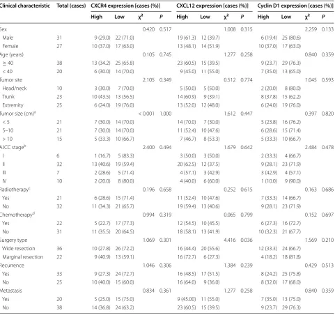
Table 1 Relationships between CXCR4, CXCL12, and Cyclin D1 expression levels and clinicopathologic characteristics of sporadic MPNST patients
MPNST malignant peripheral nerve sheath tumor, CXCR4 C-X-C motif chemokine receptor 4, CXCL12 C-X-C motif chemokine ligand 12, AJCC American Joint Committee on Cancer
a Tumor size information is missing for 1 patient b AJCC stage information is missing for 3 patients c Radiotherapy information is missing for 5 patients d Chemotherapy information is missing for 5 patients
Clinical characteristic Total (cases) CXCR4 expression [cases (%)] CXCL12 expression [cases (%)] Cyclin D1 expression [cases (%)] High Low χ2 P High Low χ2 P High Low χ2 P
Sex 0.420 0.517 1.008 0.315 2.259 0.133
Male 31 9 (29.0) 22 (71.0) 19 (61.3) 12 (39.7) 6 (19.4) 25 (80.6)
Female 27 10 (37.0) 17 (63.0) 13 (48.1) 14 (51.9) 10 (37.0) 17 (63.0)
Age (years) 0.105 0.745 1.277 0.258 0.840 0.359
≥ 40 38 13 (34.2) 25 (65.8) 23 (60.5) 15 (39.5) 9 (23.7) 29 (76.3)
< 40 20 6 (30.0) 14 (70.0) 9 (45.0) 11 (55.0) 7 (35.0) 13 (65.0)
Tumor site 2.105 0.349 0.512 0.774 1.045 0.593
Head/neck 10 3 (30.0) 7 (70.0) 5 (50.0) 5 (50.0) 2 (20.0) 8 (80.0)
Trunk 23 10 (43.5) 13 (56.5) 14 (60.9) 9 (39.1) 8 (37.8) 15 (62.2)
Extremity 25 6 (24.0) 19 (76.0) 13 (52.0) 12 (48.0) 6 (24.0) 19 (76.0)
Tumor size (cm)a < 0.001 1.000 1.612 0.447 0.397 0.820
< 5 21 7 (30.0) 14 (70.0) 14 (70.0) 7 (30.0) 5 (23.8) 16 (76.2)
5–10 21 7 (30.0) 14 (70.0) 11 (52.4) 10 (47.6) 6 (28.6) 15 (71.4)
> 10 15 5 (33.3) 10 (66.7) 7 (46.7) 8 (53.3) 5 (33.3) 10 (66.7)
AJCC stageb 2.400 0.494 1.679 0.642 2.484 0.478
I 6 1 (16.7) 5 (83.3) 3 (50.0) 3 (50.0) 2 (33.3) 4 (66.7)
II 32 13 (40.6) 19 (59.4) 20 (62.5) 12 (37.5) 9 (28.1) 23 (71.9)
III 7 2 (28.6) 5 (71.4) 4 (57.1) 3 (42.9) 3 (42.9) 4 (57.1)
IV 10 2 (20.0) 8 (80.0) 4 (40.0) 6 (60.0) 1 (10.0) 9 (90.0)
Radiotherapyc 0.196 0.658 0.252 0.615 0.163 0.686
Yes 21 6 (28.6) 15 (71.4) 11 (52.4) 10 (47.6) 7 (33.3) 14 (66.7)
No 32 11 (34.3) 21 (65.7) 19 (59.4) 13 (40.6) 9 (28.1) 23 (71.9)
Chemotherapyd 0.994 0.319 0.065 0.799 0.152 0.697
Yes 22 5 (22.7) 17 (77.3) 12 (54.5) 10 (45.5) 6 (27.3) 16 (72.7)
No 31 11 (35.5) 20 (64.5) 18 (58.1) 13 (41.9) 10 (32.3) 21 (67.7)
Surgery type 1.069 0.301 4.416 0.036 1.569 0.210
Wide resection 36 10 (27.8) 26 (72.2) 16 (44.4) 20 (55.6) 12 (33.3) 24 (66.7)
Marginal resection 22 9 (40.9) 13 (59.1) 16 (72.7) 6 (27.3) 4 (18.2) 18 (81.8)
Recurrence 1.046 0.306 1.384 0.239 0.429 0.513
Yes 33 9 (27.3) 24 (72.7) 16 (48.5) 17 (51.5) 8 (24.2) 25 (75.8)
No 25 10 (40.0) 15 (60.0) 16 (64.0) 9 (36.0) 8 (32.0) 17 (68.0)
Metastasis 0.834 0.361 1.277 0.258 0.840 0.359
Yes 20 5 (25.0) 15 (75.0) 9 (45.00) 11 (55.0) 7 (35.0) 13 (75.0)
Results
Clinicopathologic characteristics of sporadic MPNST patients
A total of 58 patients with sporadic MPNST, 31 males and 27 females with an average age of 46.5 years (median, 46 years; range 6–86 years), were selected (Table 1). At the end of follow-up, 22 patients showed no evidence of tumor recur-rence or metastasis, whereas 36 had disease progression as determined with imaging or pathologic evidence of recur-rence or metastasis. Thirty-one patients died of MPNST. The median DFS was 29 months (range 0–149 months). The 5- and 10-year DFS rates were 32% and 28%. The median OS was 109 months (range 0–150 months). The 5- and 10-year OS rates were 59% and 29%.
CXCR4, CXCL12, and Cyclin D1 protein expression and their correlations
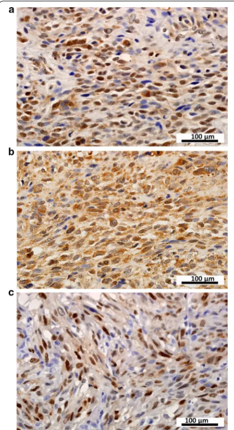
IHC analysis of MPNST samples revealed that CXCR4 was mainly expressed in the cytoplasm and membrane (Fig. 1a), CXCL12 was present in the cytoplasm and plasma membrane (Fig. 1b), and Cyclin D1 was pre-dominantly localized in the nucleus (Fig. 1c). CXCR4, CXCL12, and Cyclin D1 were expressed at high lev-els in 19 (32.8%), 32 (55.2%), and 16 (27.6%) samples, respectively. Correlation analysis revealed positive correlations between CXCR4 expression and that of CXCL12 (r = 0.334, P = 0.010) and Cyclin D1 (r = 0.309, P = 0.018). However, there was no correlation between
CXCL12 and Cyclin D1 expression (r = 0.091, P = 0.497). Relationships between CXCR4, CXCL12, and Cyclin D1 protein expression and clinicopathologic variables
CXCL12 expression was significantly associated with sur-gery type (χ2 = 4.416, P = 0.036); no significant
associa-tions were observed between clinicopathologic variables and CXCR4 or Cyclin D1 expression levels (Table 1).
Relationships between CXCR4, CXCL12, and Cyclin D1 protein expression and survival
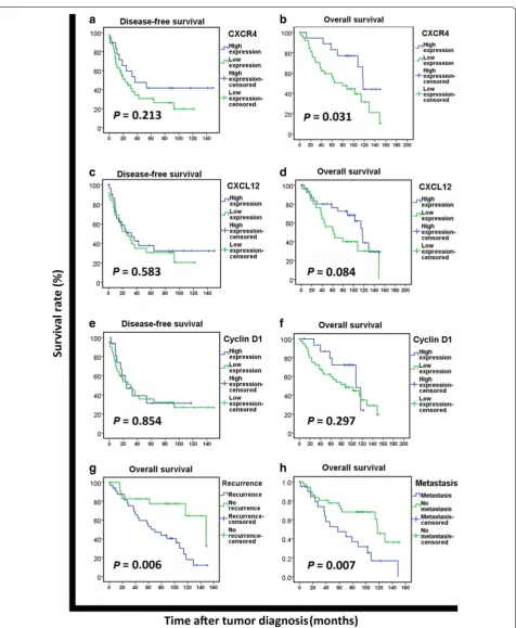
Kaplan–Meier survival analysis indicated that high CXCR4 expression was not significantly associated with DFS (χ2 = 1.550, P = 0.213; Fig. 2a), but was
associ-ated with long OS (χ2 = 4.642, P = 0.031; Fig. 2b). High
expression of CXCL12 and Cyclin D1 were not signifi-cantly associated with either DFS or OS (all P > 0.05, Fig. 2c–f). Patients who experienced recurrence or metastasis had significantly shorter OS than those who did not (both P < 0.01, Fig. 2g, h).
Univariate analysis indicated that tumor site, AJCC stage, recurrence, and metastasis were significant prog-nostic factors for DFS (all P < 0.05), whereas CXCR4 expression, recurrence, and metastasis were significant prognostic factors for OS (all P < 0.05; Table 2). However,
neither CXCL12 nor Cyclin D1 had prognostic value as determined with univariate analysis (Table 2). Multivariate analysis revealed that recurrence and metastasis, but not CXCR4 expression, were independent prognostic factors for patients with sporadic MPNST (both P < 0.05; Table 3).
Discussion
In the present study, we identified a significant increase in CXCL4, CXCL12, and Cyclin D1 expression in tumor samples from Chinese patients with sporadic MPNST.
CXCR4 expression was positively correlated with that of CXCL12 and Cyclin D1. Most importantly, patients with high CXCR4 expression had longer OS than did patients with low expression. High CXCR4 expression might define a specific sporadic MPNST subtype.
Our present data about the prognostic value of CXCR4 in sporadic MPNSTs was quite different from the reported data from NF1-associated MPNSTs [16], which showed that high CXCR4 expression was associ-ated with poor prognosis. In NF1-associassoci-ated MPNSTs, it was reported that CXCR4/CXCL12 signaling stimulated Cyclin D1 expression and enhanced MPNST cell prolif-eration which indicated poor prognosis [16]. Suppres-sion of CXCR4 inhibited the growth of MPNST cell lines and tumorigenesis in allografts and spontaneous genetic mouse models of NF1-associated MPNST [16]. There are
at least two possible explanations for the different prog-nostic values of CXCR4 in different subtypes of MPNST. One is that the pathogenesis of sporadic MPNST and NF1-associated MPNST may differ as a result of the quite different frequency of NF1 mutations. While neurofibro-min inhibits tumor cell proliferation through blockade of RAS-mediated signal transduction, it should also be con-sidered a modulator of cell motility and cell adhesion [6]. Through interfacing with the cytoskeleton and membrane structures, neurofibromin acts as a negative regulator of the Rhodopsin/Rhodopsin-associated coiled-coil con-taining protein kinase (RHO/ROCK) signaling pathway involved in cytoskeletal dynamics that are instrumental in proper neuronal development [6]. The mutations of
NF1 cause neurofibromin to lose its functions, leading to poor prognosis of NF1-associated MPNSTs. However,
Table 2 Univariate analysis of the prognostic values of clinicopathologic characteristics and protein expression in spo-radic MPNST patients
Italic values indicate statistically significant difference
MPNST malignant peripheral nerve sheath tumor, AJCC American Joint Committee on Cancer, CXCR4 C-X-C motif chemokine receptor 4, CXCL12 C-X-C motif chemokine ligand 12, HR hazard ratio, CI confidence interval
Parameter Disease-free survival Overall survival
HR 95% CI P HR 95% CI P
Gender 0.791 0.409–1.530 0.486 1.203 0.585–2.472 0.616
Age group 1.003 0.508–1.983 0.992 0.628 0.280–1.407 0.258
Tumor site 1.606 1.004–2.571 0.048 0.882 0.540–1.440 0.615
Tumor size 1.199 0.792–1.814 0.391 0.972 0.610–1.548 0.905
AJCC stage 1.465 1.028–2.086 0.034 1.265 0.857–1.867 0.236
Radiotherapy 0.526 0.264–1.044 0.066 0.754 0.356–1.598 0.461
Chemotherapy 0.778 0.386–1.568 0.483 0.627 0.297–1.324 0.221
Surgical type 1.323 0.672–2.604 0.419 1.347 0.624–2.909 0.449
Recurrence 0.051 0.015–0.174 < 0.001 0.324 0.139–0.755 0.009
Metastasis 0.295 0.150–0.580 < 0.001 0.388 0.191–0.789 0.009
CXCR4 1.586 0.762–3.303 0.218 2.472 1.057–5.782 0.037
CXCL12 1.200 0.624–2.308 0.584 1.854 0.910–3.779 0.089
Cyclin D1 1.071 0.515–2.226 0.854 1.610 0.652–3.972 0.302
Table 3 Multivariate analysis of the prognostic values of clinicopathologic characteristics and protein expression in spo-radic MPNST patients
Italic values indicate statistically significant difference
MPNST malignant peripheral nerve sheath tumor, AJCC American Joint Committee on Cancer, CXCR4 C-X-C motif chemokine receptor 4, HR hazard ratio, CI confidence interval
Parameters Disease-free survival Overall survival
HR 95% CI P HR 95% CI P
Tumor site 1.145 0.673–1.951 0.617 – – –
AJCC stage 0.942 0.597–1.488 0.799 – – –
Recurrence 0.062 0.018–0.215 < 0.001 0.378 0.159–0.899 0.028
Metastasis 0.558 0.222–1.400 0.214 0.447 0.215–0.928 0.031
sporadic MPNSTs show no or less frequent NF1 muta-tions. Different pathogenesis might affect the role of CRCR4 in different subtypes of MPNSTs. A second pos-sibility is that high CXCR4 expression might define a spe-cific sporadic MPNST subtype. In this MPNST subtype, the typical phenotype is high CXCR4 expression which is associated with good prognosis. However, whether this phenotype represents a cause or a result is still unknown. Clearly, a more complete understanding of the role of CXCR4 in sporadic MPNSTs must await further study.
In the present study, we found that CXCR4 expres-sion was positively correlated with that of CXCL12 and Cyclin D1, suggesting that CXCL12 and CXCR4 may act synergistically and interdependently to activate the down-stream factor, Cyclin D1. However, although we found that CXCR4 expression was associated with OS of our patients, Cyclin D1 expression showed no apparent prog-nostic value. As a regulatory subunit of cyclin-dependent kinase 4 (CDK4) or cyclin-dependent kinase 6 (CDK6), Cyclin D1 activity is required for cell cycle G1/S transition,
and many factors, including CXCR4, would regulate the expression and function of Cyclin D1. It is unclear whether CXCL12/CXCR4 signaling regulates Cyclin D1 in sporadic MPNSTs, as is the case in NF1-associated MPNSTs. We will use the human sporadic MPNST cell lines ST88-14 and T265p21 to investigate the role of CXCL12/CXCR4/ Cyclin D1 signaling in this disease in our future study.
There are several limitations in our study. First, we did not examine the function of the CXCR4/CXCL12 sign-aling pathway, which is likely to shed light on the prog-nostic role of CXCR4 in sporadic MPNSTs. A functional study using sporadic MPNST cell lines might supply more data and clues. Second, we did not investigate the expression pattern or clinical significance of the CXCR4/ CXCL12 signaling pathway in NF1-associated or radi-otherapy-induced MPNST. However, these are rare dis-ease subtypes, and we hope to perform such an analysis in the future when more samples are collected. Third, we did not investigate the influence of genetic/epigenetic factors on CXCL12 and CXCR4 expression.
In conclusion, CXCR4 expression is positively cor-related with that of CXCL12 and Cyclin D1 and asso-ciated with OS in Chinese sporadic MPNST patients. High CXCR4 expression may define a specific sporadic MPNST subtype which is associated with better progno-sis as compared with NF1-assciated MPNSTs.
Abbreviations
AJCC: American Joint Committee on Cancer; CXCL12: chemokine (C‑X‑C motif ) ligand 12; CXCR4: chemokine (C‑X‑C motif ) receptor 4; DFS: disease‑ free survival; IHC: immunohistochemistry; MPNST: malignant peripheral nerve sheath tumor; NF1: neurofibromatosis type 1; OS: overall survival; TMA: tissue microarray.
Authors’ contributions
CZ, FYC, and WYZ performed the experiments, wrote and revised the manu‑ script, and analyzed the data. JLY guided the experimental design and revised the manuscript. All authors read and approved the final the manuscript.
Author details
1 Department of Bone and Soft Tissue Tumor, Tianjin Medical University Cancer Institute & Hospital, Tianjin 30060, P. R. China. 2 National Clinical Research Center for Cancer, Tianjin Medical University Cancer Institute & Hospital, Tianjin 300060, P. R. China. 3 Key Laboratory of Cancer Prevention and Therapy, Tianjin Medical University Cancer Institute & Hospital, Tianjin 300060, P. R. China.
Acknowledgements
We thank Anne M. O’Rourke, Ph.D., from Liwen Bianji, Edanz Group China (http://www.liwenbianji.cn/ac), for editing this manuscript.
Competing interests
The authors declare that they have no competing interests.
Availability of data and materials
All datasets supporting the conclusions in this article are included in the article.
Declarations
Tissues and patient information were collected with the approval of the Research Ethics Committee of the Tianjin Medical University Cancer Institute and Hospital (China). All patients provided informed written consent.
Funding
This study was partly supported by the National Nature Science Foundation of China (81372872 to Jilong Yang, 81672650 to Ze Zhu, and 81402215 to Xiaoling Du) and funds from the University Cancer Foundation via the Sister Institution Network Fund (to Jilong Yang).
Received: 30 November 2016 Accepted: 20 July 2017
References
1. Alaggio R, Turrini R, Boldrin D, Merlo A, Gambini C, Ferrari A, Rosato A, et al. Survivin expression and prognostic significance in pediat‑ ric malignant peripheral nerve sheath tumors (MPNST). PLoS ONE. 2013;8(11):e80456.
2. Gupta G, Mammis A, Maniker A. Malignant peripheral nerve sheath tumors. Neurosurg Clin N Am. 2008;19(4):533–43.
3. Wang T, Yin H, Han S, Yang X, Wang J, Huang Q, Xiao J, et al. Malignant peripheral nerve sheath tumor (MPNST) in the spine: a retrospective analysis of clinical and molecular prognostic factors. J Neurooncol. 2015;122(2):349–55.
4. Evans DG, Baser ME, McGaughran J, Sharif S, Howard E, Moran A. Malig‑ nant peripheral nerve sheath tumours in neurofibromatosis 1. J Med Genet. 2002;39(5):311–4.
5. Koshiba T, Hosotani R, Miyamoto Y, Ida J, Tsuji S, Nakajima S, Imamura M, et al. Expression of stromal cell‑derived factor 1 and CXCR4 ligand recep‑ tor system in pancreatic cancer: a possible role for tumor progression. Clin Cancer Res. 2000;6(9):3530–5.
6. Rad E, Tee AR. Neurofibromatosis type 1: fundamental insights into cell signalling and cancer. Semin Cell Dev Biol. 2016;52:39–46.
7. Trovo‑Marqui AB, Tajara EH. Neurofibromin: a general outlook. Clin Genet. 2006;70(1):1–13.
8. Hirbe AC, Kaushal M, Sharma MK, Dahiya S, Pekmezci M, Perry A, Gutmann DH. Clinical genomic profiling identifies tyk2 muta‑ tion and overexpression in patients with neurofibromatosis type 1‑associated malignant peripheral nerve sheath tumors. Cancer. 2017;123(7):1194–201.
• We accept pre-submission inquiries
• Our selector tool helps you to find the most relevant journal • We provide round the clock customer support
• Convenient online submission • Thorough peer review
• Inclusion in PubMed and all major indexing services • Maximum visibility for your research
Submit your manuscript at www.biomedcentral.com/submit
Submit your next manuscript to BioMed Central
and we will help you at every step:
expression and homozygous deletion of cdkn2a/p16: a case report showing progression from a neurofibroma to a high‑grade mpnst. Int J Clin Exp Pathol. 2015;8(5):5113–20.
10. Rodriguez FJ, Folpe AL, Giannini C, Perry A. Pathology of peripheral nerve sheath tumors: diagnostic overview and update on selected diagnostic problems. Acta Neuropathol. 2012;123(3):295–319.
11. Zhou W, Du X, Song F, Zheng H, Chen K, Zhang W, Yang J. Prognostic roles for fibroblast growth factor receptor family members in malignant peripheral nerve sheath tumor. Oncotarget. 2016;7(16):22234–44. 12. Nagasawa T. Cxcl12/sdf‑1 and cxcr4. Front Immunol. 2015;6:301. 13. Nagasawa T. Cxc chemokine ligand 12 (cxcl12) and its receptor cxcr4. J
Mol Med (Berl). 2014;92(5):433–9.
14. Oh JW, Drabik K, Kutsch O, Choi C, Tousson A, Benveniste EN. Cxc chemokine receptor 4 expression and function in human astroglioma cells. J Immunol. 2001;166(4):2695–704.
15. Righi E, Kashiwagi S, Yuan J, Santosuosso M, Leblanc P, Ingraham R, Poznansky MC, et al. Cxcl12/cxcr4 blockade induces multimodal antitu‑ mor effects that prolong survival in an immunocompetent mouse model of ovarian cancer. Cancer Res. 2011;71(16):5522–34.
16. Mo W, Chen J, Patel A, Zhang L, Chau V, Li Y, Parada LF, et al. Cxcr4/cxcl12 mediate autocrine cell‑cycle progression in nf1‑associated malignant peripheral nerve sheath tumors. Cell. 2013;152(5):1077–90.
17. Sengupta R, Dubuc A, Ward S, Yang L, Northcott P, Woerner BM, Rubin JB, et al. Cxcr4 activation defines a new subgroup of sonic hedgehog‑driven medulloblastoma. Cancer Res. 2012;72(1):122–32.
18. Begum NA, Shibuta K, Mori M, Barnard GF. Reduced expression of the cxcr4 receptor mrna in hepatocellular carcinoma and lack of inducibility of its ligand alpha‑chemokine hirh/sdf1alpha/pbsf in vitro. Int J Oncol. 1999;14(5):927–34.
19. Luker KE, Luker GD. Functions of cxcl12 and cxcr4 in breast cancer. Cancer Lett. 2006;238(1):30–41.
20. Li YM, Pan Y, Wei Y, Cheng X, Zhou BP, Tan M, Hung MC, et al. Upregula‑ tion of cxcr4 is essential for her2‑mediated tumor metastasis. Cancer Cell. 2004;6(5):459–69.
21. Kim MS, Song WS, Cho WH, Lee SY, Jeon DG. Ezrin expression pre‑ dicts survival in stage iib osteosarcomas. Clin Orthop Relat Res. 2007;459:229–36.
22. Liang S, Ren Z, Han X, Yang J, Shan L, Li L, Wang G, et al. Pla2g16 expres‑ sion in human osteosarcoma is associated with pulmonary metastasis and poor prognosis. PLoS ONE. 2015;10(5):e0127236.