R E S E A R C H
Open Access
Baseline factors identified for the
prediction of good responders in patients
with end-stage diffuse coronary artery
disease undergoing intracoronary CD34+
cell therapy
Pei-Hsun Sung
1,2, Hsin-Ju Chiang
3,4, Yi-Chen Li
1, John Y. Chiang
5,6, Chi-Hsiang Chu
7,8, Pei-Lin Shao
9,
Fan-Yen Lee
10,11, Mel S. Lee
12*†and Hon-Kan Yip
1,2,13,14,15,9*†Abstract
Background:Treating patients with end-stage diffuse coronary artery disease (EnD-CAD) unsuitable for coronary intervention remains a clinical challenge. They usually express refractory angina and have a high risk of mortality. Although growing data have indicated cell therapy is an alternative solution to medical or invasive therapy, there are still lacking useful markers to predict whether heart function will improve in the EnD-CAD patients who underwent circulatory-derived CD34+ cell therapy. By utilizing the baseline variables and results from our previous phase I/II clinical trials, the aim of this study tried to elucidate the variables predictive of the“good response”to CD34+ cell therapy.
Methods:This retrospective study included 38 patients in phase I clinical trial (2011–2014), and 30 patients in phase II clinical trial (2013–2017). These patients were categorized into“good responders”and“non-responders”according to their 1-year improvement of LVEF≥7.0% or < 7.0% after intracoronary CD34+ cell therapy. Univariate and multivariate logistic regression models were performed to identify potential independent predictors of a good responder to cell therapy, followed by Hosmer–Lemeshow (H-L) test for goodness of fit and prediction power.
(Continued on next page)
© The Author(s). 2020Open AccessThis article is licensed under a Creative Commons Attribution 4.0 International License, which permits use, sharing, adaptation, distribution and reproduction in any medium or format, as long as you give appropriate credit to the original author(s) and the source, provide a link to the Creative Commons licence, and indicate if changes were made. The images or other third party material in this article are included in the article's Creative Commons licence, unless indicated otherwise in a credit line to the material. If material is not included in the article's Creative Commons licence and your intended use is not permitted by statutory regulation or exceeds the permitted use, you will need to obtain permission directly from the copyright holder. To view a copy of this licence, visithttp://creativecommons.org/licenses/by/4.0/. The Creative Commons Public Domain Dedication waiver (http://creativecommons.org/publicdomain/zero/1.0/) applies to the data made available in this article, unless otherwise stated in a credit line to the data.
* Correspondence:mellee@cgmh.org.tw;han.gung@msa.hinet.net
†Mel S. Lee and Hon-Kan Yip have an equal contribution to correspondent
author.
12Department of Orthopedics, College of Medicine, Kaohsiung Chang Gung
Memorial Hospital and Chang Gung University, Kaohsiung 83301, Taiwan
1Division of Cardiology, Department of Internal Medicine, College of
(Continued from previous page)
Results:Among baseline data, multivariate analysis demonstrated that the history of a former smoker was independently predictive of good responders (p= 0.006). On the other hand, male gender, the baseline Canadian Cardiovascular Society angina score≥3, and grades of LV diastolic dysfunction≥2 were significantly negative
predictors of good responders (allp< 0.01). After administration of subcutaneous granulocyte-colony stimulating factor (G-CSF), a higher post-G-CSF neutrophil count in addition to the above four baseline variables also played crucial roles in early prediction of good response to CD34+ cell therapy for EnD-CAD (allp< 0.03). The H-L test displayed a good prediction power with sensitivity 83.3%, specificity 85.3%, and accuracy 84.4%.
Conclusions:Using the results of our phase I/II clinical trials, previous smoking habit, female sex, lower grades of angina score, and diastolic dysfunction were identified to be independently predictive of“good response”to CD34+ cell therapy in the patients with EnD-CAD.
Trial registration:This is a retrospective analysis based on phase I (ISRCTN72853206) and II (ISRCTN26002902) clinical trials.
Keywords:CD34+ cell therapy, Good responders, Diffuse coronary artery disease, Refractory angina, Left ventricular ejection fraction
Background
Despite the state-of-the-art pharmacomodulation [1, 2], mature and skillful techniques in a coronary intervention such as percutaneous coronary intervention (PCI) [3, 4] and coronary artery bypass surgery (CABG) [5], continu-ous education [6], and renewal of guideline for the treat-ment of coronary artery disease (CAD) [7], the atherosclerotic cardiovascular disease, especially CAD, re-mains the leading cause of death worldwide. Accordingly, the treatment of CAD remains regrettably an unmet need currently. In light of the aforementioned observation, sci-entists and physicians are encouraged to seek some poten-tially therapeutic strategy for patients with complex or severe diffuse CAD who are not only non-candidates for surgical or percutaneous coronary intervention but also refractory to aggressive medical therapy. In fact, previous studies [8–10] in addition to our research [11, 12] have found that a significant number of patients with so-called end-stage diffuse CAD (EnD-CAD) suffered from a refrac-tory symptom of dyspnea or angina and had rather high adverse clinical events and mortality.
Cell therapies for tissue and organ regeneration, in-cluding endothelial progenitor cells (EPCs) and mesen-chymal stem cells (MSCs), have been extensively investigated in both animal studies and clinical trials in the past two decades [11–17]. Looking closer at these re-ports, a majority of these investigations done to date fo-cused on the themes of EPC or MSC therapy in improving the ischemia-related heart dysfunction or as-sociated symptoms [11–16]. Additionally, these reported studies [11–13,18] have demonstrated cell therapies are attractive and promising with favorable clinical out-comes, including improvements in heart failure (HF) symptoms, angina, and left ventricular (LV) systolic function. Most this research [11–13, 18] found the
improvement of LV ejection fraction (LVEF)≥5.0 to 7.0% was considered as a “great responder” after cell therapy for refractory angina or EnD-CAD. Surprisingly, while this consensus of “great response” to cell therapy is widely adopted, factors that can be used for the pre-diction of LVEF improvement≥5.0 to 7.0% have not yet been fully investigated. This could be attributed to the relatively small sample size in previous clinical trials ad-versely distorting the statistical significance. Currently, the quality assessment of efficacy and the evaluation of potency play key roles in determining the therapeutic success and acceptable quality of cell products [19, 20]. Early prediction of good or great response to cell-based therapy not only is cost-effective but also provides inves-tigators or participants useful information regarding ex-pected beneficial outcomes.
In fact, the patient number in our phase I and phase II clinical trials was actually relatively small so that a statis-tical significance of outcomes might be affected by the small sample size. Accordingly, this study was designed to identify which baseline or early factors have the po-tential to predict the great response in LVEF improve-ment after the circulatory-derived autologous CD34+ cell therapy for the treatment of EnD-CAD by aggregat-ing the datasets of our phase I and II clinical trials.
Material and methods Study population
clinical trial entitled “intracoronary injection of autolo-gous CD34+ cells improves 1-year left ventricular sys-tolic function in patients with diffuse coronary artery disease and partially preserved cardiac performance un-suitable for coronary intervention—a randomized, open-label, controlled phase II clinical trial (registration num-ber: ISRCTN26002902) [21].
A total of 38 patients with EnD-CAD receiving intracor-onary CD34+ cell therapy in phase I trial between Decem-ber 2011 and March 2014, and 30 patients undergoing CD34+ cell therapy in phase II trial between December 2013 and November 2017, were selected in the present study. The inclusion and exclusion criteria have been thoroughly described in our clinical trials [11,12,21]. The written informed consent was obtained from all partici-pants before enrollment. All variables in phase I/II trials were collected and subjected to detailed analysis.
The data acquisitions during the study period, includ-ing the clinical and laboratory parameters and imaginclud-ing studies such as echocardiographic and coronary angio-graphic findings, have been approved by the Taiwan Food and Drug Administration (TFDA) (IRB No: 99-3985A [phase I], 1066062944 [phase II]) and the Institu-tional Review Committee on Human Research at Chang Gung Memorial Hospital (IRB No: 96-1381A [phase I], 201003985A0 [phase II]). Both phase I and II clinical tri-als were conducted at Kaohsiung Chang Gung Memorial Hospital, a tertiary referral center. Additionally, a long-term 5-year follow-up for phase I study was also permit-ted to perform in the same institute [12].
Definition of“good responder”after cell therapy for EnD-CAD
The good responder was defined as a 1-year improvement of LVEF≥7.0% after CD34+ cell therapy for EnD-CAD. A cut-off value of LVEF≥7.0% or < 7% was calculated by the aver-age change of LVEF from baseline to 1-year measurement among all participants in our previous phase I [11,12] and phase II trials [21]. This cutoff value was also considered rea-sonable after taking several previous reports [13, 22,23] as references in which the great responder was defined as the mean improvement of at least 5% in LVEF after cell therapy.
Definition of end-stage diffuse CAD (EnD-CAD)
The definition of EnD-CAD has been described in detail in our previous phase I clinical trial [11]. Briefly, the EnD-CAD was confirmed by coronary angiographic findings which showed more than or equal to one ob-structive CAD with severe diffuse morphological feature (defined as the diffuse lesion≥50.0 mm in lengths, espe-cially in relatively distal portion) or total occlusion of the vessel with an unclarified length of the obstruction and non-candidates for PCI or CABG (i.e., since vessel
involvement was too diffuse and the diameter was too small for intervention).
Retrospective collection of the variables and 1-year clinical follow-up
Enrolled patients who successfully underwent stem cell therapy were followed-up for 1 year in the previous phase I/II studies [11, 21]. The baseline characteristics, laboratory data, bench-work results, and imaging find-ings of coronary angiography (CAG), transthoracic echo-cardiography, and cardiac magnetic resonance imaging were retrospectively retrieved from our stem cell re-search database that entered in computers during the previous phase I and II trials. The clinical and preclinical measures at baseline, after granulocyte colony-stimulating factor (G-CSF) administration but prior to CD34+ cell therapy, as well as 1, 3, 6, 9, and 12 months after cell delivery were collected to ensure a thorough analysis. Each patient was regularly followed up at our outpatient clinic, and the relevant clinical information including presentation of symptoms, presence or ab-sence of adverse clinical events, and drug prescription was recorded by research nurses with case report forms, as well as telephone interviews on an irregular basis.
Procedure and protocol for CD34+ cell isolation
The procedure and protocol were based on our previous report [11]. In detail, the number of CD34+ cells in the mononuclear cell preparation isolated during leukapher-esis was enriched by utilizing a commercially available device [COBE Spectra 6.1 (Terumo BCT, INC.)] at 8:00 a.m. through a double lumen catheter inserted into the right femoral vein.
After a time-interval about four hours, an adequate amount of circulatory-derived CD34+ cells was collected and well prepared for intra-coronary infusion. According to the International Society of Hematotherapy and Grafting Engineering (ISHAGE) Guidelines for CD34+ cell determination with flow cytometric measurement of circulating CD34+ cells, hematological stem cells are characterized by the presence of surface markers CD34high/CD45dim/SSClow that were used to quantify the number of isolated CD34+ cells. The formula for the number of circulation-derived CD34+ cells was: number of CD34+ cells = (percentage of CD34+ cells) × WBC count × 103× peripheral-blood stem cell (PBSC) volume (mL). The flow cytometric analysis followed the current guidelines of the College of American Pathology with a performance coefficient of variation (CV) < 4.0% (3.4 ± 2.5) (by definition, CV < 10.0% is acceptable).
Laboratory assessment of circulating levels of soluble angiogenesis factors
Circulating levels of vascular endothelial growth factor (VEGF), angiopoietin, epithelial growth factor (EGF), hepatocyte growth factor (HGF), transforming growth factor (TGF)-β, and stromal cell-derived growth factor (SDF)-1α, six indicators of soluble angiogenesis bio-markers were measured by duplicated determination with a commercially available ELISA method (R&D Sys-tems, Minneapolis, MN, USA). Intra-observer variability of the measurements was also assessed, and the mean intra-assay coefficients of variance were all < 4.5%.
Imaging studies
Cardiac magnetic resonance image (MRI) was performed (i.e., prior to and at 6 months after cell therapy) by a radiologist blinded to the treatment allocation of the pa-tients using the current standard evaluation method. In addition, 2D and 3D transthoracic echocardiography were performed by an experienced cardiologist blinded to the patient grouping. The procedure and protocol of 3D transthoracic echocardiography were previously de-scribed [12].
Circulatory-derived mononuclear cells for EPC culture and Matrigel assay for evaluating angiogenesis
The protocol and procedure of EPC culture and the as-sessment of angiogenesis were based on our previous re-port [24]. In brief, mononuclear cells (MNCs) were isolated cells and cultivated in differential endothelial cell culture medium (endothelial cell basal medium-2, Cambrex) with 10% fetal bovine serum (FBS), 50 U/mL penicillin, 50 g/mL streptomycin, and 2 mmol/LL -glu-tamine (Invitrogen) with vascular endothelial growth fac-tor (VEGF) and basic fibroblast growth facfac-tor (10 ng/ mL) plated on gelatcoated tissue culture flasks and in-cubated at 37 °C with 5% CO2 for 21 days. The culture medium was changed every 48 h. By day 21, cells with spindle-shaped and cobblestone-like phenotype typical of endothelial cells were found attached on the plate.
The cells with endothelial cell phenotype were then plated in 96-well plates at 1.0 × 10⁴ cells/well in 150μL serum-free M199 culture medium mixed with 50μL cold Matrigel (Chemicon International, Inc. Temecula, CA, USA) for 24 h using passages 3 to 4 EPCs incubated at 37 °C in 5% CO2. Three random microscopic images (200×) were taken from each well to count cluster, tube, and network formations and the mean values were de-rived. Both cumulative and mean tube lengths were cal-culated by Image-Pro Plus software (Media Cybernetics, Bethesda, MD, USA).
Cardiopulmonary exercise testing
Cardiopulmonary exercise testing (CPET) was used to objectively assess the patients’ functional capacity. The result of peak oxygen update (peak VO2) at maximal ex-ercise was recorded as metabolic equivalents, i.e., the best index of aerobic capacity and cardiorespiratory function.
Statistical analysis
All variables are expressed as mean ± standard deviation or number with a percentage. Independenttand Mann-WhitneyUtests were used to compare the difference in continuous variables between two groups as appropriate. For categorical variables between groups, the variables were compared with chi-square analysis with Fisher’s exact test. Logistic regression models with univariate and multivariate analyses were performed to identify po-tential independent predictors of a good responder to cell therapy, followed by Hosmer–Lemeshow (H-L) test for goodness of fit in the logistic regression model. In addition, those variables withpvalue < 0.08 in univariate analysis were chosen into multivariate analysis for ad-justment. Finally, a nomogram was drawn based on the identified predictors to facilitate the calculation of the probability rate of good response to cell therapy. Statis-tical analysis was performed using SPSS statisStatis-tical soft-ware for Windows version 22 (SPSS for Windows, version 22; SPSS, IL, USA). Apvalue < 0.05 was consid-ered statistically significant.
Results
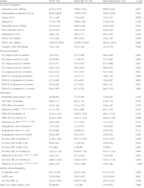
Baseline characteristics among the EnD-CAD patients (Table1)
Among 68 subjects receiving CD34+ cell therapy, three patients in the phase II trial were excluded, including one case died of brain stem hemorrhage at 1 month, an-other one was expired of traumatic cervical spine injury with hypoxia at 1 month, and the other case refused to continue follow-up 1 week after cell delivery. A total of 65 study patients followed up for at least 6 months were selected for the analyses. All EnD-CAD patients expressed high-risk baseline profiles such as 100% for one of the atherosclerotic risk factors, > 76% for diabetes mellitus, > 90% for hypertension, > 87% for dyslipidemia, 70% for PCI history, 100% for multi-vessel CAD, and > 50% for chronic total occlusion (CTO) at left anterior descending (LAD) artery. The age, gender, and rates of body mass index, old stroke, old myocardial infarction (MI), and CABG did not differ between 30 responders and 35 non-responders.
Table 1Baseline characteristics and variables at enrollment
Variable All (N= 65) Responder (N= 30) Non-responder (N= 35) Pvalue
Clinical feature
Age, year 64.51 ± 8.33 65.47 ± 8.86 63.69 ± 7.89 0.395
Male sex,n(%) 52 (80.0%) 21 (70.0%) 31 (88.6%) 0.062
Body height (cm) 161.95 ± 9.97 162.4 ± 7.8 161.7 ± 11.6 0.781
Body weight (kg) 69.68 ± 10.93 70.02 ± 11.96 69.38 ± 10.13 0.814
Body mass index (kg/m2) 26.65 ± 4.36 26.50 ± 3.71 26.77 ± 4.89 0.990
Former smoker,n(%) 26 (40.0%) 16 (53.3%) 10 (28.6%) 0.042
Hypertension,n(%) 59 (90.8%) 26 (86.7%) 33 (94.3%) 0.403
Diabetes mellitus,n(%) 50 (76.9%) 23 (76.7%) 27 (77.1%) 0.964
Dyslipidemia,n(%) 57 (87.7%) 25 (83.3%) 32 (91.4%) 0.455
Old stroke,n(%) 26 (23.1%) 8 (26.7%) 7 (20.0%) 0.525
Old myocardial infarction,n(%) 16 (24.6%) 5 (16.7%) 11 (31.4%) 0.168
Chronic hepatitis B or C,n(%) 5 (7.7%) 1 (3.3%) 4 (11.4%) 0.363
History of CABG,n(%) 22 (33.8%) 7 (23.3%) 15 (49.2%) 0.097
History of PCI,n(%) 45 (69.2%) 21 (70.0%) 24 (68.6%) 0.901
Left main involvement,n(%) 19 (29.2%) 7 (23.3%) 12 (34.3%) 0.333
Multi-vessel CAD,n(%) 68 (100%) 40 (100%) 28 (100%) 1.000
CTO at LAD,n(%) 35 (53.8%) 16 (53.3%) 19 (54.3%) 0.939
Laboratory data
Leukocyte count, 1000/μL 7.00 ± 2.00 6.50 ± 2.07 7.44 ± 1.86 0.017
Hemoglobin, g/dL 13.24 ± 1.87 13.14 ± 2.04 13.32 ± 1.73 0.707
Platelet count, 1000/μL 201.88 ± 57.11 193.1 ± 44.3 209.4 ± 65.9 0.254
Serum creatinine, mg/dL 1.28 ± 0.52 1.25 ± 0.56 1.31 ± 0.48 0.116
eGFR, ml/min/1.73 m2 61.33 ± 21.53 62.19 ± 22.73 60.60 ± 20.74 0.770
Serum sodium, mEq/L 140.11 ± 2.69 140.30 ± 2.89 139.94 ± 2.54 0.531
Serum potassium, mEq/L 4.28 ± 0.42 4.35 ± 0.40 4.22 ± 0.44 0.231
Alanine aminotransferase, U/L 27.02 ± 23.33 27.67 ± 28.88 26.46 ± 17.68 0.594
Total cholesterol, mg/dL 163.49 ± 42.01 167.03 ± 36.86 160.46 ± 26.30 0.378
Low-density lipoprotein, mg/dL 96.23 ± 36.34 99.03 ± 34.73 93.83 ± 38.00 0.569
High-density lipoprotein, mg/dL 43.74 ± 9.79 43.97 ± 11.29 43.54 ± 8.45 0.863
Triglyceride, mg/dL 143.20 ± 79.20 132.13 ± 49.16 152.69 ± 97.71 0.989
Medication
Antiplatelet,n(%) 65 (100.0%) 30 (100.0%) 35 (100.0%) 1.000
Anticoagulant,n(%) 2 (3.1%) 0 (0.0%) 2 (5.7%) 0.495
Beta-blocker,n(%) 58 (89.2%) 20 (100.0%) 28 (80.0%) 0.013
RAS inhibitor,n(%) 54 (83.1%) 23 (76.7%) 31 (88.6%) 0.202
Calcium channel blocker,n(%) 31 (47.7%) 12 (40.0%) 19 (54.3%) 0.250
Diuretic,n(%) 22 (33.8%) 6 (20.0%) 16 (45.7%) 0.029
Lipid lowering agent,n(%) 45 (69.2%) 20 (66.7%) 25 (71.4%) 0.678
Vasodilator,n(%) 2 (3.1%) 0 (0.0%) 2 (5.7%) 0.495
Notes: Responder was defined as 1-year improvement of LVEF≥7.0% after cell-based therapy for EnD-CAD. Data are expressed as mean ± standard deviation or number (percentage).Abbreviation:CABGcoronary artery bypass grafting surgery,PCIpercutaneous coronary intervention,CADcoronary artery disease,CTO
creatinine, total cholesterol, high-density lipoprotein, low-density lipoprotein, and triglyceride did not differ between the two groups.
In addition, responders had a significantly higher pre-scription of beta-blocker and lower diuretic use than the non-responders. All patients took antithrombotic agents and a majority of them received guideline-directed med-ical therapy.
Laboratory parameters after G-CSF injection, clinical presentations, and results of examinations during follow-up, and 1-year outcomes (Table2)
The post-G-CSF laboratory findings demonstrated that the circulating levels of leukocyte count, hematopoietic stem cells, young cells, neutrophils, CD34+ cells, CD45+ cells, and stem cell percentage measured by flow cyto-metric analysis, as well as troponin-I did not differ be-tween the two groups.
The responders had significantly lower angina severity and insignificantly less dyspnea compared with the non-responders prior to CD34+ cell therapy. Within the follow-up period, the responders had better clinical symptomatic improvement in both angina and HF as compared with the non-responders at the time points of every 3 months, indicating the improvement of cardiac systolic function (i.e., LVEF improvement ≥7.0%) was correlated with the relief of clinical symptoms.
Regarding objective evaluations for functional capacity, angiogenesis, chamber sizes, and cardiac/valvular func-tions, there were no significant differences between groups at baseline. However, the responders had higher coronary angiogenesis score and less echocardiographic grade 2 or 3 diastolic dysfunction than the non-responders. Notably, the difference of LVEF on 3D echo-cardiography between the follow-up period and baseline began to be significant at 3 months after stem cell ther-apy, implicating good clinical, and subclinical responses could be observed as early as 3 months since delivery of CD34+ cells. After 1-year follow-up, composite end-points occurred in nearly one half of EnD-CAD patients but did not differ between the two groups. Around 1 in 5 patients needed hospitalization for acute decompen-sated HF, and nearly 1 in 4 patients received salvage myocardial revascularization strategy for relief of refrac-tory angina in both groups.
Identification of“predictors of good responder”to CD34+ cell therapy from baseline characteristics or presentations (Table3, Figs.1and2)
To understand which baseline variable could be predict-ive of a good responder prior to CD34+ cell therapy in patients with EnD-CAD, logistic regression analysis was performed. In univariate analysis, male gender, higher baseline leukocyte count, CCS angina score≥3, and
grade of diastolic dysfunction≥2 were identified as po-tentially poor responders to the cell therapy. On the contrary, a former smoker and higher baseline angiogen-esis score could be used to predict good response to the cell therapy. After multivariate adjustment for the above potential variables, the“presence” of the former smoker and “absence” of the male gender, Canadian Cardiovas-cular Society (CCS) angina score≥3, and grade of dia-stolic dysfunction ≥2 on the initial survey were identified as independent predictors of good responder after stem cell therapy.
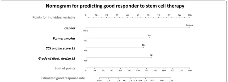
H-L test shown in Fig. 1a demonstrated sensitivity 86.7%, specificity 70.6%, and accuracy 78.1% after con-sidering the above four predictors (P= 0.777, which was higher than the cutoff value of 0.5). To facilitate an effi-cient evaluation in clinical practice, the nomogram in Fig. 2 was utilized to calculate the estimated good re-sponse rate of CD34+ cell therapy for EnD-CAD. Sum-mation of individual points available from gender, former smoker, CCS angina score, and grade of diastolic dysfunction helps researchers to assess the probability of good responder when EnD-CAD patients are enrolled as candidates for cell therapy.
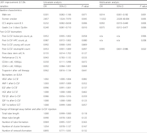
Identification of“early predictors of good responder” after receiving G-CSF injection or CD34+ cell therapy (Table4, Fig.1and Supplemental Table1)
Owing to a lot of useful information available after ad-ministering G-CSF or transfusing CD34+ cells, those variables viable to be recognized as“early predictors” of a good responder, e.g., the vasculogenic activity of stem cells, biomarkers before and after G-CSF injection, and detailed imaging for angiogenesis (refer to Supplemental Table1), were collected and added to the logistic regres-sion analysis. Table 4 shows in addition to the four aforementioned baseline characteristics, the elevation of post G-CSF-treated neutrophil count was found nega-tively associated with good response to the cell therapy after multivariate adjustment. After adding this newly-identified independent predictor to the H-L test, a better predictor power was obtained with sensitivity 83.3%, specificity 85.3%, and accuracy 84.4% (p= 0.881, which was much higher than the cutoff value of 0.5) (Fig. 1b). Regrettably, we did not further identify other potential predictors of good responder from remaining variables after G-CSF, prior to, or after stem cell therapy.
G-Table 2Variables within follow-up period and clinical outcomes
Variable All (N= 65) Responder (N= 30) Non-responder (N= 35) Pvalue
Post-GCSF biomarkers
Leukocyte count, 1000/μL 42.29 ± 10.76 39.49 ± 10.34 44.69 ± 10.67 0.051
Hematopoietic progenitor cell,μL 49.78 ± 46.68 37.00 ± 29.01 59.56 ± 55.05 0.105
Young cell, % 7.21 ± 5.06 7.10 ± 4.93 7.30 ± 5.23 0.859
Segment, % 77.14 ± 7.86 76.86 ± 7.60 77.39 ± 8.18 0.790
Neutrophil count, 1000/μL 32.76 ± 9.31 30.66 ± 9.44 34.56 ± 8.94 0.093
Flow data: stem cell, % 0.35 ± 0.24 0.30 ± 0.18 0.40 ± 0.28 0.226
Performance CV, % 4.68 ± 2.67 4.46 ± 2.71 4.87 ± 2.65 0.540
CD34+ cell, 1000/μL 1.27 ± 0.90 0.87 ± 0.50 1.49 ± 1.00 0.076
CD45+ cell, 1000/μL 343.05 ± 100.35 297.66 ± 103.38 366.74 ± 92.24 0.082
Troponin-I after cell therapy 1.24 ± 3.30 1.02 ± 2.64 1.42 ± 3.78 0.430
Clinical presentation
CCS angina score at baseline 2.55 ± 0.75 2.27 ± 0.64 2.80 ± 0.58 0.003
CCS angina score≥3,n(%) 36 (55.4%) 11 (36.7%) 25 (71.4%) 0.005
CCS angina score at 3 months 0.57 ± 0.77 0.33 ± 0.55 0.77 ± 0.88 0.021
CCS angina score at 6 months 0.50 ± 0.74 0.38 ± 0.62 0.60 ± 0.66 0.282
CCS angina score at 12 months 0.33 ± 0.57 0.17 ± 0.47 0.47 ± 0.62 0.021
NYHA Fc of dyspnea at baseline 1.71 ± 1.13 1.43 ± 1.17 1.94 ± 1.06 0.086
NYHA Fc of dyspnea at 3 months 0.71 ± 0.88 0.37 ± 0.62 1.00 ± 0.97 0.003
NYHA Fc of dyspnea at 6 months 0.53 ± 0.87 0.17 ± 0.47 0.83 ± 1.01 0.001
NYHA Fc of dyspnea at 12 months 0.54 ± 0.90 0.21 ± 0.56 0.82 ± 1.03 0.002
Examination
Endothelial dysfunction*,n(%) 38 (64.4%) 15 (57.7%) 23 (69.7%) 0.339
CPET METs at baseline 5.08 ± 1.17 4.83 ± 1.19 5.24 ± 1.16 0.255
CPET METs at 6 months 5.33 ± 1.44 5.22 ± 1.44 5.38 ± 1.46 0.750
Difference of METs6 months vs. baseline 0.17 ± 1.20 0.47 ± 0.86 0.01 ± 1.33 0.252
CMR LVEF at baseline, % 50.31 ± 14.52 52.69 ± 13.28 48.21 ± 15.42 0.214
CMR LVEF at 6 months, % 52.93 ± 14.08 56.25 ± 12.33 50.03 ± 15.05 0.098
Difference of LVEF6 months vs. baseline 2.02 ± 4.96 2.71 ± 3.87 1.41 ± 5.74 0.312
Angiogenesis score at baseline 1.57 ± 0.63 1.73 ± 0.57 1.43 ± 0.66 0.048
Angiogenesis score≥2,n(%) 36 (56.3%) 20 (66.7%) 16 (47.1%) 0.115
Angiogenesis score at 9 months 2.60 ± 0.83 2.65 ± 0.70 2.56 ± 0.94 0.955
3D echo LVEF at baseline, % 51.30 ± 10.62 49.41 ± 10.38 52.91 ± 10.71 0.188
3D echo LVEF≥50%,n(%) 40 (61.5%) 17 (56.7%) 23 (65.7%) 0.455
3D echo LVEF≤40%,n(%) 9 (13.8%) 6 (20.0%) 3 (8.6%) 0.282
3D echo LVEF at 3 months, % 55.01 ± 11.46 56.18 ± 11.36 54.11 ± 11.63 0.494
Difference of 3D LVEF3 months vs. baseline 3.24 ± 5.91 5.71 ± 5.60 1.35 ± 5.49 0.002
3D echo LVEF at 6 months, % 56.45 ± 12.56 57.40 ± 13.74 55.67 ± 11.64 0.359
Difference of 3D LVEF6 months vs. baseline 4.89 ± 7.94 7.47 ± 10.06 2.76 ± 4.80 < 0.001
Baseline echocardiography
LA diameter, mm 42.17 ± 5.39 42.70 ± 4.94 41.71 ± 5.79 0.369
LVEDD, mm 53.25 ± 8.42 53.22 ± 8.37 53.27 ± 8.59 0.981
2D echo LVEF, % 54.32 ± 13.03 53.85 ± 13.82 54.72 ± 12.51 0.790
CSF injections as well as after CD34+ cell therapy. Add-itionally, SDF-1αlevels in coronary sinus checked at dif-ferent time points were also significantly lower in the former group than in the latter one. However, coronary angiogenesis on Wimasis analysis, angiogenic capacity on Matrigel assay, and levels of angiopoietin (ANP)-1, epidermal growth factor (EGF), and transforming growth factor (TGF)-β1 (i.e., three soluble angiogenesis factors) on enzyme-linked immunosorbent assay (ELISA) did not differ between the two groups, suggesting prediction of a good responder to cell therapy was mainly dependent on baseline characteristics ra-ther than on those variables collected after G-CSF or CD34+ cell therapy. These findings were very useful and practical on the screening for potential good responders to the cell ther-apy in the early stage of the trial.
Discussion
This study which utilized and analyzed the parameters from our phase I/II clinical trials delineated several fun-damental clinical-relevant information. First, the baseline variables of female gender and former smoker were sig-nificantly and positively predictive of, whereas the ad-vanced angina score (i.e., CCS angina score≥3) and moderate to severe LV diastolic dysfunction (i.e., grade≥ 2) were negatively predictive of, good response to CD34+ cell therapy for EnD-CAD. Second, increased neutrophil count after G-CSF treatment not only was negatively predictive of good response to the cell therapy but also augmented the predictive power when it was
considered in addition to the four aforementioned base-line variables. Third, most predictors of a good re-sponder were identified based on patient’s baseline characteristics and laboratory/examination findings, sug-gesting that we are able to expect the probability of ef-fectiveness and responsiveness before conducting expensive cell-based therapy.
It is well recognized that regenerative medicine is cur-rently of paramount importance for organ dysfunction, especially for those patients with ischemia-related LV dysfunction. Abundant data [11–16, 18–20] have also supported that cell therapy is an alternative to conven-tional anti-ischemic treatment for the patients with in-tractable ischemic cardiomyopathy, complex diffuse coronary stenotic lesions, and refractory angina. How-ever, not all the patients with EnD-CAD who received cell therapy had satisfactory clinical outcomes and prominent improvement of LV function. This issue drives the investigators [13, 22, 23] trying to integrate the consensus of improvement of LVEF≥5.0 to 7.0% to be recognized as the“acceptable or great response”after cell therapy. Surprisingly, how many (i.e., percentage) or what kind of EnD-CAD patients met the criteria of “great response”to cell therapy have not yet been identi-fied. One novel finding in the present study was that the analytical results of pooled data from our phases I/II tri-als showed that nearly 50% of CD34+ cell treated pa-tients met the criteria of “good responders”, i.e., LVEF improved ≥7.0%. Our finding, in addition to extending Table 2Variables within follow-up period and clinical outcomes(Continued)
Variable All (N= 65) Responder (N= 30) Non-responder (N= 35) Pvalue
TVPG, mmHg 18.32 ± 11.66 19.52 ± 13.16 17.31 ± 10.33 0.568
Moderate to severe MR,n(%) 9 (13.8%) 5 (16.7%) 4 (11.4%) 0.722
3D echo LVEDV, mm3 87.11 ± 27.14 86.60 ± 26.43 87.51 ± 28.08 0.896
Systolic dyssynchrony index, % 7.75 ± 7.25 8.48 ± 8.81 7.10 ± 5.54 0.850
Outcome at 1 year
Composite endpoints†,n(%) 31 (47.7%) 14 (46.7%) 17 (48.6%) 0.878
All-cause mortality,n(%) 10 (15.4%) 4 (13.3%) 6 (17.1%) 0.742
Cardiovascular death,n(%) 1 (1.5%) 1 (3.3%) 0 (0.0%) 0.462
Acute myocardial infarction,n(%) 3 (4.6%) 1 (3.3%) 2 (5.7%) 1.000
Hospitalization for HF,n(%) 13 (20.0%) 6 (20.0%) 7 (20.0%) 1.000
Revascularization,n(%) 16 (24.6%) 9 (30.0%) 7 (20.0%) 0.351
Sepsis,n(%) 6 (12.3%) 2 (6.7%) 6 (17.1%) 0.270
Notes: Responder was defined as 1-year improvement of LVEF≥7.0% after cell-based therapy for EnD-CAD. Data are expressed as mean ± standard deviation or number (percentage)
Abbreviation:GCSFgranulocyte-colony stimulating factor,CVcoefficient of variability,CCSCanadian Cardiovascular Society,NYHA FcNew York Heart Association functional classification,CPETcardiopulmonary exercise testing,METmetabolic equivalent of task,CMRcardiovascular magnetic resonance imaging,LVEFleft ventricular ejection fraction,3D echothree-dimensional echocardiography,2D echotwo-dimensional echocardiography,LAleft atrium,LVEDDleft ventricular end-diastolic diameter,diast. dysfxndiastolic dysfunction,TVPGtrans-tricuspid valve pressure gradient,MRmitral regurgitation,LVEDVleft ventricular end-diastolic volume,HFheart failure
*Endothelial dysfunction was defined as post-nitroglycerin flow-mediated dilatation of brachial artery < 300%
†Composite endpoints were comprised of all-cause mortality, major adverse cardiac or cerebrovascular events (defined as cardiovascular death, acute myocardial
Table 3Baseline predictors of“good responder”before IC CD34+ therapy for EnD-CAD
LVEF improvement≥7.0% Univariate analysis Multivariate analysis
Variables OR 95% CI Pvalue OR 95% CI Pvalue
Baseline characteristics
Age per year 1.027 0.967–1.090 0.389
Age≥65 years 1.524 0.571–4.065 0.400
Male sex 0.301 0.082–1.106 0.071 0.028 0.003–0.261 0.002
Body mass index 0.985 0.879–1.104 0.799
Former smoker 2.857 1.024–7.970 0.045 8.898 1.891–41.858 0.006
Diabetes mellitus 1.102 0.357–3.405 0.866
Atherosclerotic risk factor* n/a n/a n/a
Old stroke or old MI 0.706 0.263–1.893 0.489
Chronic hepatitis B or C 0.267 0.028–2.533 0.250
History of CABG 0.406 0.138–1.194 0.101
Left main involvement 0.583 0.195–1.747 0.335
CTO at LAD 0.962 0.362–2.560 0.939
Leukocyte count 0.765 0.576–1.018 0.066 0.860 0.632–1.170 0.077
Hemoglobin 0.950 0.729–1.237 0.702
Serum creatinine 0.776 0.292–2.060 0.610
eGFR 1.003 0.981–1.027 0.766
Total Cholesterol 1.004 0.992–1.016 0.528
Beta blocker n/a n/a n/a
RAS inhibitor 0.424 0.111–1.622 0.210
Lipid lowering agent 0.800 0.278–2.299 0.679
Presentation and exams at baseline
CCS angina score≥3 0.232 0.082–0.658 0.006 0.138 0.033–0.576 0.007
NYHA Fc of dyspnea≥3 0.664 0.220–2.009 0.468
Endothelial dysfunction 0.593 0.202–1.738 0.341
CPET-METs 0.732 0.429–1.246 0.250
CMR LVEF 1.022 0.987–1.059 0.225
Angiogenesis score≥2 2.250 0.816–6.207 0.117
3D echo LVEF 0.968 0.923–1.106 0.187
3D echo LVEF ≥50% 0.682 0.250–1.863 0.456
3D echo LVEF≤40% 2.667 0.605–11.756 0.195
Baseline echocardiography
LA diameter 1.035 0.944–1.136 0.461
LVEDD 0.999 0.943–1.059 0.980
2D echo LVEF 0.995 0.958–1.033 0.786
Grade 2 or 3 diast. dysfxn 0.240 0.081–0.710 0.010 0.104 0.022–0.505 0.005
TVPG 1.017 0.972–1.064 0.469
Moderate to severe MR 1.550 0.376–6.390 0.544
3D echo LVEDV 0.999 0.980–1.017 0.894
Systolic dyssynchrony index 1.028 0.955–1.106 0.466
Notes:Abbreviation:ICintracoronary,EnD-CADend-stage diffuse coronary artery disease,LVEFleft ventricular ejection fraction,ORodds ratio,CIconfidence interval,n/anot applicable,MImyocardial infarction,CABGcoronary artery bypass grafting surgery,CTOchronic total occlusion,LADleft anterior descending artery,eGFRestimated glomerular filtration rate,RASrenin-angiotensin system,CCSCanadian Cardiovascular Society,NYHA FcNew York Heart Association functional classification,CPETcardiopulmonary exercise testing,METmetabolic equivalent of task,CMRCardiovascular magnetic resonance imaging,LVEFleft ventricular ejection fraction,3D echothree-dimensional echocardiography,LAleft atrium,LVEDDleft ventricular end-diastolic diameter,2D echotwo-dimensional echocardiography,diast. dysfxndiastolic dysfunction,TVPGtrans-tricuspid valve pressure gradient,MRmitral regurgitation,LVEDVleft ventricular
end-diastolic volume
the findings of the previous studies, [13, 22, 23] is the first one to highlight the early prediction of “good re-sponders” and certainly would provide useful informa-tion in future clinical cell-based researches for the treatment of refractory angina or EnD-CAD.
Despite the important issue of good response after cell therapy has been extensively investigated and 5.0 to 7.0% of improvement in LVEF is viewed as a well acceptable value [13, 22, 23], so far, no study has reported which variables could be served as independent predictors of good response to cell-based therapy in the field of ische-mic heart disease. In fact, there is only one similar study [25] which identified age, blood fibrinogen, arterial
occlusion level, transcutaneous pressure of oxygen, and the transplanted CD34+ cell count were significant prog-nostic factors of the responders in patients with no-option critical limb ischemia. Somewhat different from the analytic results in the former study [25], our findings in the present study emphasized the importance of base-line patients’characteristics rather than laboratory vari-ables or lesion complexities. As for the patients with EnD-CAD, we successfully identified that the baseline variables of female gender and former smoker were in-dependently predictive of good responders of CD34+ cell therapy, whereas the advanced angina score and diastolic dysfunction were two independent predictors of
responders of CD34+ cell therapy. Of distinctive finding was that an increase in the neutrophil count after G-CSF treatment was another negative predictor of non-responder to cell therapy. Our findings, therefore, pro-vide extremely useful research information on the screening and enrollment of suitable candidates for re-ceiving CD34+ cell therapy, especially for those EnD-CAD patients who are refractory to conventional therapy.
We found discrepancies among improvements of LVEF, presentation of HF symptoms, and unfavorable clinical outcomes. In many clinical circumstances, LVEF is not consistent with the New York Heart Association functional class because the latter is namely dependent on patient’s physiological adaptation, metabolic status, oxygen consumption, and myocardial oxygen balance determined by the ratio of oxygen supply to oxygen de-mand [26]. Therefore, besides N-terminal pro-brain natriuretic peptide level, LVEF, 6-min walk distances, and CPET, well-designed questionnaires are suggested for functional evaluation and risk stratification in HF pa-tients [27].
There is a close association between an increase in the circulatory level of soluble angiogenesis factors and en-hancement of microvasculature/microcirculation, result-ing in an improvement of LVEF [11, 12]. In the present study, circulating angiogenesis factors, such as VEGF and HGF, were also notably higher in the responders than in the non-responders (refer to Supplemental Table 1). Accordingly, our findings were consistent with those of our previous studies [11,12].
SDF-1α, a chemokine protein of CXCL12, not only plays a beneficial role in the mobilization of EPCs from
the bone marrow to circulation and homing of EPC to the ischemic area for angiogenesis through CXCR4-dependent mechanism [28, 29], but also is associated with an increased risk of CAD via leukocyte activation and proinflammatory stimulation [30]. An interesting finding of ELISA in the present study was that the levels of SDF-1α in the circulation and coronary sinus at dif-ferent time points were consistently and notably lower in the responders than in the non-responders (refer to Supplemental Table 1), suggesting the non-responders presented with more severe coronary lesions (i.e., higher rates of old MI, CABG history, left main involvement and CTO at LAD shown in Table1) and a more domin-ant intrinsic response to ischemic stimulation (i.e., higher levels of SDF-1α) than the responders.
Regrettably, this study did not measure the circulatory level of thrombospondin-1 (TSP1), a natural inhibitor of neovascularization, notably increased in the platelet-poor plasma of aged adults, and those with cardiovascu-lar and metabolic disease [31]. Additionally, TSP1 has been identified to inhibit endothelial function, stimulates apoptosis, and suppresses nitric oxide, vascular endothe-lial growth factor, and the pluripotent transcription fac-tors Oct3/4, Sox2, Klf4, and cMyc signaling [32]. These aforementioned issues raise the hypothesis that the low circulatory TSP1 level, perhaps, could be an extremely useful biomarker for predictive of responder and good outcome in our study.
The other interesting finding of the present study was former smokers closely linked to a good responder of stem cell therapy in the EnD-CAD patients. This associ-ation was compatible with the previous misleading phenomenon of “smoker’s paradox” [33, 34], indicating
Fig. 2Nomogram for scaling a good response rate to stem cell therapy for EnD-CAD. The nomogram was designed for scaling and calculating the probability of good response rate to cell therapy for patients with EnD-CAD by using individual baseline variable. The scales could help investigators to calculate the response rate and further predict who will be a“good responder”before applying cell infusion to treat EnD-CAD or refractory angina. Abbreviation: EnD-CAD = end-stage diffuse coronary artery disease; CCS = Canadian Cardiovascular Society; diast.
better clinical outcomes were observed among the smokers suffering from acute myocardial infarction. Re-cent clinical researches [35, 36] have debunked that bi-zarre “smoker’s paradox” was mainly affected by age factor because the majority of smokers presenting to the hospital were 10 years younger than nonsmokers. In addition, further analysis in our study revealed that the former smokers not only were younger, taller, and heav-ier but also had better eGFR and less history of CABG (refer to Supplemental Table2). As a result, our findings and recent deep investigations could be explained, at least in part, for the close relationship of “former smoker”to“good responder”of cell therapy.
In the current study, 20% of patients in both groups suffered from acute decompensated HF with a need for hospitalization for further management. If there had been no evidence of myocardial injury, i.e., an elevated level of cardiac troponin-I, the patient was discharged without CAG after HF symptoms were improved. Other-wise, those with elevated troponin-I would undergo CAG and receive bailout coronary stenting at culprit le-sion to increase myocardial perfule-sion. By the 9th month, all study subjects received follow-up CAG for the assess-ment of angiogenesis. Majority of patients in both groups underwent diagnostic CAG only. However, if the coronary flow had been limited by critical or progressive
Table 4Early predictors of“good responder”after receiving G-CSF injection or stem cell therapy
LVEF improvement≥7.0% Univariate analysis Multivariate analysis
Variables OR 95% CI Pvalue OR 95% CI Pvalue
Baseline characteristics
Male sex 0.301 0.082–1.106 0.071 0.014 0.001–0.182 0.001
Former smoker 2.857 1.024–7.970 0.045 11.032 2.028–60.004 0.005
CCS angina score≥3 0.232 0.082–0.658 0.006 0.092 0.019–0.449 0.003
Grade 2 or 3 diast. Dysfxn 0.240 0.081–0.710 0.010 0.075 0.012–0.477 0.006
Post G-CSF biomarkers
Post G-CSF leukocyte count,μL 0.952 0.905–1.002 0.058 n/a n/a 0.906
Post G-CSF HPC count,μL 0.987 0.972–1.002 0.080 n/a n/a 0.208
Post G-CSF young cell count 0.992 0.900–1.093 0.869
Post G-CSF neutrophil count 0.953 0.901–1.009 0.097 0.905 0.831–0.986 0.022
Flow data: stem cell, % 0.155 0.014–1.702 0.127
Performance CV, % 0.943 0.783–1.135 0.533
CD34+ cell, 1000/μL 0.350 0.111–1.098 0.072
CD45+ cell, 1000/μL 0.992 0.984–1.001 0.068
Troponin-I after cell therapy 0.962 0.816–1.134 0.641
Biomarkers on ELISA
VEGF after G-CSF 1.002 1.000–1.004 0.082
ANP-1 after G-CSF 1.003 0.997–1.009 0.352
EGF after G-CSF 0.996 0.991–1.001 0.137
HGF after G-CSF 1.000 1.000–1.000 0.462
TGF-β1 after G-CSF 0.986 0.956–1.016 0.359
SDF-1αafter G-CSF 1.000 1.000–1.000 0.123
SDF-1αbefore SCT 1.000 0.999–1.000 0.057
Change of Matrigel assay before and after G-CSF injection
Total tube length 1.000 0.999–1.000 0.118
Mean tube length 0.990 0.978–1.003 0.125
Number of tube formation 0.969 0.905–1.037 0.363
Number of cluster formation 1.054 0.978–1.137 0.167
Number of network formation 0.893 0.771–1.035 0.132
Notes:Abbreviation:G-CSFgranulocyte-colony stimulating factor,LVEFleft ventricular ejection fraction,ORodds ratio,CIconfidence interval,n/anot applicable,MI
myocardial infarction,CCSCanadian Cardiovascular Society,diast. dysfxndiastolic dysfunction,WBCwhite blood cell,HPChematopoietic progenitor cell,CV
stenotic lesions as compared with baseline CAG findings as well as the vessel larger and suitable for intervention after cell therapy, myocardial revascularization with PCI would be performed as a bailout therapeutic strategy.
This study has limitations. First, despite the four vari-ables were identified to be strongly predictive of either good responder or non-responder in the present study, these variables were not valid factors predictive of 1-year untoward clinical outcomes (refer to Supplemental Table 3) after CD34+ cell therapy. A prospective study to test predictors of good outcomes such as longer sur-vival or better functional/echocardiographic improve-ment is encouraged. However, there are still several issues to be addressed in a prospective case-control study that will be conducted, e.g., difficulty in patient’s enrollment and high event rate in such high-risk EnD-CAD patients. Maybe, a retrospective study with a lon-ger follow-up period or increase in study case number would be a better solution to answer the question. At that time, we will be able to apply cell-based therapy in highly selected patients or specific patient population with cost-effectiveness. Second, the long-term clinical follow-up (i.e., more than 1 to 5 years follow-up) is con-tinuously conducted in phase II clinical trial. Accord-ingly, whether the currently identified variables will be able to predict 5-year “good responder” or “ non-re-sponder”remains to be answered.
Conclusions
The results of this study identified four baseline variables for the prediction of good responders prior to CD34+ therapy for the patients with EnD-CAD. These findings may provide essential and useful information on screen-ing suitable candidates in a clinical cell-based research.
Supplementary information
Supplementary informationaccompanies this paper athttps://doi.org/10. 1186/s13287-020-01835-z.
Additional file 1 : Table S1. Angiogenesis, biomarkers, and cell migration function before and after stem cell therapy.
Additional file 2 : Table S2.Variables of interest compared between smokers versus non-smokers.
Additional file 3 : Table S3. To test whether the new 4 identified factors can be used to predict adverse clinical events.
Abbreviations
PCI:Percutaneous coronary intervention; CABG: Coronary artery bypass grafting surgery; CAD: Coronary artery disease; EnD-CAD: End-stage diffuse coronary artery disease; EPC: Endothelial progenitor cell; MSC: Mesenchymal stem cell; LV: Left ventricular; LVEF: Left ventricular ejection fraction; G-CSF: Granulocyte-colony stimulating factor; H-L test: Hosmer–Lemeshow test; CTO: Chronic total occlusion; LAD: Left anterior descending; MI: Myocardial infarction; eGFR: Estimated glomerulus filtration rate; CCS: Canadian Cardiovascular Society; VEGF: Vascular endothelial growth factor; HGF: Hepatocyte growth factor; SDF-1α: Stromal cell-derived factor-1 alpha; ANP: Atrial natriuretic peptide; EGF: Epidermal growth factor;
TGF: Transforming growth factor; ELISA: The enzyme-linked immunosorbent assay
Acknowledgements None.
Authors’contributions
PHS, MSL, and HKY designed the study and performed a catheterization procedure for cell delivery. HJC, YCL, PLS, and FYL gathered the data and recorded clinical events. YCL and PLS prepared cell products and did bench work. PHS, HJC, CHC, and HKY analyzed and interpreted the data. PHS and HKY wrote the manuscript. The authors read and approved the final manuscript.
Funding
This study grant was funded by a program grant from the National Science Council, Taiwan, Republic of China (Grant number: 100-2314-B-182A-077 for the phase I trial; NMRPG8C0291, NMRPG8E0011, and NMRPG8F0011 for the phase II trial). It was also supported by a grant from Chang Gung Memorial Hospital, Chang Gung University (Grant number: CMRPG8B0901 and CMRP G8B0902 for the phase I trial).
Availability of data and materials
The datasets generated and analyzed are not publicly available due to consideration of patient privacy, but are available from the corresponding author on reasonable request for academic purpose.
Ethics approval and consent to participate
Ethics approval for this study was obtained from the Institutional Review Committee on Human Research at Chang Gung Memorial Hospital. All study subjects were informed about the study design and provided their informed consent.
Consent for publication
All patients signed a consent for use of their research data and publication.
Competing interests
The authors declare no competing interests.
Author details
1Division of Cardiology, Department of Internal Medicine, College of
Medicine, Kaohsiung Chang Gung Memorial Hospital and Chang Gung University, No. 123, Ta Pei Road, Niao Sung District, Kaohsiung 83301, Taiwan.
2
Center for Shockwave Medicine and Tissue Engineering, Kaohsiung Chang Gung Memorial Hospital, Kaohsiung 83301, Taiwan.3Department of
Obstetrics and Gynecology, College of Medicine, Kaohsiung Chang Gung Memorial Hospital and Chang Gung University, Kaohsiung 83301, Taiwan.
4
Chung Shan Medical University School of Medicine, Taichung 40201, Taiwan.5Department of Computer Science and Engineering, National Sun
Yat-sen University, Kaohsiung 80424, Taiwan.6Department of Healthcare
Administration and Medical Informatics, Kaohsiung Medical University, Kaohsiung 80708, Taiwan.7Department of Statistics, National Cheng Kung University, Tainan 70101, Taiwan.8Institute of Statistics, National University of
Kaohsiung, Kaohsiung 80708, Taiwan.9Department of Nursing, Asia
University, Taichung 41354, Taiwan.10Division of Thoracic and Cardiovascular
Surgery, Department of Surgery, Kaohsiung Chang Gung Memorial Hospital and Chang Gung University College of Medicine, Kaohsiung 83301, Taiwan.
11Division of Cardiovascular Surgery, Department of Surgery, Tri-Service
General Hospital, National Defense Medical Center, Taipei 11490, Taiwan.
12
Department of Orthopedics, College of Medicine, Kaohsiung Chang Gung Memorial Hospital and Chang Gung University, Kaohsiung 83301, Taiwan.
13Institute for Translational Research in Biomedicine, College of Medicine,
Kaohsiung Chang Gung Memorial Hospital and Chang Gung University, Kaohsiung 83301, Taiwan.14Department of Medical Research, China Medical University Hospital, China Medical University, Taichung 40402, Taiwan.
15Division of Cardiology, Department of Internal Medicine, Xiamen Chang
Received: 11 June 2020 Revised: 8 July 2020 Accepted: 15 July 2020
References
1. Held C, Asenblad N, Bassand JP, Becker RC, Cannon CP, Claeys MJ, et al. Ticagrelor versus clopidogrel in patients with acute coronary syndromes undergoing coronary artery bypass surgery: results from the PLATO (Platelet Inhibition and Patient Outcomes) trial. J Am Coll Cardiol. 2011;57(6):672–84. 2. Yasuda S, Kaikita K, Ogawa H, Akao M, Ako J, Matoba T, et al. Atrial
fibrillation and ischemic events with rivaroxaban in patients with stable coronary artery disease (AFIRE): protocol for a multicenter, prospective, randomized, open-label, parallel group study. Int J Cardiol. 2018;265:108–12. 3. Ho YC, Tsai TH, Sung PH, Chen YL, Chung SY, Yang CH, et al. Minimizing
door-to-balloon time is not the most critical factor in improving clinical outcome of ST-elevation myocardial infarction patients undergoing primary percutaneous coronary intervention. Crit Care Med. 2014;42(8):1788–96. 4. Sung PH, Wu CJ, Yip HK. Is extracorporeal membrane oxygenator a new
weapon to improve prognosis in patients with profound cardiogenic shock undergoing primary percutaneous coronary intervention? Circ J. 2016;80(3): 572–8.
5. Liu H, Wilton SB, Southern DA, Knudtson ML, Maitland A, Hauer T, et al. Automated referral to cardiac rehabilitation after coronary artery bypass grafting is associated with modest improvement in program completion. Can J Cardiol. 2019;35(11):1491–8.
6. Lin YS, Fang HY, Hussein H, Fang CY, Chen YL, Hsueh SK, et al. Predictors of contrast-induced nephropathy in chronic total occlusion percutaneous coronary intervention. EuroIntervention. 2014;9(10):1173–80.
7. Knuuti J, Wijns W, Saraste A, Capodanno D, Barbato E, Funck-Brentano C, et al. 2019 ESC guidelines for the diagnosis and management of chronic coronary syndromes. Eur Heart J. 2020;41(3):407–77.
8. Gegouskov V, Tochtermann U, Badowski-Zyla D, Thomas G, Hagl S, Osswald B. Long-term results after coronary artery reconstructive surgery. Thorac Cardiovasc Surg. 2007;55(5):293–7.
9. Jolicoeur EM, Cartier R, Henry TD, Barsness GW, Bourassa MG, McGillion M, et al. Patients with coronary artery disease unsuitable for revascularization: definition, general principles, and a classification. Can J Cardiol. 2012;28(2 Suppl):S50–9.
10. Henry TD, Satran D, Jolicoeur EM. Treatment of refractory angina in patients not suitable for revascularization. Nat Rev Cardiol. 2014;11(2):78–95. 11. Lee FY, Chen YL, Sung PH, Ma MC, Pei SN, Wu CJ, et al. Intracoronary
transfusion of circulation-derived CD34+ cells improves left ventricular function in patients with end-stage diffuse coronary artery disease unsuitable for coronary intervention. Crit Care Med. 2015;43(10):2117–32. 12. Sung PH, Lee FY, Tong MS, Chiang JY, Pei SN, Ma MC, et al. The five-year clinical and angiographic follow-up outcomes of intracoronary transfusion of circulation-derived CD34+ cells for patients with end-stage diffuse coronary artery disease unsuitable for coronary intervention-phase I clinical trial. Crit Care Med. 2018;46(5):e411–8.
13. Losordo DW, Henry TD, Davidson C, Sup Lee J, Costa MA, Bass T, et al. Intramyocardial, autologous CD34+ cell therapy for refractory angina. Circ Res. 2011;109(4):428–36.
14. Dill T, Schachinger V, Rolf A, Mollmann S, Thiele H, Tillmanns H, et al. Intracoronary administration of bone marrow-derived progenitor cells improves left ventricular function in patients at risk for adverse remodeling after acute ST-segment elevation myocardial infarction: results of the Reinfusion of Enriched Progenitor cells And Infarct Remodeling in Acute Myocardial Infarction study (REPAIR-AMI) cardiac magnetic resonance imaging substudy. Am Heart J. 2009;157(3):541–7.
15. Fisher SA, Doree C, Brunskill SJ, Mathur A, Martin-Rendon E. Bone marrow stem cell treatment for ischemic heart disease in patients with no option of revascularization: a systematic review and meta-analysis. PLoS One. 2013; 8(6):e64669.
16. Leu S, Sun CK, Sheu JJ, Chang LT, Yuen CM, Yen CH, et al. Autologous bone marrow cell implantation attenuates left ventricular remodeling and improves heart function in porcine myocardial infarction: an
echocardiographic, six-month angiographic, and molecular-cellular study. Int J Cardiol. 2011;150(2):156–68.
17. Chen KH, Chen CH, Wallace CG, Yuen CM, Kao GS, Chen YL, et al. Intravenous administration of xenogenic adipose-derived mesenchymal stem cells (ADMSC) and ADMSC-derived exosomes markedly reduced brain
infarct volume and preserved neurological function in rat after acute ischemic stroke. Oncotarget. 2016;7(46):74537–56.
18. Quyyumi AA, Vasquez A, Kereiakes DJ, Klapholz M, Schaer GL, Abdel-Latif A, et al. PreSERVE-AMI: a randomized, double-blind, placebo-controlled clinical trial of intracoronary administration of autologous CD34+ cells in patients with left ventricular dysfunction post STEMI. Circ Res. 2017;120(2):324–31. 19. Bravery CA, Carmen J, Fong T, Oprea W, Hoogendoorn KH, Woda J, et al. Potency
assay development for cellular therapy products: an ISCT review of the requirements and experiences in the industry. Cytotherapy. 2013;15(1):9–19. 20. Porat Y, Abraham E, Karnieli O, Nahum S, Woda J, Zylberberg C. Critical elements in the development of cell therapy potency assays for ischemic conditions. Cytotherapy. 2015;17(7):817–31.
21. Sung PH, Li YC, Lee MS, Hsiao HY, Ma MC, Pei SN, et al. Intracoronary injection of autologous CD34+ cells improves one-year left ventricular systolic function in patients with diffuse coronary artery disease and preserved cardiac performance-a randomized, open-label, controlled phase II clinical trial. J Clin Med. 2020;9(4):1043.
22. Wang S, Cui J, Peng W, Lu M. Intracoronary autologous CD34+ stem cell therapy for intractable angina. Cardiology. 2010;117(2):140–7.
23. Povsic TJ, Junge C, Nada A, Schatz RA, Harrington RA, Davidson CJ, et al. A phase 3, randomized, double-blinded, active-controlled, unblinded standard of care study assessing the efficacy and safety of intramyocardial autologous CD34+ cell administration in patients with refractory angina: design of the RENEW study. Am Heart J. 2013;165(6):854–61 e852. 24. Tsai T-H, Chai H-T, Sun C-K, Yen C-H, Leu S, Chen Y-L, et al. Obesity
suppresses circulating level and function of endothelial progenitor cells and heart function. J Transl Med. 2012;10(1):1–12.
25. Pan T, Liu H, Fang Y, Wei Z, Gu S, Fang G, et al. Predictors of responders to mononuclear stem cell-based therapeutic angiogenesis for no-option critical limb ischemia. Stem Cell Res Ther. 2019;10(1):15.
26. Bredy C, Ministeri M, Kempny A, Alonso-Gonzalez R, Swan L, Uebing A, et al. New York Heart Association (NYHA) classification in adults with congenital heart disease: relation to objective measures of exercise and outcome. Eur Heart J. 2018;4(1):51–8.
27. Caraballo C, Desai NR, Mulder H, Alhanti B, Wilson FP, Fiuzat M, et al. Clinical implications of the New York Heart Association Classification. J Am Heart Assoc. 2019;8(23):e014240.
28. Yeh KH, Sheu JJ, Lin YC, Sun CK, Chang LT, Kao YH, et al. Benefit of combined extracorporeal shock wave and bone marrow-derived endothelial progenitor cells in protection against critical limb ischemia in rats. Crit Care Med. 2012;40(1):169–77.
29. Leu S, Lu HI, Sun CK, Sheu JJ, Chen YL, Tsai TH, et al. Retention of endothelial progenitor cells in bone marrow in a murine model of endogenous tissue plasminogen activator (tPA) deficiency in response to critical limb ischemia. Int J Cardiol. 2014;170(3):394–405.
30. Mega JL, Stitziel NO, Smith JG, Chasman DI, Caulfield M, Devlin JJ, et al. Genetic risk, coronary heart disease events, and the clinical benefit of statin therapy: an analysis of primary and secondary prevention trials. Lancet. 2015;385(9984):2264–71.
31. Isenberg JS, Roberts DD. Thrombospondin-1 in maladaptive aging responses: a concept whose time has come. Am J Physiol Cell Physiol. 2020; 319(1):C45–63.
32. Bornstein P. Thrombospondins function as regulators of angiogenesis. J Cell Commun Signal. 2009;3(3–4):189–200.
33. Kunz F, Pechlaner C, Hortnagl H, Pfister R. The smoker’s paradox and the real risk of smoking. Eur J Epidemiol. 2005;20(2):161–7.
34. Venkatason P, Salleh NM, Zubairi Y, Hafidz I, Ahmad WA, Han SK, et al. The bizzare phenomenon of smokers’paradox in the immediate outcome post acute myocardial infarction: an insight into the Malaysian National Cardiovascular Database-Acute Coronary Syndrome (NCVD-ACS) registry year 2006-2013. Springerplus. 2016;5:534.
35. Redfors B, Furer A, Selker HP, Thiele H, Patel MR, Chen S, et al. Effect of smoking on outcomes of primary PCI in patients with STEMI. J Am Coll Cardiol. 2020;75(15):1743–54.
36. White HD. Deconstructing the paradox of smoking and improved short-term cardiovascular outcomes after myocardial infarction. J Am Coll Cardiol. 2020;75(15):1755–7.
Publisher’s Note