Complete genome sequence of
Desulfomicrobium
baculatum
type strain (X
T)
Alex Copeland1, Stefan Spring2, Markus Göker2, Susanne Schneider2, Alla Lapidus1,Tijana Glavina Del Rio1, Hope Tice1, Jan-Fang Cheng1, Susan Lucas1, Feng Chen1, Matt Nolan1, Da-vid Bruce1,3, Lynne Goodwin1,3, Sam Pitluck1, Natalia Ivanova1, Konstantinos Mavromatis1, Galina Ovchinnikova 1, Amrita Pati1, Amy Chen4, Krishna Palaniappan4, Miriam Land1,5, Lo-ren Hauser1,5, Yun-Juan Chang1,5, Cynthia C. Jeffries1,5, Linda Meincke3 , David Sims3, Tho-mas Brettin1,3, John C. Detter1,3, Cliff Han1,3, Patrick Chain1,6, Jim Bristow1, Jonathan A. Ei-sen1,7, Victor Markowitz4, Philip Hugenholtz1, Nikos C. Kyrpides1, Hans-Peter Klenk2, and Susan Lucas1*
1 DOE Joint Genome Institute, Walnut Creek, California, USA
2 DSMZ - German Collection of Microorganisms and Cell Cultures GmbH, Braunschweig, Germany
3 Los Alamos National Laboratory, Bioscience Division, Los Alamos, New Mexico USA 4 Biological Data Management and Technology Center, Lawrence Berkeley National
Labora-tory, Berkeley, California, USA
5 Oak Ridge National Laboratory, Oak Ridge, Tennessee, USA
6 Lawrence Livermore National Laboratory, Livermore, California, USA 7 University of California Davis Genome Center, Davis, California, USA
*Corresponding author:
Keywords: Sulfate reducer, Gram-negative, free-living, non-pathogenic, freshwater, anae-robe, mesophile, Desulfomicrobiaceae
Desulfomicrobium baculatum is the type species of the genus Desulfomicrobium, which is the type genus of the family Desulfomicrobiaceae. It is of phylogenetic interest because of the isolated location of the family Desulfomicrobiaceae within the order Desulfovibrionales. D. baculatum strain XT is a Gram-negative, motile, sulfate-reducing bacterium isolated from
wa-ter-saturated manganese carbonate ore. It is strictly anaerobic and does not require NaCl for growth, although NaCl concentrations up to 6% (w/v) are tolerated. The metabolism is respi-ratory or fermentative. In the presence of sulfate, pyruvate and lactate are incompletely oxi-dized to acetate and CO2. Here we describe the features of this organism, together with the
complete genome sequence and annotation. This is the first completed genome sequence of a member of the deltaproteobacterial family Desulfomicrobiaceae, and this 3,942,657 bp long single replicon genome with its 3494 protein-coding and 72 RNA genes is part of the Genomic Encyclopedia of Bacteria and Archaea project.
Introduction
Strain X
T(DSM 4028 = CCUG 34229 = VKM
B-1378) is the type strain of the species
Desulfomi-crobium baculatum
, which is the type species of
the genus
Desulfomicrobium
. Strain X
Twas first
described as
Desulfovibrio baculatus
by
Rozano-va and Nazina [1,2], and later transferred to the
novel genus
Desulfomicrobium
(currently
1998 the species epithet was corrected to
D.
ba-culatum
[4]. Three accompanying strains have
been described in addition to strain X
T: Strain
H.L21 (DSM 2555) was isolated from anoxic
in-tertidal sediment at the Ems-Dollard Estuary,
Netherlands (16S rRNA gene accession
AJ277895) [5], strain 5174 (DSM 17142) was
isolated from a forest pond near Braunschweig,
Germany (16S rRNA gene accession AJ277896)
[6], and strain 9974 (DSM 17143) was isolated
as a contaminating chemotrophic bacterium
from a culture of a green sulfur bacterium
des-ignated
’Chloropseudomonas ethylica
’ N2 [6].
These strains were tentatively affiliated with
the species
D. baculatum
based on some
pheno-typic traits. Although 16S rRNA gene sequence
data are now available for two strains, a
defini-tive affiliation of strains to the species
Desulfo-microbium
requires supplementary DNA-DNA
hybridization experiments due to the observed
high similarity values of 16S rRNA gene
se-quences among distinct species of this genus
[7]. Other isolates and clones related to the
spe-cies were isolated from production waters of a
low-temperature biodegraded oil reservoir in
Canada [8], and wastewater from penicillin G
production in China (clone B19 EU234202).
Screening of environmental genomic samples
and surveys reported at the NCBI BLAST server
indicated no closely related phylotypes that can
be linked to the species. Here we present a
summary classification and a set of features for
D. baculatum
strain X
T(Table 1), together with
the description of the complete genomic
se-quencing and annotation.
Classification and features
Cells of
D. baculatum
strain X
Tare short rods
with rounded ends of 0.6 x 1-2 µm (Figure 2).
Cells stain Gram-negative, are motile by a single
polar flagellum, and do not form endospores.
The metabolism is strictly anaerobic and can be
respiratory or fermentative [3,9]. Temperature
range for growth is 2-41°C (optimum 28-37°C)
and NaCl concentrations of 0-6% (w/v) are
to-lerated (optimum 1% w/v). Sulfate, sulfite and
thiosulfate are used as electron acceptors and
are reduced to H
2S. Nitrate is not reduced.
Sim-ple organic compounds are incomSim-pletely
oxi-dized to acetate [3]. Malate, fumarate and
pyru-vate can be fermented with succinate and
ace-tate as end products. Carbohydrates are not
growth [3].
D. baculatum
strain 9974 (DSM
1743) is also able to use ethanol as a substrate
[10] and sulfur as an electron acceptor [6]. The
use of ethanol as an electron donor for sulfate
respiration depends on supplementing the
me-dium with the trace elements tungstate or
mo-lybdate [10]. Sulfate uptake in symport with
so-dium ions has been shown in strain 9974,
un-like in other fresh water sulfate reducers which
use protons [11]. Distinctive features of
D.
bacu-latum
strain X
Tare: (i) NaCl is not required for
growth [3], (ii) fermentation of fumarate and
malate to succinate and acetate is preferred
against utilization of these substrates as
elec-tron donors for sulfate reduction [9], (iii) sulfur
is not used as an electron acceptor and (IV)
mo-lecular nitrogen can be assimilated [3].
A desulfoviridin-type dissimilatory sulfite
re-ductase, which is a hallmark feature of the
ge-nus Desulfovibrio, is absent in strain X
T,
howev-er a sulfite reductase of the desulforubidin-type
was reported for strain 9974 [12]. Cells of D.
baculatum strain X
Tcontain c- and b-type
cy-tochromes [3]. The tetraheme cytochrome c
3of
strain 9974, which is thought to play a role in
sulfur reduction and the coupling of electron
transfer to hydrogenases, has been analyzed in
some detail using advanced biophysical
me-thods [13-15]. Strain 9974 also contains several
distinct [NiFeSe] hydrogenases that are located
in different cellular compartments [16]. The
crystal structure of the periplasmic [NiFeSe]
hydrogenase of this strain has been determined
[17] and it is proposed that the selenium ion in
the active center plays a role in the
oxygen-tolerant hydrogen production of this enzyme,
which distinguishes it from most [NiFe]
hydro-genases [18]. An active selenocysteine system
for usage of the 21
stamino acid has been
stu-died in detail for D. baculatum strain 9974
[19-21]. Pyridoxal-5’-phosphate, the prosthetic
group of selenocysteine synthases, is bound to a
distinct lysine residue (Lys295) within the
ac-tive site of the enzyme of this strain [20].
Figure 1 shows the phylogenetic neighborhood
of
D. baculatum
strain X
Tin a 16S rRNA based
tree. Analysis of the two 16S rRNA gene
se-quences in the genome of strain X
Tindicated
Figure 1.
Phylogenetic tree of
D. baculatum
strain X
Tand all type strains of species within members
of the family
Desulfomicrobiaceae
, inferred from 1,457 aligned characters [22, 23] of the 16S
rRNA gene sequence under the maximum likelihood criterion [24]. The tree was rooted with all
members from the
Desulfonatronaceae
, another family in the order
Desulfovibrionales
.
The
branches are scaled in terms of the expected number of substitutions per site. Numbers above
branches are support values from 1,000 bootstrap replicates if larger than 60%. Strains with a
ge-nome-sequencing project registered in GOLD [25] are printed in blue; published genomes in bold.
Figure 2.
Scanning electron micrograph of
D. baculatum
X
T(Manfred Rohde, Helmholtz Centre for
Chemotaxonomy
The cellular fatty acid patterns of
D. baculatum
strain X
Tand the accompanying strains 5174,
9974 and H.L21 [26] were found to be dominated
by anteiso- (ai) and iso-methyl branched
unsatu-rated and satuunsatu-rated fatty acids. The most
abun-dant fatty acid is iso-17:1 cis7 (24.2-28.6%),
fol-lowed by 18:1 cis11 (6.4-12.2%), iso-15:0
(8.2-11.6%), ai-17:0 (4.5-8.3%), ai-15:0 (5.2-7.7%),
18:0 (3.9-7.1%) and 16:0 (3.6-5.7%). Less
abun-dant fatty acids are iso-15:1 (3.1–4.0%), 16:1 cis7
(2.2–5.0%), ai-17:1 (2.4–4.1%), 18:1 cis9 (2.6–
4.3%), iso-16:1 (0.5–2.2%), and 17:0 (0.2-0.3%).
Branched chain, hydroxylated fatty acids are also
present, 3-OH iso-15:0 (1.4–2.4%), 3-OH ai-15:0
(0.7–1.2%), and 3-OH iso-17:0 (1.2–2.2%), which
may be derived from a lipopolysaccharide. The
po-lar lipid composition of
D. baculatum
strain X
Thas
not been investigated. The respiratory quinone
composition of
D. baculatum
strain X
Thas also not
been investigated, but the presence of MK-6 has
been reported in
D. macestii
and
D. norvegicum
[7,
27].
Table 1. Classification and general features of D. baculatum XT in accordance to the MIGS recommendations [28]
MIGS ID Property Term Evidence code
Current classification
Domain Bacteria
Phylum Proteoobacteria
Class Deltaproteobacteria
Order Desulfovibrionales Family Desulfomicrobiaceae
Genus Desulfomicrobium
Species Desulfomicrobium baculatum
Type strain X
TAS [29] TAS [29] TAS [29] TAS [1] TAS [1] TAS [1]
Gram stain negative TAS [1]
Cell shape rod-shaped TAS [1]
Motility motile, single polar flagellum TAS [1]
Sporulation non-sporulating TAS [1]
Temperature range mesophilic TAS [1]
Optimum temperature 28-37°C TAS [1]
Salinity 10 g NaCl/l TAS [1]
MIGS-22 Oxygen requirement strictly anerobic TAS [1]
Carbon source lactate, pyruvate, malate, fumarate TAS [1, 8]
Energy source formate, H2 TAS [1]
MIGS-6 Habitat freshwater to brackish anoxic sediments TAS [1]
MIGS-15 Biotic relationship free-living NAS
MIGS-14 Pathogenicity none NAS
Biosafety level 1 TAS [30]
Isolation water-saturated manganese carbonate ore TAS [1] MIGS-4 Geographic location not reported
MIGS-5 Sample collection time 1975 or earlier IDA
MIGS-4.1
MIGS-4.2 Latitude – Longitude not reported
MIGS-4.3 Depth not reported
MIGS-4.4 Altitude not reported
Genome sequencing and annotation
Genome project history
This organism was selected for sequencing on the
basis of its phylogenetic position, and is part of
the
Genomic Encyclopedia of Bacteria and Ar
c-haea
project. The genome project is deposited in
the Genomes OnLine Database [25] and the
com-plete genome sequence (CP001629) is deposited
in GenBank. Sequencing, finishing and annotation
were performed by the DOE Joint Genome
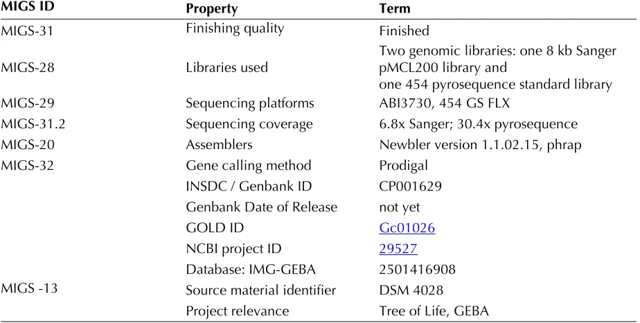
Insti-tute (JGI). A summary of the project information
is shown in Table 2.
Table 2. Genome sequencing project information
MIGS ID Property Term
MIGS-31 Finishing quality Finished
MIGS-28 Libraries used
Two genomic libraries: one 8 kb Sanger pMCL200 library and
one 454 pyrosequence standard library
MIGS-29 Sequencing platforms ABI3730, 454GS FLX
MIGS-31.2 Sequencing coverage 6.8x Sanger; 30.4x pyrosequence
MIGS-20 Assemblers Newbler version 1.1.02.15, phrap
MIGS-32 Gene calling method Prodigal
INSDC / Genbank ID CP001629 Genbank Date of Release not yet
GOLD ID
NCBI project ID
Database: IMG-GEBA 2501416908 MIGS -13 Source material identifier DSM 4028
Project relevance Tree of Life, GEBA
Growth conditions and DNA isolation
D. baculatum
strain X
T(DSM 4028) was grown in
1-1.5 g of cell paste using Qiagen Genomic 500
DNA Kit (Qiagen, Hilden, Germany) with a
modi-fied protocol for cell lysis, adding 100 µl lysozyme;
500 µl achromopeptidase, lysostaphin,
mu-tanolysin, each, to standard lysis solution, but
re-ducing proteinase K to 160µl, only. Incubation
over night at 35°C.
Genome sequencing and assembly
The genome was sequenced using a combination
of Sanger and 454 sequencing platforms. All
gen-eral aspects of library construction and
sequenc-ing performed at the JGI can be found at the
bled using the Newbler assembler version
1.1.02.15 (Roche). Large Newbler contigs were
broken into 4,375 overlapping fragments of 1,000
bp and entered into assembly as pseudo-reads.
The sequences were assigned quality scores based
on Newbler consensus q-scores with
modifica-tions to account for overlap redundancy and to
ad-just inflated q-scores. A hybrid 454/Sanger
sembly was made using the parallel phrap
as-sembler (High Performance Software, LLC).
Possi-ble mis-assemblies were corrected with
Dupfi-nisher or transposon bombing of bridging clones
[32]. Gaps between contigs were closed by editing
in Consed, custom primer walk or PCR
amplifica-tion. 731 Sanger finishing reads were produced to
close gaps, to resolve repetitive regions, and to
raise the quality of the finished sequence. The
er-ror rate of the completed genome sequence is less
than 1 in 100,000. Together all sequence types
provided 37.2 x coverage of the genome.
Genome annotation
Uni-Prot, TIGRFam, Pfam, PRIAM, KEGG, COG, and
In-terPro databases. Additional gene prediction
anal-ysis and functional annotation were performed
within the
ER) platform [35].
Genome properties
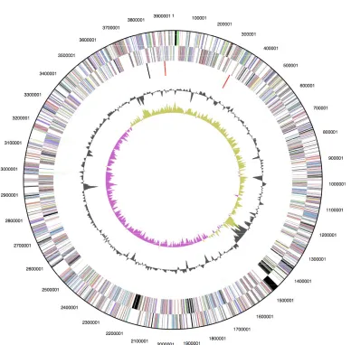
The genome is 3,942,657 bp long and comprises
one circular chromosome with a 58.7% GC
con-tent (Table 3 and Figure 3). Of the 3,565 genes
predicted, 3,494 were protein coding genes, and
71 RNAs; 58 pseudogenes were also identified.
74.9% of the genes were assigned a putative
func-tion while the remaining ones were annotated as
hypothetical proteins. The properties and the
sta-tistics of the genome are summarized in Table 3.
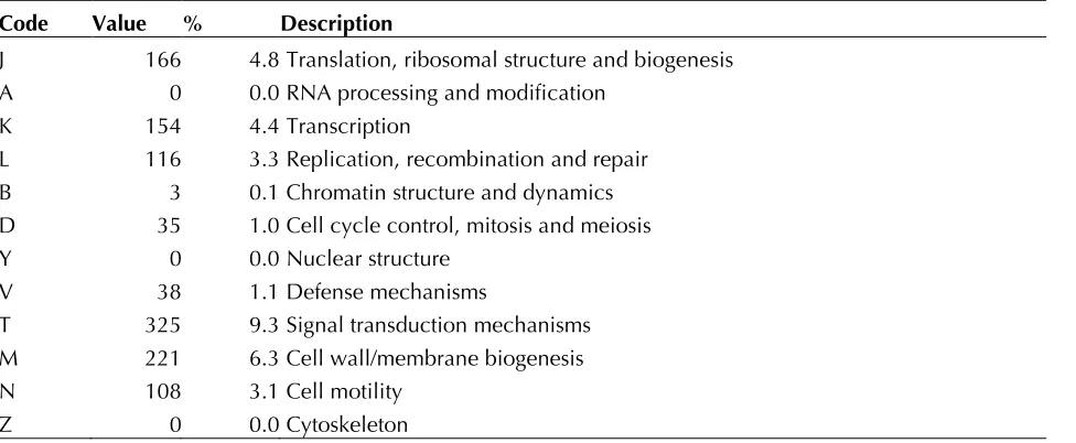
The distribution of genes into COGs functional
cat-egories is presented in Table 4.
Table 3. Genome Statistics
Attribute Value % of Total
Genome size (bp) 3,942,657
DNA Coding region (bp) 3,572,336 90.61%
DNA G+C content (bp) 2,312,250 58.65%
Number of replicons 1
Extrachromosomal elements 0
Total genes 3565
RNA genes 71 2.02%
rRNA operons 2
Protein-coding genes 3494 97.98%
Pseudo genes 58 1.63%
Genes with function prediction 2675 75.01%
Genes in paralog clusters 357 12.82%
Genes assigned to COGs 2689 75.41%
Genes assigned Pfam domains 2688 75.38%
Genes with signal peptides 723 20.27%
Genes with transmembrane helices 897 25.15%
CRISPR repeats 0
Table 4. Number of genes associated with the 21 general COG functional categories Code Value % Description
J 166 4.8 Translation, ribosomal structure and biogenesis A 0 0.0 RNA processing and modification
K 154 4.4 Transcription
L 116 3.3 Replication, recombination and repair B 3 0.1 Chromatin structure and dynamics D 35 1.0 Cell cycle control, mitosis and meiosis
Y 0 0.0 Nuclear structure
V 38 1.1 Defense mechanisms
T 325 9.3 Signal transduction mechanisms M 221 6.3 Cell wall/membrane biogenesis
N 108 3.1 Cell motility
Table 4. Number of genes associated with the 21 general COG functional categories (cont.) Code Value % Description
W 0 0.0 Extracellular structures
U 82 2.3 Intracellular trafficking and secretion
O 122 3.5 Posttranslational modification, protein turnover, chaperones C 241 6.9 Energy production and conversion
G 126 3.6 Carbohydrate transport and metabolism E 266 7.6 Amino acid transport and metabolism F 68 1.9 Nucleotide transport and metabolism H 135 3.9 Coenzyme transport and metabolism I 52 1.5 Lipid transport and metabolism
P 137 3.9 Inorganic ion transport and metabolism
Q 34 1.0 Secondary metabolites biosynthesis, transport and catabolism R 319 9.1 General function prediction only
S 221 6.3 Function unknown
- 805 23.0 Not in COGs
Acknowledgements
We would like to gratefully acknowledge the help of
Maren Schroeder (DSMZ) for growing D. baculatum
cultures. This work was performed under the auspices of the US Department of Energy Office of Science, Bio-logical and Environmental Research Program, and by the University of California, Lawrence Berkeley
National Laboratory under contract No. DE-AC02-05CH11231, Lawrence Livermore National Laboratory under Contract No. DE-AC52-07NA27344, and Los Alamos National Laboratory under contract No. DE-AC02-06NA25396 as well as German Research Founda-tion (DFG) INST 599/1-1.
References
1. Rozanova EP, Nazina TN. A mesophilic, sulfate-reducing, rod-shaped, nonsporeforming bacte-rium. Mikrobiologiya 1976; 45:825-830 2. List Editor. Validation of the publication of new
names and new combinations previously effec-tively published outside the ISJB. Int J Syst Bacte-riol 1984; 34:355-357
3. Rozanova EP, Nazina TN, Galushko AS. Isolation of a new genus of sulfate-reducing bacteria and description of a new species of this genus, Desul-fomicrobium apsheronum gen. nov., sp. nov. Mi-krobiologiya 1988; 57:634-641
4. Euzeby JP. Taxonomic note: necessary correction of specific and subspecific epithets according to Rules 12c and 13b of the International Code of Nomenclature of Bacteria (1990 Revision). Int J
Syst Bacteriol 1998; 48:1073-1075
5. Laanbroek HJ, Pfennig N. Oxidation of short-chain fatty acids by sulfate-reducing bacteria in freshwater and in marine sediments. Arch
Micro-biol 1981; 128:330-
6. Biebl H, Pfennig, N.. Growth of sulfate-reducing bacteria with sulfur as electron acceptor. Arch
Microbiol 1977; 112:115-117
7. Grabowski A, Nercessian O, Fayolle F, Blanchet D, Jeanthon C. Microbial diversity in production waters of a low-temperature biodegraded oil re-servoir. FEMS Microbiol Ecol 2005; 54:427-443
8. Hippe H, Vainshtein M, Gogotova GI, Stacke-brandt E. Reclassification of Desulfobacterium macestii as Desulfomicrobium macestii comb. nov. Int J Syst Evol Microbiol 2003; 53:1127-1130
9. Sharak-Genthner BR, Friedman SD, Devereux R. Reclassification of Desulfovibrio desulfuricans
comb. nov. and confirmation of Desulfomicro-bium escambiense (corrig., formerly "escam-bium") as a new species in the genus Desulfomi-crobium. Int J Syst Bacteriol 1997; 47:889-892 10. Hensgens CMH, Nienhuis-Kuiper ME, Hansen TA. Effect of tungstate on the growth of Desulfo-vibrio gigas NCIMB 9332 and other sulfate-reducing bacteria with ethanol as a substrate.
Arch Microbiol 1994; 162:143-147
11. Cypionka H, Kreke B. Energetics of sulfate trans-port in Desulfomicrobium baculatum. Arch Mi-crobiol 1995; 163:307-309
12. Lee JP, Yi CS, LeGall J, Peck HD, Jr. Isolation of a new pigment, desulforubidin, from Desulfovibrio desulfuricans (Norway strain) and its role in sulfite reduction. J Bacteriol 1973; 115:453-4
13. Fauque G, Herve D, Le Gall J. Structure-function relationship in hemoproteins: the role of cytoch-rome c3 in the reduction of colloidal sulfur by sulfate-reducing bacteria. Arch Microbiol 1979;
121:261-2
14. Coutinho IB, Turner DL, Legall J, Xavier AV. NMR studies and redox titration of the tetraheme cy-tochrome c3 from Desulfomicrobium baculatum. Identification of the low-potential heme. Eur J
Bi-ochem 1995; 230:1007-101
15. Correia IJ, Paquete CM, Coelho A, Almeida CC, Catarino T, Louro RO, Frazao C, Saraiva LM, Car-rondo MA, Turner DL, et al. Proton-assisted two-electron transfer in natural variants of tetraheme cytochromes from Desulfomicrobium sp. J Biol
Chem 2004; 279:52227-5223
Nickel-[iron-sulfur]-selenium-culatus (DSM 1743). Redox centers and catalytic properties. Eur J Biochem 1987; 167:47-5
17. Garcin E, Vernede X, Hatchikian EC, Volbeda A, Frey M, Fontecilla-Camps JC. The crystal structure of a reduced [NiFeSe] hydrogenase provides an image of the activated catalytic center. Structure
1999; 7:557-5
18. Parkin A, Goldet G, Cavazza C, Fontecilla-Camps JC, Armstrong FA. The difference a Se makes? Oxygen-tolerant hydrogen production by the [Ni-FeSe]-hydrogenase from Desulfomicrobium bacu-latum. J Am Chem Soc 2008; 130:13410-13416
19. Tormay P, Wilting R, Heider J, Bock A. Genes coding for the selenocysteine-inserting tRNA spe-cies from Desulfomicrobium baculatum and Clo-stridium thermoaceticum: structural and evolutio-nary implications. J Bacteriol 1994; 176
:1268-1274
20. Tormay P, Wilting R, Lottspeich F, Mehta PK, Christen P, Bock A. Bacterial selenocysteine syn-thase - structural and functional properties. Eur J
Biochem 1998; 254:655-66
21. Kromayer M, Wilting R, Tormay P, Bock A. Do-main structure of the prokaryotic selenocysteine-specific elongation factor SelB. J Mol Biol 1996;
262:413-4
22. Lee C, Grasso C, Sharlow MF. Multiple sequence alignment using partial order graphs.
Bioinformat-ics 2002; 18:452-46
23. Castresana J. Selection of conserved blocks from multiple alignments for their use in phylogenetic analysis. Mol Biol Evol 2000; 17:540-5
24. Stamatakis A, Hoover P, Rougemont J. A rapid bootstrap algorithm for the RAxML Web servers.
Syst Biol 2008; 57:758-771
25. Liolios K, Mavromatis K, Tavernarakis N, Kyrpides NC. The Genomes On Line Database (GOLD) in 2007: status of genomic and metagenomic
projects and their associated metadata. Nucleic
Acids Res 2008; 36:D475-4
26. Vainshtein M, Hippe H, Kroppenstedt RM. Cellu-lar fatty acid composition of Desulfovibrio species and its use in classification of sulfate-reducing bacteria. Syst Appl Microbiol 1992; 15:554-566 27. Collins MD, Widdel F. Respiratory quinones of
sulphate-reducing and sulphur-reducing bacteria: a systematic investigation. Syst Appl Microbiol
1986; 8:8-18
28. Field D, Garrity G, Gray T, Morrison N, Selengut J, Sterk P, Tatusova T, Thomson N, Allen MJ, An-giuoli SV, et al. The minimum information about a genome sequence (MIGS) specification. Nat
Biotechnol 2008; 26:541-547
29. Kuever J, Rainey FA, Widdel F. Family II Desul-fomicrobiaceae fam. nov. NR Krieg JS, GM Garri-ty, editor. New York: Springer; 2005. 944 30. Anonymous. Biological Agents: Technical rules
for biological agents. <www.baua.de>. 31. Ashburner M, Ball CA, Blake JA, Botstein D,
But-ler H, Cherry JM, Davis AP, Dolinski K, Dwight SS, Eppig JT, et al. Gene ontology: tool for the un-ification of biology. The Gene Ontology Consor-tium. Nat Genet 2000; 25:25-
32. Sims D, Brettin T, Detter JC, Han C, Lapidus A, Copeland A, Del Rio TG, Nolan M, Chen F, Lucas S, et al. Complete genome of Kytococcus seden-tarius type strain (541T). SIGS 2009; 1:12-20
33. Anonymous. Prodigal Prokaryotic Dynamic Pro-gramming Genefinding Algorithm. Oak Ridge National Laboratory and University of Tennessee 2009 <http://compbio.ornl.gov/prodigal>. 34. Pati A. GenePRIMP: A Gene Prediction
Improve-ment Pipeline for microbial genomes; 2009 (In press)
![Figure 1. Phylogenetic tree of D. baculatum strain XT and all type strains of species within members of the family Desulfomicrobiaceae, inferred from 1,457 aligned characters [22, 23] of the 16S rRNA gene sequence under the maximum likelihood criterion [24](https://thumb-us.123doks.com/thumbv2/123dok_us/664784.2065582/3.612.52.561.298.696/phylogenetic-baculatum-desulfomicrobiaceae-inferred-characters-sequence-likelihood-criterion.webp)
![Table 1. Classification and general features of D. baculatum XT in accordance to the MIGS recommendations [28]](https://thumb-us.123doks.com/thumbv2/123dok_us/664784.2065582/4.612.50.557.264.652/table-classification-general-features-baculatum-accordance-migs-recommendations.webp)