R E S E A R C H
Open Access
Normal mammary development and function in
mice with
Ift88
deleted in MMTV- and K14-Cre
expressing cells
Elizabeth H Mitchell and Rosa Serra
*Abstract
Background:Primary cilia (PC) are non-motile microtubule based organelles present on almost every cell type and are known to serve as critical organizing centers for several signaling pathways crucial to embryonic and postnatal development. Alterations in the Hh pathway, the most studied signaling pathway regulated by PC, affect mammary gland development as well as maintenance of the stem and progenitor cell populations.
Results:We developed mouse models with deletion of PC in mammary luminal epithelial, basal epithelial, and stromal cells for evaluation of the function of PC in mammary development via MMTV-Cre, K14-Cre, and Prx1-Cre mediated deletion, respectively. The activity of Cre was confirmed using ROSA26 reporters. Mammary stem and progenitor cells were enriched through growth as mammospheres. Adenovirus-Cre mediated deletion ofIft88was used to determine a role for PC in this population of cells. Disruption ofIft88and PC were confirmed in using PCR and immunofluorescent methods. Prx1-Cre;Ift88Delmice demonstrated defects in terminal end buds during puberty. However, theseIft88Delglands exhibited typical terminal end bud formation as well as normal ductal histology when transplanted into wild type hosts, indicating that the phenotype observed was not intrinsic to the mammary gland. Furthermore, no discernable alterations to mammary development were observed in MMTV-Cre- or K14-Cre;Ift88Dellines. These mice were able to feed and support several litters of pups even though wide spread depletion of PC was confirmed.
Cells grown in mammosphere culture were enriched for PC containing cells suggesting PC are preferentially expressed on mammary stem and progenitor cells. Deletion ofIft88in mammary epithelial cells resulted in a significant reduction in the number of primary mammospheres established; however, there was no effect on outgrowth of secondary mammospheres in PC-depleted cells.
Conclusions:PC regulate systemic factors that can affect mammary development in early puberty. PC on MMTV- or K14-expressing epithelial cells are not required for normal mammary development or function. PC are expressed at high levels on cells in mammosphere cultures. PC may be required for cells to establish mammospheres in culture; however, PC are not required for renewal of the cultures.
Keywords:Primary cilia, Intraflagellar transport, Mammosphere, Stroma, Orpk
* Correspondence:[email protected]
Department of Cell, Developmental, and Integrative Biology, University of Alabama at Birmingham, 1918 University Blvd., 660 MCLM, Birmingham, AL 35294-0005, USA
© Mitchell and Serra; licensee BioMed Central Ltd. This is an Open Access article distributed under the terms of the Creative Commons Attribution License (http://creativecommons.org/licenses/by/2.0), which permits unrestricted use, distribution, and reproduction in any medium, provided the original work is properly credited. The Creative Commons Public Domain Dedication waiver (http://creativecommons.org/publicdomain/zero/1.0/) applies to the data made available in this article, unless otherwise stated.
Background
Mammary gland development is unique in that most growth occurs post-natal with terminal end bud (TEB) formation and ductal extension beginning at puberty. In adulthood, the gland undergoes cyclical restructuring during pregnancy, lactation, and involution. Mammary development is highly controlled with all cell types working together. Coordinated crosstalk between epithe-lial cells and stroma allows for proper terminal end bud formation, ductal extension, and side branching [1,2].
Primary cilia (PC) are organelles that were once thought insignificant but are now renowned as regulators of devel-opment and homeostasis [3-6]. These non-motile, micro-tubule based organelles are present one per cell on many cell types and project into the microenvironment where they serve as a signaling center for the cell, functioning as chemical and mechanical sensors. PC are formed and maintained by intraflagellar transport (IFT), a sophisti-cated process with large protein complexes employed in carrying cargo from the ciliary base to tip (anterograde) and tip to base (retrograde) [7]. Disruption of IFT prevents the formation, maintenance, and function of PC. Defects in genes important for the formation or function of PC have been linked to a wide array of human diseases and syndromes now termed ciliopathies [8].
The primary cilium is accepted as a critical regulator of several pathways including, Hedgehog (Hh), Wnt, PDGF, and von-Hippel Lindau tumor suppressor. The most studied of these is the Hh pathway, which is also known to be key in embryonic development and tis-sue homeostasis [4,7,9,10]. Hh signaling begins by the binding of one of three hedgehog secreted ligands; Sonic hedgehog (Shh), Indian hedgehog (Ihh) or Desert Hedgehog (Dhh), to its receptor patched (Ptch1) reliev-ing its inhibition of smoothened (Smo). Smo is then able to move into the PC and mediate activation of Gli transcription factors, thus PC are required for ligand-mediated Hh signaling [7,11-14]. In the absence of Hh ligand, Gli3 is processed to a repressor form (Gli3R) that inhibits expression of transcriptional targets of Hh. PC are also required for processing Gli3 to this repressor form, therefore, PC are required for both dependent Hh signal activation as well as ligand-independent repression.
Markers of active Hh signaling are not present in nor-mal mammary epithelium and active Hh signal in the epithelial compartment does not appear to be required for normal mammary development [15]. However, there is evidence that activation of Hh signaling in the stromal compartment is required for normal development [16]. During embryonic stages of mammary development it is likely that repression of Hh signaling is required to pro-mote development of mammary glands rather than hair follicles, which require Hh signaling [17]. The Gli3R
function is critical for embryonic mammary develop-ment and activation of Hh signaling via loss of Gli3R re-sults in loss of mammary buds [15]. In post-natal mice, inappropriate activation of Hh signaling in mammary epithelium results in impaired mammary development and ductal dysplasia suggesting suppression of Hh sig-naling is critical throughout mammary development and activation could participate in formation of breast cancer [18]. A role for primary cilia in basal cell carcinoma and medulloblastoma have been demonstrated [19,20]. Fur-thermore, PC are differentially presented in numerous carcinomas, including; basal cell, brain, pancreas, kidney, and breast [19-23]. In the breast, PC are more frequently found on normal epithelial cells than on cancer cells. PC are also more frequent on epithelial cell lines derived from benign breast than those from breast cancer re-gardless of the level of proliferation in those cells sug-gesting PC could act as tumor suppressors [21].
In this study, we generated mice with depletion of PC via deletion of Ift88, an IFT protein required for forma-tion and maintenance of PC, in specific cell types within the mammary gland. Luminal, basal, and stromal cells were targeted using MMTV-, K14-, and Prx1-Cre lines, respectively. As previously reported, we found that dele-tion ofIft88in Prx1-Cre expressing mice resulted in loss of TEB and delayed extension of ducts through the fat pad during early puberty [24]. In this study, whole gland transplant indicated that the phenotype was not inherent to the mammary gland suggesting the involvement of systemic factors. Surprisingly, alterations in mammary de-velopment or function associated with depletion of PC in MMTV-Cre or K14-Cre expressing cells were not found. Using mammosphere cultures to select for stem and pro-genitor cells, we found that PC are enriched on these cell populations. Depletion of primary cilia on mammary epi-thelial cells resulted in reduced ability of the cells to form primary mammospheres; however, formation of secondary mammospheres was not affected. We conclude that PC do not play a major role in regulating normal post-natal mammary development or function.
Methods Animals
Ift88orpk
, Ift88LoxP/LoxP, K14-Cre, and MMTV-Cre mice have previously been described [25-28]. MMTV-Cre mice were obtained from the NCI mouse repository (strain 01XA9). K14-Cre, Gt(ROSA)26Sortm4(ACTB-tdTomato,-EGFP)
Luo
/J (ROSA26mTmG), and Gt(ROSA)26Sortm1Sor/J (Rosa26
LacZ
) mice were obtained from (Jackson Laboratories (stock 004782, 007576, and 003474), Ift88orpk and Ift88LoxP/LoxP mice were generously provided by Dr. Bradley Yoder University of Alabama at Birmingham. MMTV-Cre and K14-Cre mice were on a C57Bl/6 genetic background.
Ift88LoxP/LoxP
We used two sets ofIft88Orpkmice; either on a C57Bl/6 or a Balb/c background. Experimental crosses were set up as MMTV- or K14-Cre;Ift88LoxP/Wt×Ift88LoxP/LoxPto gener-ate Cre-positive Ift88LoxP/LoxP mice (hereafter called Ift88
Del
). MMTV- or K14-Cre; Ift88LoxP/Wt and Cre-negative;
Ift88LoxP/LoxP
mice were used as controls. Age matched or littermate controls were used. All mice utilized in this study were maintained following the guidelines of the Institu-tional Animal Care and Use Committee of the University of Alabama at Birmingham. All animal usage in this study was approved by the Institutional Animal Care and Use Committee of the University of Alabama at Birmingham.
Beta-galactosidase staining
Mammary glands were removed and fixed in 4% parafor-maldehyde (PFA) for 1 hour. Afterwards they were washed 3 times in rinse buffer (2 mM MgCl2, 0.01%Na
Deoxycho-late, 0.02%NP-40, in PBS) and stained overnight at room temperature in 1 mg/ml 4-chloro-5-bromo-3-indoyl-β -D-galactopyranoside (X-gal), 5 mM K3Fe(CN)6,5 mM K4FE
(CN)6H20, 2 mM MgCl2, in PBS. Mammary glands were
rinsed in PBS and post fixed for one hour with 4% PFA, then dehydrated through graded ethanol and cleared in 80% glycerol before mounting. For sections, glands were equilibrated with 30% sucrose and then equilibrated with Optimal Cutting Temperature (O.C.T.) Compound (Sakura, Torrance, Ca). Glands were flash frozen in O.C.T. by liquid nitrogen in a 2-methyl butane bath. Sections were cut at 20-40μm.
Immunofluorescence and cilia staining
Mammary glands were removed and fixed in 4% parafor-maldehyde (PFA) for 1 hour at room temperature then they were placed in 30% sucrose overnight and equili-brated with an equal amount O.C.T. After equilibration, glands were flash frozen in O.C.T by liquid nitrogen in a 2-methyl butane bath. Sections were cut at 20-40 μm and fixed in ice-cold methanol for 20 minutes. Sections from ROSA26mTmG mice were stained with DAPI. Im-munofluorescent staining of PC was done as described previously [29]. Slides were blocked for 1 hour with PBS with 0.1% Triton-x-100, 3% BSA, 1% Normal goat serum (Vector labs, S-1000). Primary antibodies were anti-Arl13b used at 1:2000 (a gift of Dr. Tamara Caspary, Emory University, Atlanta, GA, USA) staining was done as previously described [30] anti-actinα-smooth muscle-Cy3 (Sigma, C6198) used at 1:1000, primary antibodies incubations were overnight at 4°C. Anti-Rabbit Alexa Fluor-488 (Life Technologies, A-11008) secondary was used at 1:1000. Nuclei were stained with DAPI.
Cilia counting
20-40 μm sections were imaged using a Hamamatsu C9100-50 EM-CCD camera (Hamamatsu Photonics K.K.,
Hamamatsu City, Japan) on an inverted Nikon TE2000-U microscope equipped with a 60× Plan Apochromat oil-immersion TIRFM objective (numerical aperture (NA), 1.49; Nikon Instruments Inc., Melville, NY), and a Perkin Elmer Ultraview-ERS 6FE spinning disk confocal module controlled by Volocity 6.2 software (Perkin Elmer, Shelton, CT, USA). Cells positive for smooth muscle actin were considered myoepithelial cells and cells negative for smooth muscle actin but on the luminal side of the duct were considered luminal epithelial cells. Only cilia larger than 1 μm were counted. Cilia were counted on >600 luminal cells (n = 3 separate mice each for MMTV-Cre;
Ift88Del and controls) with at least 200 cells counted
per mouse. Cilia were counted on >500 myoepithelial cells (n = 2 K14-Cre; Ift88Del and n = 3 controls) with at least 150 cells counted per mouse. T-Test statistics were done using Microsoft Excel.
PCR
Mammary epithelial cells were isolated as described pre-viously [31]. Mammary glands minus lymph nodes were minced and digested with 1 mg/ml Collagenase Type I (Sigma- Aldrich, C9891, St. Louis, MO, USA) and 0.1 mg/ml Pronase (Roche Diagnostics, 1149643001, Mannheim, Germany) at 37°C in Hanks Balanced Salt Solution (HBSS) for 2.5 hours with agitation. Digests were pipetted 10 times every hour. Cells were pelleted at 1500 rpm for 5 min and resuspended in PBS. Cells were pelleted at 1500 rpm for 1 min and resuspened in PBS two more times the remaining cells were used for DNA extraction. PCR was carried out under standard condi-tions with three primers to detect the floxed, wildtype and deleted allele as described previously in [26].
Whole mount staining
Whole mount staining was done as previously described [32]. The inguinal #4 mammary glands were removed and placed in Carnoy’s fixative for 1 hour and stained with Carmine Aluminum Stain overnight then dehy-drated through graded ethanol, cleared with xylene, and mounted between two microscope slides.
Histology
Mammosphere cultures
Mammary epithelial cells for mammosphere culture were isolated similarly to others [33]. Briefly, digested cells were pelleted at 1500 rpm for 5 min and resus-pened in 8 ml .8% NH4Cl 10 mM EDTA and 2 ml HBSS
supplemented with 2%FBS with a final concentration of 15 mM Hepes (HF) and centrifuged again for 5 min. Cells were then resuspended for 90 seconds with constant pipetting in 0.25% Trypsin EDTA and then pel-leted by centrifugation. Pellets were then resuspended for 90 seconds with constant pipetting in 5 mg/ml Dispase (Gibco) and centrifuged. Finally, cells were diluted in 10 ml cold HF and filtered through 40μm mesh. The cells were then plated in 6 well ultra low attachment cell cul-ture plates at a concentration of 1,000-50,000 cells/well.
Adenovirus infection
Single cells isolated for mammosphere cultures were plated at 25,000 cells/ml in monolayer or in suspension and infected 24 hours after plating with either Adeno-virus containing Cre Recombinase with an IRES GFP and or a control Adenovirus GFP at an MOI of 200. 90-95% of cells were infected after 48 hours as assessed by presence of GFP.
Whole gland transplants
Scid mice were used as transplant recipients. A small in-cision was made on fat pad in the interscapular region, a 25G needle was used to score the fat to stimulate blood flow and encourage grafting [34,35]. The #4 mammary glands were removed from Prx1-Cre; Ift88Del and con-trols and placed on the scored area and stitched at the top and bottom. Glands were removed 4-6 weeks after transplantation and processed for whole mount carmine aluminum staining.
Results
Spatial activity of Cre and disruption of PC
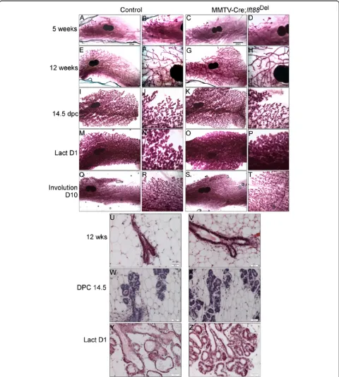
We initially proposed that depletion of PC in mammary epithelium would yield phenotypes similar to those in mice with either activating or inactivating mutations in Hedgehog signaling. To test this hypothesis, we gener-ated MMTV-Cre; Ift88Del and K14-Cre; Ift88Del mice. Before we characterized the mice, we confirmed that the Cre-models were working and that PC were de-pleted in the mammary gland. First, the spatial activity of Cre recombinase in the MMTV-Cre and K14-Cre lines was determined by crossing to Cre reporter strains, Gt(ROSA)26Sortm1Sor/J (Rosa26LacZ) and Gt(ROSA) 26Sortm4(ACTB-tdTomato,-EGFP)Luo/J (ROSA26mTmG) respect-ively (Figure 1A-D). MMTV-Cre showed activity, as dem-onstrated by blue beta-galactosidase staining, throughout the mammary ducts. Luminal cells as well as body cells of the TEBs were more efficiently targeted than basal
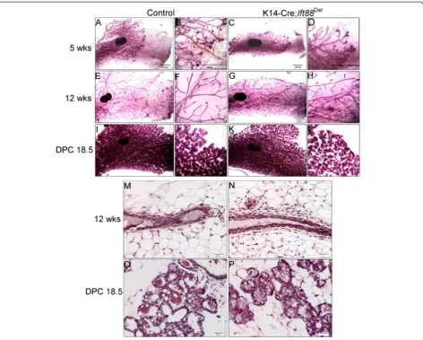
cells in mice at 5-weeks of age (Figure 1A, B) as de-scribed previously [27]. K14-Cre demonstrated activity (green membrane fluorescence) throughout the ducts of adult mice with both basal and luminal cells targeted (Figure 1C, D). We could not detect cells within ducts, basal or luminal, with red membrane staining indicating virtually every epithelial cell was targeted by K14-Cre (Figure 1D). Stromal cells were stained red and thus were not targeted by K14-Cre as expected (Figure 1D).
Deletion of theIft88gene in MMTV-Cre and K14-Cre mice was confirmed by PCR of genomic DNA isolated from mammary epithelial cells from adult K14-Cre;
Ift88Del
and MMTV-Cre; Ift88Del mice as well negative and positive controls (data not shown). Disruption of PC in Ift88Del mammary glands was determined by im-munofluorescent staining of PC in cryosections from adult mammary glands using the cilia specific antibody Arl13B (Figure 1E-P; green). Myoepithelial cells were stained using an antibody to alpha smooth muscle actin (αSMA, red). The percentage of ciliated luminal epithe-lial cells (negative for αSMA) and the percentage of ciliated myoepithelial cells (positive for αSMA) were determined in control and MMTV-Cre and K14-Cre;
Ift88Del
mammary glands, respectively (Figure 1Q, R). In agreement with previous results [36], PC were enriched on myoepithelial cells relative to luminal epithelial cells in control mice. Forty-four percent of myoepithelial cells observed contained a PC whereas only 9% of luminal cells had a PC. PC were significantly depleted in both MMTV-Cre and K14-Cre; Ift88Del glands. The percent-age of luminal cells that contained PC dropped from 9% in controls to 0.9% in MMTV-Cre; Ift88Del mice (p < 0.01). The percentage of myoepithelial cells that contained PC dropped from 44% in controls to 5% in K14-Cre; Ift88Del mice (p < 0.01). The results indicate that our Cre models are functional and that PC are sig-nificantly depleted in mammary epithelial cells.
Characterization of mammary development in PC depleted mice
including retired breeders (n = 9 controls and 10 mutants; data not shown).
Recently, it was shown that Hedgehog responding cells in the mammary gland are basally located, contain
primary cilia, and express K14 [37]. Nevertheless, K14-Cre; Ift88Del glands were not distinguishable from con-trol glands through puberty and adulthood (Figure 3; n = 5 controls and 4 mutants) or during pregnancy and Figure 2Loss ofIft88in MMTV-Cre expressing cells does not affect mammary development.Whole mount carmine staining of #4
lactation (Figure 3; n = 3 controls and n = 2 mutants). These mice were also able to lactate and fully support multiple litters. Palpable tumors were not detected in any mice over 9 months of age. These mice did not demonstrate any histological phenotypes typical of mam-mary glands from mice with mutations in Hedgehog as-sociated genes including Gli2-/-, Ptch1Δ-/+, Ptch1mes/
Ptch1 mes
, MMTVrtA-Gli1, MMTV, or SmoM2 MMTV
Cre knockin; which resulted in at least one or more defects including ductal dysplasia, distended ducts, hyperplasia, altered differentiation, excessive budding, or aberrant termini [16,38-40,47] (Figure 3 M-P and data not shown). The results indicate PC in MMTV- and K14- expressing lineages are not required for normal mammary development or function and loss of PC does not phenocopy mice with alterations in Hedgehog signaling proteins.
PC are enriched in mammospheres
contrast to what we observed in whole mammary gland, where PC are found on a limited number of cells, virtu-ally every cell in the mammosphere demonstrated a PC (Figure 4A, B) suggesting PC are enriched on mammary stem and progenitor cells although culture conditions could contribute to the formation of PC. Next, we grew
mammary cells from MMTV-Cre; Ift88Del and
Cre-negative controls in mammosphere culture and measured
the number of mammospheres that grew out from 4 × 10⁴ cells over 10 days. Only spheres over 50 microns were scored. No statistical difference was detected in the number of mammospheres in MMTV-Cre;Ift88Delversus control cultures (Figure 4C n = 3). To determine if MMT V-Cre was active in the stem or progenitor cells that make-up the mammospheres, we grew mammary epithe-lial cells isolated from Cre-negative and MMTV-Cre;
Rosa 26LacZfemales in mammosphere culture and stained for beta-galactosidase activity (blue, Figure 4D-F). Mam-mosphere staining was mosaic suggesting that MMTV-Cre may have become active after the mammosphere formed. In this case the initiating stem or progenitor cell may not have been targeted by MMTV-Cre in vivo.
Next, to achieve more widespread depletion of PC in mammosphere cultures, we used an adenovirus express-ing Cre with IRES controlled GFP (Ad-CRE) or a control GFP only virus (Ad-GFP) to infect mammary epithelial cells isolated from Ift88LoxP/LoxP mice. Infected cells were then placed in mammosphere culture at 5 × 10⁴ cells per well for 10 days (Figure 4G). Deletion of
Ift88 was confirmed by PCR of genomic DNA isola-ted from the Ad-Cre and Ad-GFP infecisola-ted cells (Figure 4H). Widespread depletion of PC was con-firmed using immunofluorescent staining (Figure 4I,J). There was a significant 50% decrease in the formation of primary mammospheres in the PC-depleted cul-tures (Figure 4G; n = 4 separate experiments p-value < 0.05 paired T-test). However, when the mammo-spheres were passaged into secondary mammosphere cultures (1 × 10⁴ cells/well) to test for self-renewal we did not detect statistically significant differences in outgrowth of mammospheres (Figure 4K n = 2 sep-arate experiments) although PC were still clearly de-pleted from the secondary cultures when compared to the controls (Figure 4L,M). PC may affect the initial outgrowth of mammospheres, but they are not re-quired for renewal of the cultures.
The mammary phenotype in Prx-1-Cre; Ift88Delmice is not inherent to the mammary gland
Mammary development is a highly controlled event with all cell types working together. Coordinated crosstalk between epithelial cells and stroma allows for proper terminal end bud formation, ductal extension, and side branching [2,41]. Mice with LacZ knocked in to the
Ift88 locus were found to have high Ift88 expression in the mammary stroma and studies with ROSA26LacZ re-porter mice showed that Prx1-Cre is active in the mam-mary stroma as well as ovaries and other tissues [24]. We showed previously and confirm here that Prx1-Cre;
Ift88Del
mice have defective TEB formation and delayed ductal elongation [24]; Figure 5A,B). We proposed that the mammary phenotype was indirect and mediated by al-terations in the ovary since exogenous estrogen injection could restore end bud activity [24]. To test whether the loss of PC in the mammary stroma affects mammary de-velopment without the confounding effects of disrupted ovarian hormone levels, we transplanted whole mammary glands from 3 week-old Prx1-Cre; Ift88Del and littermate controls mice onto the highly vascularized region between the shoulder blades of Scid hosts similar to what was de-scribed in [34]. Transplants were removed after 4 weeks and stained with carmine; sections were stained with H&E (Figure 5C, D); n = 3 controls and 4 mutants). Transplants exhibited typical terminal end bud formation (Figure 5C-F) as well as normal ductal histology (Figure 5G, H), in-dicating that the phenotype observed in Prx1-Cre; Ift88Del mice is not inherent to the mammary gland.
Figure 5The mammary phenotype in Prx1-Cre;Ift88Delmice is likely due to systemic factors.Whole mount Carmine staining of #4
TEBs and mammary branching are normal in Ift88Orpk/Orpk female mice on Bl/6 or Balbc background
In an effort to study mice that had reduced ciliary func-tion in epithelial and stromal compartments of the mammary gland, we examined mammary glands in the well-characterized Oak Ridge Polycystic kidney disease (Ift88Orpk) mouse model. Orpk is a hypomorphic allele of
Ift88 and thus Ift88 has some function in these mice.
Ift88Orpk
females from both Bl/6 and Balb/c backgrounds showed no obvious developmental deviations from controls as determined by whole mount staining (Figure 6A-H; n = 6 control, 4 mutant). This is in contrast to a previous report ofIft88Orpk mice on a mixed C57Bl/6 × C3H/FVB background, in which a decrease in ductal extension and branching was noted [36]. Together the results suggest that there may be a strong genetic modifier effect on PC func-tion in the mammary gland.
Male Bl/6 and Balb/c mice do not generally develop a mammary ductal tree. During embryonic development, mesenchymal cells, in response to testosterone, con-dense around the stalk of the developing mammary bud, severing the connection of the bud to the surface epider-mis. The epithelial cells subsequently undergo apoptosis and thus a ductal tree is not formed [42]; and Figure 6I). In contrast, male Ift88Orpk developed a complete ductal tree by 8 weeks of age in both Bl/6 and Balb/c back-grounds (Figure 6I,J; n = 3 controls and 5 mutants). The results suggest thatIft88is somehow required to prevent mammary ductal development in male mice.
Discussion
We developed mouse models with deletion of PC in mammary epithelial or myoepithelial cells for evaluation of the function of PC in mammary development. We
confirmed that Ift88 was deleted in Cre-positive mice and that PC were disrupted in the expected cell types. Nevertheless, minimal disruption to normal mammary development was observed and mice were able to feed and support several litters of pups. In addition, tumors were not detected in mice aged out to 18 months. Mam-mary glands from virgin mice with a hypomorphic allele of Ift88, Ift88Orpk, did not display defects in branching morphogenesis in adult mice; however, males from this line of mice demonstrated uncharacteristic development of a full ductal tree. While characterizing the PC deleted mice, we discovered that PC were enriched on cells grown in mammospheres. Deletion ofIft88in these cells using an Adenovirus Cre resulted in reduced expansion of cells into primary mammospheres; however, out-growth of secondary mammospheres was not affected. We also used whole gland transplants to show that de-layed mammary development observed in Prx1-Cre;
Ift88Del
mice, in which the mammary stroma and other tissues are targeted, is likely due to systemic factors and not inherent to the mammary gland.
Our original hypothesis when initiating these studies was that deletion of Ift88 and subsequent depletion of PC would phenocopy alterations in mammary develop-ment observed in mice with disruptions to Hh signaling. It is well known that PC mediate signaling by Hh pro-teins in many cell types [9]. In addition to regulating ligand dependent signaling, PC are required to process Gli3 to a repressor form. Active Hh signaling in the epi-thelium does not appear to be required for normal mammary development although there is evidence to suggest Hh signaling in the stroma may be required. De-letion of Gli2 in mice results in ductal dysplasia and ex-tended ducts; however, when Gli2 null epithelium was
Figure 6Mammary phenotype inIft88Orpkmice on Bl/6 and Balb/c background.Mammary glands from female mice(A-H). Whole mount
transplanted in to a normal host fat pad development was normal suggesting a role for Hh signaling in the stroma [16]. We previously showed that Prx1-Cre has some activity in the mammary stroma. Unfortunately, Prx1 is also active in additional tissues throughout the mouse including the ovary [24]. To alleviate the con-founding effects of PC loss in the ovary, we transplanted mammary glands from Prx1-Cre; Ift88Del mice to wild type hosts. The formation of TEBs and branching ap-peared normal in the transplanted tissue suggesting the effects seen in the Prx1-Cre; Ift88Del mice were due to systemic factors.
While loss of Hh signaling in mammary epithelium does not appear to affect normal development, inappro-priate activation of Hh signaling via an activated smoothened protein in mammary epithelium results in excessive budding in TEBs, increased proliferation of epithelial cells, and alterations in luminal cell differenti-ation suggesting that repression of Hh signaling plays an important role in normal mammary development [18,43,44]. Based on these data we predicted that loss of PC, which would be expected to inhibit the formation of the Gli3 repressor, would mimic phenotypes seen in mice with inappropriate activation of Hh signals in epi-thelial cells; however, we did not detect any significant alterations in development of MMTV-Cre or K14-Cre;
Ift88Del
mammary glands. One explanation for normal development in absence of PC is that there may be a constant level of Hh signal activation in the mammary gland that is continuously repressed by Gli3 to allow normal development and prevent tumor formation. In the absence of cilia, this positive signal would not exist so Gli3 repressor not required. Loss of Gli3 results in failure in the formation of specific mammary buds, but the role of the Gli3 repressor in post-natal mammary de-velopment was not addressed [15].
Research involving PC and their relationship with can-cer has become an area of intense interest. Recently, the involvement of PC in basal cell carcinoma and medullo-blastoma were investigated [19,20]. It was shown that PC can work to prevent tumorigenesis in some contexts but promote it in others. If the hedgehog pathway was activated at the level of Smoothened, the presence of PC accelerated tumor formation; however, with the loss of PC, tumor formation was suppressed. Suppression was likely due to the fact that PC are required for Smooth-ened function. On the other hand, if tumors were initi-ated with a constitutively active form of Gli2, which would be downstream of Smoothened function, loss of cilia accelerated tumor growth [19,20]. Accelerated growth was due to loss of the Gli3 repressor, which would counteract the functions of Gli2. For breast can-cer, it has been shown that the incidence of PC de-creases during the progression of normal to cancerous
cells, independent of the levels of proliferation, leading to the hypothesis that PC act as tumor suppressors [21,45]. Even though we did not detect significant alter-ations in mammary development or spontaneous tumors in aged mice, our results do not rule out an effect of PC on breast cancer in combination with other signals. For example, loss of PC in breast cancers initiated by Gli1 may promote tumor formation whereas tumors initiated by excess Hh ligand, mutations in Ptc1, or activated Smoothened could be inhibited.
A role for Hh signaling in mammary stem cells has been suggested. Mammary stem cells cultured as mam-mospheres demonstrate high levels of markers for Hh signaling including Ptch1 and Gli1 [46]. Furthermore, activated Smoothened resulted in increased mammo-sphere formation suggesting Hh may positively regulate stem cell populations in the mammary gland [47]. Mis-expression of Shh in the mammary glands of transgenic mice results in ductal dysplasia after pregnancy [37]. Hh responding cells were localized by Ptch1-LacZ staining to the basal layer within the dysplastic areas. Hh responding cells were ciliated and expressed markers of basal and stem/progenitor cells suggesting PC may have a positive role in regulation of mammary stem and pro-genitor populations. In agreement, we show enrichment of ciliated cells in mammosphere cultures. Although It is possible culture conditions necessary for mammosphere formation may promote PC formation, this observation confirms the previous results described above in which cells in vivo expressing progenitor markers are enriched for PC [37]. We also show a decrease in outgrowth of primary mammospheres that could be due to loss of the positive Hh signal due to absence of PC in the mammo-sphere initiating cells. However, we do not see an effect of losing PC on passage of the cultures into secondary mammospheres suggesting PC do not play a role in re-newal of cells in the established mammospheres.
Recently, it was shown that mice with a germline, hypomorphic allele of Ift88, Ift88Orpk, have defects in mammary development [36]. Branching morphogenesis during puberty and alveolar differentiation during preg-nancy were inhibited. Epithelium from the Ift88Orpk transplanted into cleared fat pads also demonstrated in-hibition of branching suggesting the phenotype was due to loss of PC in the mammary epithelium and not due to systemic effects. Decreased Hh signaling and in-creased canonical Wnt signaling were noted although neither a decrease in Hh signaling nor an increase in canonical Wnt signaling would be expected to result in reduced branching. We did not observe alterations in branching morphogenesis in Ift88Orpk mice in our colony. One explanation for the different observations is the background strains used for each study. The
Ift88Orpk
C backgrounds. The mice used in the previous study were F1 hybrids generated by crossingIft88Orpk mice on the C57Bl/6 background to Ift88Orpk mice on a mixed C3H and FVB background. Together the results suggest a strong genetic modifier that affects the function of PC and their role in the mammary phenotype.
Mammary buds are formed in the embryos of both male and female mice; however, in most mouse strains, male mice do not develop a ductal tree [48]. Androgen receptors are found in the mammary mesenchyme start-ing at embryonic day 13. In response to testosterone production in the male embryos, this mesenchyme con-denses around the epithelial bud, separating it from the external ectoderm and triggering regression of the epi-thelium. The time window for this process is between embryonic day 13 and 16. While characterizing mam-mary glands in Ift88Orpk mice, we discovered that male mice carrying this hypomorphic allele of Ift88 have a fully developed mammary ductal tree. The mechanism of this alteration is not known and could involve local PC in the mammary mesenchyme or PC in other tissues that would have systemic effects on regression of the nascent mammary duct. Recently, it was shown that male embryos with a mutation in Gli3 (Gli3xt/xt) demonstrate inappropriate retention of mammary buds. Furthermore, misactivation of Hh signaling through expression of Gli1 resulted in a similar phenotype. It was suggested that repression of Hh signal through Gli3 repressor activity is required for proper specification of the mammary mesen-chyme. Abnormal mammary mesenchyme including low levels of the androgen receptor likely prevented the de-struction of the mammary bud in mutant male embryos [49]. Since PC are required for processing of the Gli3 re-pressor, it is possible that altered PC function in the mam-mary mesenchyme acts through a similar mechanisms.
PC also regulate pathways other than Hh including Wnt, PDGF, and von-Hippel Lindau tumor suppressor [4,7,9,10]. Wnt proteins are well established as key regu-lators of mammary development, misexpression of canonical Wnt/ß-catenin signaling in the mammary epi-thelium results in increased branching, lobuloalveolar hyperplasia and tumorigenesis; whereas inactivation of canonical signaling results in a reduction in branching and TEB number [50,51]. Loss of a noncanonical signal-ing Wnt, Wnt5a, resulted in rapid ductal elongation and increased side branching, while misexpression of Wnt5a resulted in a lactation defect [31,52]. Even though Wnt signaling is key to mammary development, recent data suggest PC do not directly regulate Wnt/ß-catenin signaling [53,54]. Most importantly, we did not detect any phenotypes characteristic of alterations in Wnt/ß-catenin signaling in our PC-deleted mouse models. PDGFR-alpha is localized to the PC; however studies de-scribing the loss of this receptor in the mammary gland
have not been reported [55]. We also did not detect any indication of alterations in signaling by the tumor sup-pressor von- Hippel Lindau. Its loss in the mammary epithelium causes reduced proliferation and alveolar differentiation during pregnancy but does not cause tumorigenesis [56].
Conclusion
We conclude that PC can regulate mammary develop-ment through systemic factors, but, overall, PC localized in the gland have a limited role in development or nor-mal function of the mammary gland.
Abbreviations
PC:Primary cilia; TEB: Terminal end bud; IFT: Intraflagellar transport; Hh: Hedgehog; ROSA26: Reverse oriented splice acceptorβ-galactosidase/neomycin 26; Ad: Adenovirus; PFA: Paraformaldehyde; DAPI: 4′,6-diamidino-2-phenylindole; PBS: Phosphate buffered saline; PCR: Polymerase chain reaction; Del: Deleted; WT: Wild type; GFP: Green fluorescent protein; IRES: Internal Ribosome entry site; Orpk: Oak Ridge Polycystic Kidney disease; HBSS: Hanks balanced salt solution; HEPES: 4-(2-Hydroxyethyl)piperazine-1-ethanesulfonic acid sodium salt; FBS: Fetal bovine serum; HF: HBSS plus 15 mM HEPES and 2% FBS; X-GAL: 4-chloro-5-bromo-3-indoyl-β-D-galactopyranoside; EDTA: Ethylenediaminetetraacetic acid.
Competing interests
The authors declare that they have no competing interests.
Authors’contributions
RS and EHM designed experiments. EHM performed experiments. EHM and RS wrote the manuscript. All the authors read and approved of the final version of the manuscript.
Acknowledgements
This work was supported through grants from the CDMRP: W81XWH-09-1-0344 and NCI: R01CA126942 to R.S.
Ift88OrpkandIft88LoxP/LoxPmice were obtained from the laboratory of Dr. Bradley K. Yoder, University of Alabama at Birmingham. We would like to acknowledge Dr. Tamara Caspary, Emory University, for providing us with the Arl13b antibody. We would like to acknowledge Dr. Andra Frost and Dr. Kun Yuan, Department of Pathology, University of Alabama at Birmingham, for helpful discussions and assistance in primary cilia immunofluorescence. We would also like to thank Zak Kosan, for assistance with confocal microscopy.
Received: 24 September 2013 Accepted: 14 February 2014 Published:
References
1. Wiseman BS, Werb Z (2002) Stromal effects on mammary gland development and breast cancer. Science 296:1046–1049
2. Hynes NE, Watson CJ (2010) Mammary gland growth factors: roles in normal development and in cancer. Cold Spring Harb Perspect Biol 2: a003186
3. Pedersen LB, Veland IR, Schrøder JM, Christensen ST (2008) Assembly of primary cilia. Dev Dyn 237:1993–2006
4. Berbari NF, O’Connor AK, Haycraft CJ, Yoder BK (2009) The primary cilium as a complex signaling center. Curr Biol 19:R526–R535
5. D’Angelo A, Franco B (2009) The dynamic cilium in human diseases. PathoGenetics 2:3
6. Singla V (2006) The Primary Cilium as the Cell’s Antenna: Signaling at a Sensory Organelle. Science 313:629–633
7. Veland IR, Awan A, Pedersen LB, Yoder BK, Christensen ST (2009) Primary Cilia and Signaling Pathways in Mammalian Development, Health and Disease. Nephron Physiol 111:39–53
8. Badano J, Mitsuma N, Beales P, Katsanis N (2006) The ciliopathies: an emerging class of human genetic disorders. Annu Rev Genomics Hum Genet 7:125
9. Nielsen SK, Møllgård K, Clement CA, Veland IR, Awan A, Yoder BK, Novak I, Christensen ST (2008) Characterization of primary cilia and hedgehog
signaling during development of the human pancreas and in human pancreatic duct cancer cell lines. Dev Dynam 237:2039–2052 10. Christensen ST, Pedersen SF, Satir P, Veland IR (2008) The primary cilium
coordinates signaling pathways in cell cycle control and migration during development and tissue repair. Current topics in Developmental Biology 85:261–301
11. Hatsell S, Frost AR (2007) Hedgehog signaling in mammary gland development and breast cancer. Annu Rev Genomics Hum Genet 12:163–173
12. Liu A, Wang B, Niswander LA (2005) Mouse intraflagellar transport proteins regulate both the activator and repressor functions of Gli transcription factors. Development 132:3103–3111
13. Wang Y, Zhou Z, Walsh CT, McMahon AP (2009) Selective translocation of intracellular Smoothened to the primary cilium in response to Hedgehog pathway modulation. Proc Natl Acad Sci 106:2623–2628
14. Tukachinsky H, Lopez LV, Salic A (2010) A mechanism for vertebrate Hedgehog signaling: recruitment to cilia and dissociation of SuFu-Gli protein complexes. J Cell Biol 191:415–428
15. Hatsell SJ, Cowin P (2006) Gli3-mediated repression of Hedgehog targets is required for normal mammary development. Development 133:3661–3670 16. Lewis MT, Ross S, Strickland PA, Sugnet CW, Jimenez E, Hui C, Daniel CW
(2001) The Gli2 transcription factor is required for normal mouse mammary gland development. Dev Biol 238:133–144
17. May Yin Lee LSJMV (2013) Hedgehog and Gli Signaling in Embryonic Mammary Gland Development. J Mammary Gland Biol Neoplasia 18:133 18. P Visbal A, T Lewis M (2010) Hedgehog Signaling in the Normal and
Neoplastic Mammary Gland. Curr Drug Targets 11:1103–1111
19. Wong SY, Seol AD, So P-L, Ermilov AN, Bichakjian CK, Epstein EH, Dlugosz AA, Reiter JF (2009) Primary cilia can both mediate and suppress Hedgehog pathway–dependent tumorigenesis. Nat Med 15:1055–1061
20. Han Y-G, Kim HJ, Dlugosz AA, Ellison DW, Gilbertson RJ, Alvarez-Buylla A (2009) Dual and opposing roles of primary cilia in medulloblastoma development. Nat Med 15:1062–U114
21. Yuan K, Frolova N, Xie Y, Wang D, Cook L, Kwon Y-J, Steg AD, Serra R, Frost AR (2010) Primary cilia are decreased in breast cancer: analysis of a collection of human breast cancer cell lines and tissues. J Histochem Cytochem 58:857–870 22. Seeley ES, Carriere C, Goetze T, Longnecker DS, Korc M (2009) Pancreatic
Cancer and Precursor Pancreatic Intraepithelial Neoplasia Lesions Are Devoid of Primary Cilia. Cancer Res 69:422–430
23. Moser JJ, Fritzler MJ, Rattner JB (2009) Primary ciliogenesis defects are associated with human astrocytoma/glioblastoma cells. BMC Cancer 9:448 24. Johnson ET, Nicola T, Roarty K, Yoder BK, Haycraft CJ, Serra R (2008) Role for
primary cilia in the regulation of mouse ovarian function. Dev Dyn 237:2053–2060 25. Choi YS, Chakrabarti R, Escamilla-Hernandez R, Sinha S (2009) Elf5
condi-tional knockout mice reveal its role as a master regulator in mammary alveolar development: Failure of Stat5 activation and functional differenti-ation in the absence of Elf5. Dev Biol 329:227–241
26. Haycraft CJ, Zhang Q, Song B, Jackson WS, Detloff PJ, Serra R, Yoder BK (2007) Intraflagellar transport is essential for endochondral bone formation. Development 134:307–316
27. Teissedre B, Pinderhughes A, Incassati A, Hatsell SJ, Hiremath M, Cowin P (2009) MMTV-Wnt1 and -DeltaN89beta-catenin induce canonical signaling in distinct progenitors and differentially activate Hedgehog signaling within mammary tumors. PLoS ONE 4:e4537
28. Wagner K, Ward T, Davis B, Wiseman R (2001) Spatial and temporal expression of the Cre gene under the control of the MMTV-LTR in different lines of transgenic mice. Transgenic Research 10:545–553
29. O’Connor AK, Malarkey EB, Berbari NF, Croyle MJ, Haycraft CJ, Bell P, Hohenstein P, Kesterson RA, Yoder BK (2013) An inducible CiliaGFP mouse model for in vivo visualization and analysis of cilia in live tissue. Cilia 2:8 30. Caspary T, Larkins CE, Anderson KV (2007) The Graded Response to Sonic
Hedgehog Depends on Cilia Architecture. Dev Cell 12:767–778 31. Roarty K, Serra R (2007) Wnt5a is required for proper mammary gland
development and TGF-{beta}-mediated inhibition of ductal growth. Development 134:3929–3939
32. Rasmussen S, Young LT, Smith G (2000) Preparing Mammary Gland Whole Mounts from Mice. In: Ip M, Asch B (eds) Methods in Mammary Gland Biology and Breast Cancer Research. Springer US, Boston, MA, 75–85–85. [Methods in Mammary Gland Biology and Breast Cancer Research] 33. Dontu G, Abdallah WM, Foley JM, Jackson KW, Clarke MF, Kawamura MJ,
Wicha MS (2003) In vitro propagation and transcriptional profiling of human mammary stem/progenitor cells. Genes Dev 17:1253–1270
34. Ormerod EJ, Rudland PS (1986) Regeneration of mammary glands in vivo from isolated mammary ducts. J Embryol Exp Morphol 96:229–243 35. DeOme KB, Faulkin LJ, BERN HA, BLAIR PB (1959) Development of
mammary tumors from hyperplastic alveolar nodules transplanted into gland-free mammary fat pads of female C3H mice. Cancer Res 19:515–520 36. McDermott KM, Liu BY, Tlsty TD, Pazour GJ (2010) Primary Cilia Regulate
Branching Morphogenesis during Mammary Gland Development. Current Biology 20:731–737
37. García-Zaragoza EE, Pérez-Tavarez RR, Ballester AA, Lafarga VV, Jiménez-Reinoso AA, Ramírez AA, Murillas RR, Gallego MIM (2012) Intraepithelial paracrine Hedgehog signaling induces the expansion of ciliated cells that express diverse progenitor cell markers in the basal epithelium of the mouse mammary gland. Dev Biol 372:28–44
38. Moraes RC, Chang H, Harrington N, Landua JD, Prigge JT, Lane TF, Wainwright BJ, Hamel PA, Lewis MT (2009) Ptch1 is required locally for mammary gland morphogenesis and systemically for ductal elongation. Development 136:1423–1432
39. Lewis MT, Ross S, Strickland PA, Sugnet CW, Jimenez E, Scott MP, Daniel CW (1999) Defects in mouse mammary gland development caused by conditional haploinsufficiency of Patched-1. Development 126:5181–5193 40. Fiaschi M, Rozell B, Bergström A, Toftgard R, Kleman MI (2007) Targeted
expression of GLI1 in the mammary gland disrupts pregnancy-induced maturation and causes lactation failure. J Biol Chem 282:36090–36101 41. Polyak K, Kalluri R (2010) The role of the microenvironment in mammary
gland development and cancer. Cold Spring Harb Perspect Biol 2:a003244 42. Cowin P, Wysolmerski J (2010) Molecular mechanisms guiding embryonic mammary gland development. Cold Spring Harb Perspect Biol 2:a003251 43. Lewis MT (2001) Hedgehog signaling in mouse mammary gland
development and neoplasia. J Mammary Gland Biol Neoplasia 6:53–66 44. Lewis MT, Veltmaat JM (2004) Next stop, the twilight zone: hedgehog
network regulation of mammary gland development. J Mammary Gland Biol Neoplasia 9:165–181
45. Yuan K, Serra R, Frost AR (2011) Primary Cilia in the Breast and Breast Cancer. Open Breast Cancer J 2:101–107
46. Liu S, Dontu G, Mantle ID, Patel S, Ahn N-S, Jackson KW, Suri P, Wicha MS (2006) Hedgehog signaling and Bmi-1 regulate self-renewal of normal and malignant human mammary stem cells. Cancer Res 66:6063–6071 47. Moraes RC, Zhang X, Harrington N, Fung JY, Wu M-F, Hilsenbeck SG,
Allred DC, Lewis MT (2007) Constitutive activation of smoothened (SMO) in mammary glands of transgenic mice leads to increased proliferation, altered differentiation and ductal dysplasia. Development 134:1231–1242 48. Parmar H, Cunha GR (2004) Epithelial-stromal interactions in the mouse and
human mammary gland in vivo. Endocr Relat Cancer 11:437–458 49. Chandramouli A, Hatsell SJ, Pinderhughes A, Koetz L, Cowin P (2013) Gli
activity is critical at multiple stages of embryonic mammary and nipple development. PLoS ONE 8:e79845
50. Tsukamoto AS, Grosschedl R, Guzman RC, Parslow T, Varmus HE (1988) Expression of the int-1 gene in transgenic mice is associated with mammary gland hyperplasia and adenocarcinomas in male and female mice. Cell 55:619–625
51. Jardé T, Dale T (2011) Wnt signalling in murine postnatal mammary gland development. Acta Physiol (Oxf) 204:118–127
52. Baxley SE, Jiang W, Serra R (2011) Misexpression of Wingless-Related MMTV Integration Site 5A in Mouse Mammary Gland Inhibits the Milk Ejection Response and Regulates Connexin43 Phosphorylation. Biology of Reproduction 85:907–915
53. Ocbina PJR, Tuson M, Anderson KV (2009) Primary Cilia Are Not Required for Normal Canonical Wnt Signaling in the Mouse Embryo. PLoS ONE 4(8):e6839 54. Tasouri E, Tucker KL (2011) Primary cilia and organogenesis: is Hedgehog
the only sculptor? Cell Tissue Res 345:21–40
55. Schneider L, Clement CA, Teilmann SC, Pazour GJ, Hoffmann EK, Satir P, Christensen ST (2005) PDGFRalphaalpha signaling is regulated through the primary cilium in fibroblasts. Curr Biol 15:1861–1866
56. Seagroves TN, Peacock DL, Liao D, Schwab LP, Krueger R, Handorf CR, Haase VH, Johnson RS (2010) VHL deletion impairs mammary alveologenesis but is not sufficient for mammary tumorigenesis. Am J Pathol 176:2269–2282
Cite this article as:Mitchell and Serra:Normal mammary development and function in mice withIft88deleted in MMTV- and K14-Cre expressing cells.Cilia
10.1186/2046-2530-3-4