Islamic Republic of Iran Zelghadeh 1408
Summer 1988
PERCUTANEOUS NEPHROSTOMY:
A WELL ESTABLISHED AND EFFICACIOUS
PROCEDURE
FARHOOD SEREMI M.D.
From/he Depllrll1lfllf of R(ldiolog.l'. Imlllll Khollleilli Hospilal, Tehran University of Medical Scifllccs. Tehran. Islwilic RcplIhlic of lrall.
ABSTRACT
56 percutaneous nephrostomies were evaluated in 50 patients, with a malignant underlying factor in 32 and benign conditions in 24. The clinical indication was decompression for supravesical obstruction with or without azotemia in the majority of cases. Less common indications were manage ment of ureteral fistula, ureteral stricture dilatation, and pelvic stone extraction. Serious complications were encountered in 5.4 (Yo with one fatal hemorrhage. Successful results were achieved in about 98(/';) of cases with one placement failure. The procedure offers an excellent and efficacious alterna tive to surgical nephrostomy.
MJIRI, Vol.2, No.2, /05-11 1,1988
INTRODUCTION
Percutaneous nephrostomy (PCN) was first re ported in 1955; however there was a lO-year interval before the next published paper in 1965. \·2 Since 1975, the procedure has rapidly been developed and in many hospitals and clinics has replaced surgical nephros tomy. It is the purpose of this report to decribe our experience with 56 percutaneous nephrostomy place ments, as well as its indication, failures, and complica tions.
MATERIALS AND METHODS
. 56 percutaneous nephrostomies were performed in 50 patients (46 adults and four children) at the Vali Asr, Imam Khomeini Hospital affiliated to Tehran University of Medical Sciences between April, 1986 and January, 1988. Their ages rang'ed from 1.5-68 years. Four patients underwent a bilateral procedure and two others were punctured twice. The nephrosto mies were usually performed for relief of supravesical obstruction in patients with azotemia and/or infection. Less common indications we.re uncomplicated sup ravesical obstructions, ureteral fistula and stricture, and pelvic stone extraction (Table I). The patients were evaluated preoperatively with routine coagulation
stu-dies to exclude bleeding disorders. An excretory uro gram was obtained to determine the site of puncture. In ,
patients with impaired renal function, a blind puncture was used for ante grade pyelography, based on know ledge'of the patient's renal anatomy from prior studies. In a few patients, the puncture site and it's distance from the center of the pelvocalyceal system was local ized with CT scan or real-time ultrasound.
Following skin preparation and local anesthesia, the kidney was punctured under fluoroscopic guidance from a posterolateral aspect with the patient prone oblique. After using a 16 or 18-gauge thin-walled needle for puncture, a 0.38 gauge guide-wire was introduced into the collecting system and all were
replaced with a 7 French pigtail or straigh' t angiog
raphic or nephrostomy catheter in a manner similar to that described by others.
Replacing the percutaneous 7 French catheter with a larger polyethylene catheter was performed approx imately two weeks later by tract dilatation with fascial dilators over a sturdy guide wire.
In those patients who required percutalleous stent ing, a J-shaped guide wire or Lunderquist torque wire was passed through the strictured ureter and replaced with a 6 French stent catheter and secured to the skin of the flank by a monlar disc.
Nephrostomy in children was carried out in a similar
Percutaneous nephrostomy
fashion to that described above, but a 5 or 6 French catheter was preferentially inserted into the collecting system.
RESULTS
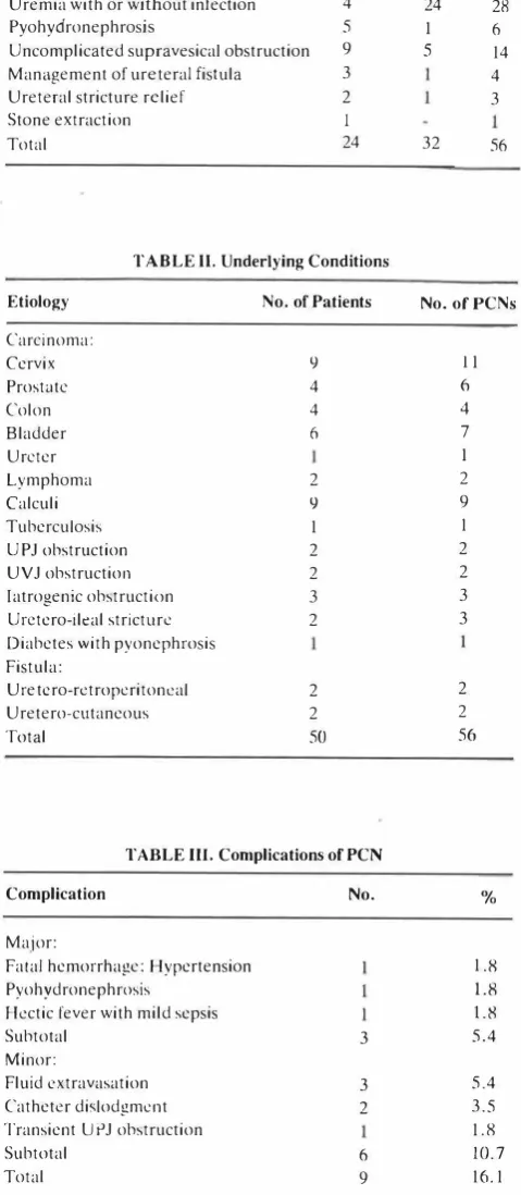
The results are summarized in Tables 1,11, and Ill.
Of 56 percutaneous nephrostomies in 50 patients. 32 procedures were performed for relief of an underlying factor in malignant disease and 24 for benign condi tions. Percutaneous nephrostomy was performed in dilated calyces and renal pelvices in all of the patients except four: one for stone extraction, one for manage ment of traumatic uretero-cutaneous fistula, and two others for relief of malignant obstructive anuria due to rectal and prostatic carcinoma.
Three major complications were encountered. One fatal hemorrhage occurred (case report). Hectic fever with mild sepsis occurred in another patient about 4R hours after a difficult nephrostomy placement. A small perirenal collection was noted on ultrasound. The patient underwent successful recovery following appropriate medical therapy.
Minor complications occurred in 10.7% of the patients. Percutaneous nephrostomy was successful in 9WYo. There was one catheter placement failure. The patient was a 3-year-old child with a mildly dilated. purulent collecting system containing multiple calnrli. Nephrostomy placement was successful only on one side.
CASE REPORT
A 25-year-old female had undergone partial bowel resection with end-to-end anastomosis for sigmoid carcinoma one year before admission. The patient admitted with uremia due to a nonfunctioning pyelonephritic right kidney and malignant obstruction
of the left ureter at the level of the pe.lvic brim. PCN was
successfully performed on the left side (Figure 4). Because of gross hematuria during the next 4R hours, a second nep
�
rostomy placement was performed on thesame side and the first was removed. This was followed. by exacerbation of hemorrhage and acute left flank pain. CT scan and ultrasound revealed perirenal and subcapsular hematomas. and hypertension developed within 24 hours due to marked compression of the kidney (Page's kidney). The patient underwent an cmergency laparotomy, and a massive subcapsular hematoma was evacuated. while the nephrostomy tube was maintained. Hcpatic metastases. peritoneal seed ing. and recurrence of tumor with invasion to left ureter was noted during surgery. The patient died 24 hours aftcr laparotomy in a clinical state of uremia and acute
TABLE L Clinical Indication For PCN
Indication Benign Malignant Total
Complicated supravesical obstruction:
Uremia with or without infection 4 24
Pyohydronephrosis 5 I
Uncomplicated supravesicaJ obstruction 9 5 Management of ureteral fistula 3
Ureteral stricture relief 2
Stone extraction I
Total 24 32
TABLE II. Underlying Conditions
28 6 14 4 3 56
Etiology No. of Patients No.ofPCNs
Carcinoma:
Cervix 9 LI
Prostate 4 6
Colon 4 4
Bladder 6 7
Ureter I
Lymphoma 2 2
Calculi 9 9
Tuberculosis I 1
UPJ obstruction 2 2
UV J obstructioll 2 2
Iatrogenic obstruction 3 3
Uretero-ileal stricture: 2 3 Diabetes with pyonephrosis
Fistula:
U re tcro-rctropcritone:al 2 2
Uretero-clltaneous 2 2
Total 50 56
TABLE Ill. Complications of PCN
Complication No. %
Major:
Fatal hemorrhage: Hypertension 1.8
Pyohydronephrosis 1.8
He:ctic fever with mild sepsis 1.8
Subtotal 3 5.4
Minor:
Fluid extravasation 3 5.4
Catheter dislodgment 2 3.5
Transient U t' J obstruction 1.8
Subtotal 6 10.7
Total 9 16.1
renal failure. Platelet count was normal, but platelet fuction studies were not performed.
DISCUSSION
The popularity of percutaneous nephrostomy has grown vastly in the last decade, and its indications have significantly broadened. Currently, the most common use of this technique is to establish an access for percutaneous nephrolithotomy and nephrolithotripsy, and
RO%
of nephrostomies are being done for stone removal."'Excluding the use of percutaneous nephrostomy for access to the upper urinary tract to treat urolithiasis, the most common indication in general practice is relief of supravesical obstruction, particularly secondary to primary or metastatic pelvic malignancies . .) The most frequent indication in the present study was compli cated supravesical obstruction due to malignant pro cesses. While azotemia may present alone, due to bilateral obstruction or obstruction of a single functioning kidney, this was frequently accompanied by infection.
Nephrostomy is indicated in those patients with malignant disease who hold some promise of survival and in whom further cancer therapy is anticipated. When proposing intervention for the patient with malignancy, medical judgment must always be tem pered by the wishes of the patient and the family.
The most gratifying response is usually evident in patients with urosepsis. This was achieved in six pa tients with pyohydronephrosis, and decompression of
A B
Figure I. (A) An excretory urogram one day after missile injury showing complete right ureteral obstruction (arrowhead) with mas sive flow via uretero-cutaneous and uretero-retroperitoneal fistulae.
the obstructed system resulted in almost immediate relief of symptoms within 24-48 hours.
Urinary leaks and fistulae may be due to trauma, surgery, or adjacent inflammatory disease. Percu taneous nephrostomy coupled with an antegrade stent catheter in these instances provides time for the fistulae and leaks to heal. 5 There are five types of fistulae: uretero-vaginal fistula, uretero-cutaneous fistula, uretero-enteric fistula, lymphatico-ureteral fistula, and uretero-retroperitoneal fistula or uriniferous pseudocyst. 6 Four patients had ureteral fistulae. A uretero-cutaneous fistula resulted from penetrating trauma in one patient (Figure
1).
In this patient, stenting was impossible because of ureteral cut off. Another uretero-cutaneous fistula developed after cystectomy with uretero-sigmoidal anastomosis for bladder carcinoma. Uretero-retroperitoneal fistulae developed in two cases after surgical repair for ureteropelvic junction obstruction. All4
patients were managed by percutaneous nephrostomy, and their fustulae healed completely. The urinomas were also aspirated coincidentally.Percutaneous antegrade stenting was carried out in three patients: one for relief of obstruction in a ureteral stricture due to prostatic carcinoma, another for stric ture by inactive urinary tract tuberculosis and the last for treating a stricture attributable to ligation of the ureter following abdominal hysterectomy (Figure 2). In the latter patient, a no. 7 French straight external
c
(8) One week after percutaneous nephrostomy placement. (C) Four weeks after urine diversion through the catheter; the fistula has completlyclosed. and the patient referred for reconstructive surgery .
Percutaneous nephrostomy
. Figure 2. Percutaneous stenting for iatrogenic distal ureteral obstruc tion one week after abdominal hysterectomy. (A) Attempts for right retrograde ureteral catheterization were unsuccessful. (8) Percu tan�us nephrostomy was performed and an angiographic guide wire
stent was placed for three months, which successfully treated the ureteral stricture. Follow up with in travenous urogram and ultrasound for another three months proved satisfactory recovery.
Numerous techinques for percutaneous nephros tomy placement have been described and each inter ventional radiologist should become comfortable with one method, but should also be familiar with alterna tive methods that can be used in specific cases. There are three basic methods of catheter placement: cathe ter over needle, catheter through cannula, and mod ified angiographic technique. The last is the most commonly employed method which requires a guide wire, dilator, and serial catheter exchanges.4
Different methods for puncture site localization can be considered in different patients. Currently,
fluoros-B
negotiated through the strictured ureter. (C) A 6 Fr straight stent catheter was passed over the wire and subsequently into the bladder. (D) Catheter was safely removed after two months. and follow-up sonographic findings were normal during the next six months.
copic guidance with or without a biplane option, seems to be the most frequently used method.7 Usually, the best position is prone oblique with the catheter entry site in a posterolateral location, so that the catheter will traverse the least vascular plane of the kidney. The intravenous administration of contrast material, with opacification of the renal pelvis, will generally obviate the need for ante grade pyelography in kidneys that do not function adequately. However, antegrade pyelography is often performed using ultrasound for needle placement. If the renal anatomy is known from prior urographic studies, ultrasound can be obviated and a blind puncture will usually be successful.
The use of sonographic methods for initial needle punture significantly reduces the number of puncture attempts. potential iatrogenic risk. and eliminates the
A
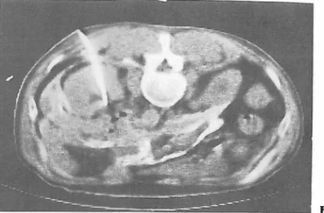
Figure 3. Bilateral obstructive nonfunctioning kidneys in a patient with rectal carcinoma. Prone CT-scan obtained for demonstrating anatomic detail and localization of puncture site. (A) Reveals slight dilatation of the collecting system on the left side. Percutaneous
need for intravenous administration of contrast mate rial. In a comparative study using sonographic and fluoroscopic guidance, the average number of needle punctures was
1.2
for sonographic punctures and more than three times for fluoroscopic insertions.8 In the present study, fluoroscopic guidance was used in almost all patients, with an average number of three needle punctures for access to the pelvocalyceal sys tem.Although the use of CT guidance for percutaneous
A
Figure 4. A 25-year-old female with chronic renal failure due to right pyelonephritic kidney and left ureteral obstruction by metastatic sigmoid carcinoma. 'percutaneous nephrostomy was performed without difficulty on the left side. (A) Nephrostomogram im mediately after catheter placement showing complete ureteral ob struction at the level of pelvic brim. (B) 48 hours later a second
B nephrostomy was performed on the left side. (B) Demonstrates catheter inserted into renal pelvis and some extravasation of Contrast material along fascial planes.
nephrostomy may seem cumbersome, time consum ing, and expensive for routine use, itis useful in patients with impaired renal function, undilated pelvocalyceal systems (Figure 3), and cross-sectional anatomic dis play. In addition to these purposes, CT accurately displays the perinephric space so that assessment of complications is possible.9
In most respects, interventional procedures do not differ in children and adults. 10 Needles, guide wires,
and catheters are generally of the same caliber as in
B c
nephrostomy was performed because of inadequate drainage due to blood clots obstructing the pelvocalyceal system. (C) Lateral decubi tus longitudinal real-time sonographic examination of the left kidney reveals perirenal hematoma at the superolateral aspect of dilated collecting system.
Percutaneous nephrostomy
Figure 5. (A) Massive extravasaion along the ureter after percu taneous catheter placement for distal ureteral obstruction in a patient
adults. In this study, four nephrostomies were per formed in three children without significant complica tion but with one placement failure involving a 3-year old boy with minimally dilated collecting systems con taining inspissated pus and multiple calculi. Percu taneous access to one of his kidneys was unsuccessful. Ureteral obstruction can lead to renal failure with out causing detectable dilatation of the pelvocalyceal system proximal to the obstruction, and ante grade pyelography is the best procedure to demtmstrate the level of obstruction. 11,12 This phenomenon occurred in two patients of the present study.
Diuresis and a decrease in azotemia occurred as a result of percutaneous nephrostomy. Years of experi ence at many centers throughout the world have con firmed the safety, efficacy, and reduced morbidity of percutaneous nephrostomy.
The mortality of this procedure has been reported as
0.2%
and the main complication has been massivehemorrhage with significant coagulopathy in the majority of cases. 13 Of our
56
patients, fatal hemor rhage occurred in one(1.8%).
The underlying causemay have been due to platelet dysfunction and/or injury to an intralobar renal artery. No bleeding diath eses other than those associated with severe uremia could be identified (Figure
4).
Injury to an intralobarrenal artery and hypertension due to massive hemor rhage in the subcapsular and perirenal space has been
with prostatic carcinoma. (B) Nephrostomogram performed 48 hours later, showing complete resolution of extravasation.
reported. 14-15 CT and ultrasound may be of value in identifying early homorrhage. In the latter patient these two procedures revealed a subcapsular and pe rirenal hematoma, and hypertension developed within
24
hours after percutaneous access, due to marked compression of the kidney (Page's kidney). It is recom mended that the radiologist be prepared for emergency interventions, including angiography and therapeutic embolization of bleeding artery. The major complica tion rate in this limited study was about5.4%,
but in large series, there are major complication rates of about1-4%,
primarily sepsis and hemorrhage.3.13 The most frequent major complication is symptomatic infection. 61t can be reduced by using sterile technique, appropriate antibiotic therapy in infected urine, and adequate drainage. Care must be taken not to overdis tend the collecting system. If obstructed systems are already infected, this increases the risk of sepsis. Transient hematuria is seen so frequently that it is considered a normal sequel rather than a complication, and usually clears within24
to48
hours. Blood clots within the renal pelvis may cause inadequate drainage. These clots usually dissolve within48
to72
hours under the action of urinary urokinase.Irrigation of the renal pelvis with normal saline can prevent heavy clot formation. Obstruction of the ureteropelvic junction occurred in one patient due to irritative edema resulting from guide wire
tions. This edema resolved within 48 hours. Perfora tion of the collecting system and extravasation of contrast media during forceful manipulation may occur (Figure
5).
However, if adequate drainage is maintained, no serious complication will occur. If extravasation con tinues longer than
72
hours, a large laceration or nephrostomy tube malplacement outside of the renal coIIecting system must be suspected. Catheter dis lodgement may occur as a result of inadequate fixation, placement of a short segment into the coIIecting sys tem, and a few weeks later because of dilatation of the nephrostomy tract. Use of monlar disc. sutures, and adequate dressing material while percutaneous cathe ters are in place wiII avoid dislodgment. Occasionally the nephrostomy catheter may coil into the perinephric space after successful nephrostomy tube placement. This may result from positional changing during move ment, respiratory excursion, and ptosis of the kidney.Reestablishment of a nephrostom y tract after cathe ter dislodgement is difficult and time consuming except in mature tracts. Failure to define a tract after contrast media injection usually suggests that the catheter has been dislodged for more than 48 hours, and requires repuncture.16
REFERENCES
1-Goodwin WE, Casey WC, WoolfW: Percu taneous trocar (needle) nephrostomy in hydronephrosis. lAMA 157: 891-4, 1955.
2-Bartly 0, Chidekel N, Radberg C: Percutaneous drainage of the renal pelvis for uremia due to obstructed urinary outflow. Acta Chir Scand 129: 443-6,1965.
3-Barbaric ZL: Percutaneous nephrostomy for urinary tract obstruc
tion. AlR 143 (4): 803-9, 1984.
4-Carson CC, Dunnick NR: Endourology. Edinburgh: Churchill Livingstone, 43-89,1985.
5-Druy EM, Gharib M, Finder CA: Percutaneous nephroureteral drainage and stenting for postsurgical ureteral leaks. AJR 141 (2): 389-94, 1983.
6- Lang EK: Diagnosis and management of ureteral fistulas by percutaneous nephrostomy and antegrade stent catheter. Radiology 138 (2): 311-7, 1981.
7-Stables DP, Ginsberg Nl. Johnson ML: Percutaneous nephros tomy: a series and review of the literature. AJR 130 (I): 75-82, 1978.
8-Zegel HG, Pollac HM, Banner MP, et al: Percutaneous nephros tQmy: comparison of sonographic and fluoroscopic guidance. AJR 137: 925-7,1981.
9-Hagga JR, Zelch MG. Alfidi RJ, et al: CT-guided antegrade pyelography and percutaneous nephrostomy. Am 1 Roentgenol 128 (4): 621-4, 1977.
10-Diament MJ, Boechat MI, KangarJoo H: Interventional radiolo gy in infants and children: Clinical and technical aspects. Radiol ogy 154 (2): 359-161,1985.
11-Naidich JB, Rackson ME, Mossey RT, Stein HL: Nondilated obstructive uropathy: percutaneous nephrostomy performed to reverse renal failure. Radiology 160 (3): 653-7,1986.
12- Maillet Pl, Pelle-Francoz D, Laville M, Gay F, Pinet A: Nondilated obstructive acute renal failure: diagnostic proce dures and therapeutic management. Radiology 160 (3): 659-62, 1986.
13- Stables DP: Percutaneous nephrostomy: techniques, indica tions, and results. Urol Clin North Am 9 (1): 15-29, 1982. 14-Harris RD, Walther PC: Renal artery injury associated with
percutaneous nephrostomy. Urology 23 (2): 215-17,1984. 15-Redman JF, Arnold WC, Smith PL, Seibert 11: Hypertension and
urino-thorax following an attempted percutaneous nephros tomy. J Urol 128 (6): 1307-8,1982.
16- Pollack HM, Banner MP: Replacing blocked or dislodged percutaneous nephrostomy and ureteral stent catheters. Ra diology 145 (1): 203-5, 1982.