www.ijper.org
Comparative Shape and Electrostatic Study of Highly
Potent and Selective CYP1B1 Inhibitor: Assessment
of Active Site of CYP1B1 by Binding Mode Analysis
Using Site Map Tool
Mohd Usman Mohd Siddique, Barij Nayan Sinha, Venkatesan Jayaprakash* Department of Pharmaceutical Sciences and Technology, Birla Institute of Technology, Mesra, Ranchi, INDIA.
ABSTRACT
Introduction: The major aim of drug design and discovery is to minimize the time and cost of drug discovery process. Various molecules which are promised to be potential candidate during computational and preclinical studies, shows the poor results during clinical trials due to less credibility of in silico results. This leads to increased burden of time and cost of drug discovery process. Methodology: A reliabel Shape and Electrostatic similarity based screening of ligands and assessment of druggability of the target protein provides a means to predict the negatives at an earlier stage of drug discovery pipeline. Two compounds (BNUA-3 & BNUB-13) reported from our lab were compared with ANF and TMS. Results and Discussion: Shape coefficient between BNUB-13 and TMS was 0.79 and electrostatic coefficient was 0.464 indicating that BNUB-13 is quite similar to
TMS. Dscore values for ANF, TMS, BNUB-13 and BNAU-3 were also found to be similar,
1.404, 1.390, 1.389 and 1.366, respectively. Conclusion: The comparative studies of two highly potent CYP1B1 inhibitors revealed minimum structural information that can modulate the potency of the inhibitors. Meanwhile assessment of the active site of CYP1B1 has shown that CYP1B1 is a druggable target.
Key words: CYP1B1, Shape electrostatic coefficient, Sitemap analysis, Druggability,
In silico ADME calculation.
DOI: 10.5530/ijper.52.1.18
Correspondence: Dr. Venkatesan Jayaprakash,
Department of Pharmaceuti-cal Sciences and Technology, Birla Institute of Technology Mesra, Ranchi, INDIA. Phone no: +91 9470137264; Email: drvenkatesanj@gmail. com
INTRODUCTION
Cytochrome P450 (CYP) enzymes are present in various organs of the human body that comprises a Formatting: leave a space
here large family of detoxification enzymes.
The cytochrome P450 1B1 isoform (CYP1B1) is a heme-thiolate monooxygenase which causes the hydroxylation of steroids, estrogens
and fatty acids. Unlike other CYPs, CYP1B1
is not present in normal healthy tissues but
significantly expressed in cancerous cells of
hormonal cancers including that of the ovary, prostate, uterus, mammary, pituitary,
regard-less of the cancer’s genetic origin. Recent
studies revealed that CYP1B1 plays a major role in the genesis of hormone-mediated
prostate and breast cancers.1,2,3 In both
can-cerous and precancan-cerous cells of mammary,
Submission Date: 21-07-2017; Revision Date: 25-09-2017; Accepted Date: 10-01-2018
prostate and ovarian tissues, the
regio-specific metabolism of estradiol produces
‘4-hydroxy estradiol (4-OHE2)’ by CYP1B1 that was reported to be one of the major
reason for oncogenesis.4 In cancer cells these
metabolite further oxidized to 3, 4-estradiol quinone that forms depurinating adducts with DNA and tubuline leading to
geno-toxic mutations.5,6,7,8,9,10,11,12 Therefore, the
rate and extent of CYP1B1 expression in endomatrium, mammary and ovarian tissues
can be considered as potential biomarker for hormonal oncogenesis.2,5,13,14,15
Recently we reported two series of com -pounds with different chemical scaffold with potent and selective inhibitory
some molecular properties are similar for these two chemical scaffolds in terms of their interaction with
the target protein. The current investigation is aimed at
unraveling them with a series of computational tech-niques that may help in designing CYP1B1 selective
inhibitors.
Active sites of CYP1 family enzymes have ≤ 40%
of amino acid sequence homology and CYP1A1 and
CYP1A2 shares ≥ 55% sequence homology. So it is quite difficult to design a highly specific and selective inhibitor against these iso-enzymes.18,19 Several attempts have been
reported on the development of computational models with the intention of predicting enhanced potency and
selectivity of inhibitors. Steric and electrostatic proper -ties of the molecules are the two most important
param-eters in predicting the binding affinity and selectivity towards the target proteins. These studies provide useful information for future drug design efforts. Comparative
Molecular Field Analysis (CoMFA) and Comparative
Molecular Similarity Indices Analysis (CoMSIA) were
employed to study the structure activity relationship of a series of molecules having same chemical scaffold by
overlaying the 3D structures of ligands. But these methods
have a limitation in the absence of protein that interacts
with them in biospace. Molecular dynamic (MD) simula -tions based methods were recently described for
study-ing ligand-protein interactions at molecular level.20,21,22
In the current study we employed Shape and Electro -static similarity assessment,23 of two highly specific and
potent CYP1B1 inhibitors TMS and BNUB-13. More
-over binding site was also evaluated by site map, which is
novel and effective algorithm for accurate and
rapidac-tive site identification. We also assessed the druggabil
-ity of active site pocket of CYP1B1 (PDB ID: 3PM0)24
by comparing the DScore values of four known and
potent inhibitors of CYP1B1, Tetramethoxy stilbene
(TMS),25 alphanaphthafalvone (ANF)26,27 BNUB-1316
and BNUA-317 Thus we believe this study will provide
useful insight in designing selective and potent CYP1B1
inhibitors.
MATERIAL AND METHODS
Shape and electrostatic analysis was performed by using VROCS (3.1.2) and EON (2.1.0) of OpenEye toolkit.28
using TMS (reference molecule) and BNUB-13 (test molecule). Molecular docking and Site map analy
-sis were done on Maestro 8.5. and SiteMap module of Schrodinger LLC suite.29 respectively, running on
RHEL5 operating system installed on DELL Precision
T3400 machine (n-series, Intel core 2 Quad processor,
8GB RAM, 500GB). Both molecules were prepared
in Glide and energy minimized using ligprep module
implemented in Maestro 8.5.111. The co-crystal structure
of CYP1B1 (PDB Code: 3PM0) was downloaded from
www.rcsb.org. For validation of software the internal ligand was extracted and redocked into the active site. Protein was prepared for docking through Protein
preparation wizard and Grid was generated through
grid preparation wizard picking ligand to specify the binding site. Docking was performed using GLIDE 5.0
with XP protocol. The docked conformers were ana
-lyzed through XP-visualizer. Default parameters were employed during all the computational studies.
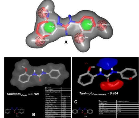
Shape and Electrostatic Study
Shape and electrostatic comparative study was done
because it gives an idea about physicochemical aspects
of molecular recognition. Thus comparing shape com -plementary and electronic features of our molecule with
BNUB-13 has revealed important structural framework which realized the binding affinity, which in turn func
-tional potency (Figure 1). For these we employed the ROCS algorithm (Rapid Overlay of Chemical Struc
-tures) and EON of OpenEye Toolkit. During the round
of ROCS analysis BNUB-13 and TMS were superim
-posed based on their shape agreement. Then they were
subjected to electrostatic correlations using EON and
results were analyzed based on their Shape and Electro
-static Tanimoto as shown in Figure 2.
Binding site of CYP1B1 was evaluated by Sitemap tool of Schrödinger. All the molecules were separately tested
for calculation of Dscore, hydorphilicty, hydrophobicity,
H bond donor and acceptor properties. These com -Figure 1: Potent CYP1B1 inhibitors used for druggability
puted properties are used to calculate the site score and
Dscore. Depending on these scores, the druggability of target was also predicted.
In-silico ADME prediction
Pharmacokinetic properties (ADME Absorption,
Distri-bution, Metabolism and Excretion) are the crucial part of new drug development procedure as many molecules
are withdrawn from the market due to their poor phar
-macokinetic profiles. We used QikProp, version 3.0, Schrödinger, LLC, New York, 2005, for prediction of ADME properties. To calculate drug likeliness properties
we consider various parameters such as molecular weight, H bond donor, H bond acceptor, polar surface area and predicted aqueous solubility and human oral
absorption.
RESULTS AND DISCUSSION
Shape and electrostatic studies
The rational in comparing shape and electrostatic prop-erties is that it is primary topological determinants of
molecule, which can be considered for better fitness and binding in the active site pocket of protein or enzyme. Recently Bostrom et al, reported the discovery of
potent fibrinolysis inhibitor by Shape and Electrostatic
Complementarity to the Drug Tranexamic Acid which has the potential for the treatment of bleeding
disor-ders.23 By the comparative shape and electrostatic study
we got good correlation between TMS and BNUB-13. Shape coefficient between two molecules was 0.769 while electrostatic coefficient was found to be 0.464 indicating that BNUB-13 was quite similar to TMS (Fig
-ure 3). Owing to the structural simiarity, the newly iden
-tified molecule could efficiently fit into the active site pocket of CYP1B1.
TMS and ANF are non-selective CYP inhibitors but
when the non-polar bridge is substituted by a polar urea
linker as in biphenyl urea the specificity of molecule was found to increase. It has been observed in
BNUB-13 with IC50 value of 69 nM and specificity of 62-fold. Hybridizing the structures of ANF and BNUB-13 we
got new scaffold which maintained its planarity and the
cyclized urea linker resembled ANF. Surprisingly the resultant molecule was highly specific and emerged as the most potent inhibitor (BNUA-3) with IC50 value of
3 nM. In-scilico and in-vitro results were in coherence with the important structural features that would guide the
design and discovery of potent and specific CYP1B1 inhibitor.
Molecular modeling studies
The X-ray crystallographic studies reveal that CYP1B1
has the rounder shape narrow slot-like substrate binding
cavity, which gets occupied by ANF and hinder the interactions between heme iron-oxo intermediate and
planner substrate.24 Moleculardocking studies revealed
the interactions between the terminal aromatic carbons
of BNUB-13 and reactive heme iron-oxo via van der
Waals’ interactions (distance is < 5 ºA). The second
aromatic ring of urea moiety positioned in such a way
that it shows the π-π interactions with hydrophobic
Figure 2: Predicted three-dimensional shape and Tanimotoelectrostatic overlays of compounds TMS (A) and
BNUB-13 (B).30
Figure 3: (A) Complete overlapping of TMS (silver color) and BNUB-13 (red color); (B) the display of shape and (C) electro-static potentials for BNUB-13 with that of TMS in their neutral
form. Blue color denotes electropositive areas, whereas red color shows electronegative areas. The calculated Tanimoto values depicted that BNUB-13 is electrostatically very similar
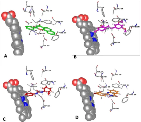
Phe231 and Phe268 residues of CYP1B1. In addi
-tion, BNUB-13 also display the polar interactions via H-bonding with Asp333 (Figure 4C). Overall, these
interactions of the urea derivative with CYP1B1 con-tribute the potent inhibition of CYP1B1 at low nano-molar IC50 value.
Similarly aromatic ring C of BNAU-3 flanks towards
the heme atom due to presence of heteroatom
contain-ing rcontain-ing B and interacts with heme via van der Waals interactions (distance is <5 ºA) in a way similar to the benzo[a]chromane ring of ANF. The geometry of BNAU-3 facilitates interaction in such a way that O atom of ring C comes nearest to iron cofactor of heme.
Moreover ring A and B display hydrophobic interaction
with Phe231. Additionally BNAU-3 also interacts with Phe134 residue via π-π interactions. These interactions
with CYP1B1 make the compound more potent inhibi
-tor of CYP1B1. All molecules occupied the rounder shape narrow slot-like substrate binding cavity and
interact with the Phe231 and Phe268, and one aromatic
ring interact with heme moiety. But in case of
BNAU-3, it has additional polar H bonding with Asp333 that
make this molecule more selective towards the CYP1B1 isoform. Similarly in case of BNUB-13 there is addi -tional hydrophobic interaction with Phe134 residue and position of C ring O atom is very close to iron of heme
moiety leading to most potent and selective inhibition. Characterization of CYP1B1 active site as druggable target
Site map tool was used to find out whether a pro
-tein can be a druggable target or not. For validation
of druggability of active site of CYP1B1 enzyme (PDB ID: 3PM0) we used four potent CYP1B1
inhibitor ANF (50 nM), TMS (6 nM), BNUB-13 (69 nM) and BNUA-3 (3 nM). The results were summarized in Table 1. Dscore as reported earlier, it
is calculated based on the sitescore, size, hydrophilicity, hydrophobicity, enclosure, exposure and H bond donor
and acceptor values.31 The Figure 5 (A) comprise of the
ANF binding site map in active site pocket of CYP1B1 while. The calculation is based on the hydrophobic
-ity of the pocket for the druggabil-ity and expresses in terms of numerical values 0-2. Highly hydrophilic
cavities are considered as undruggable target or protein
and have the value of 0, while 1 is considered difficult to target and 2 falls in druggable target category. Site
map analysis for complexes of all the four ligands with
CYP1B1 was performed to check the druggability of the target protein. The calculation using all the four com -plexes displayed that the protein is druggable because all molecules showed D-score that expressed them
as druggable site score 2 (Table 1). The hydrophobic score of ANF was found be better than the other three. BNUB-13 having the better hydrophobic interaction than BNUA-3, but it is less potent than BNUA-3 due to its polar bridge (higher hydrophilic score). We suppose
that the compounds having less polar bridges perform
well in terms of improving potency. All physicochemical
properties obtained were identical in all molecules
(Table 1).
Figure 4: 3D interaction diagram of (A) ANF, (B) TMS (C) BNUB-13 and (D) BNUA-3 at active site of CYP1B1 enzyme
and showing the interactions with amino acid residues present at the active site (PDB ID: 3PM0).
Figure 5: Binding mode analysis of ANF (A), TMS (B), BNUA-3 (C) and (D) BNUB-13. The various crucial interactions are red color denotes H Bond acceptor, blue color shows H-Bond donor, Yellow color indicates hydrophobic interaction and
prediction of druggability of CYP1B1 generate potential 3D structural information of receptor giving vital
informa-tion. Having this information of whether a given protein is druggable or not at an early stage of drug discovery therefore has the potential of saving considerable time
and expense. In silico ADMET calculations showed that
all the molecules complied with Lipinski rule of five
and hence have the highest probability to become drug
(drug-likeness).
CONFLICT OF INTEREST
Authors do not have any conflict of interest
ACKNOWLEDGMENT
MUMS acknowledge UGC for award of MANF JRF fellowship (201516-MANF-2015-17-MAH-60712).
ABBREVIATIONS USED
ANF: Alphanaphthaflavone; CYPs: Cytochrome P450;
CYP1B1: Cytochrome P450-1B1; TMS: Tetramethoxy
stilbene; ROCS: Rapid Overlay of Chemical Structures.
REFERENCES
1. McFadyen MC, Breeman S, Payne S, Stirk C, Miller ID, Melvin WT, et al. Immunohistochemical localization of cytochrome P450 CYP1B1 in breast cancer with monoclonal antibodies specific for CYP1B1. Journal of Histochemistry andCytochemistry. 1999; 47(11):1457-64.
In silico ADME prediction
In-silico ADMET calculation was performed for drug
likeness properties of molecules. The descriptors like
dipole moment, the percentage of oral absorption,
and PlogBB were analysed. Lipinski’s Rule of 5 (Ro5)
explains, in orally active molecules hydrogen bond acceptor (HBA) should not exceeds more than 10 and
molecular weight (MW) 500D. Whereas H bond donor
(HBD) groups less than 5 and it should have oil/water
partition coefficient (logPo/w) below 5.The results thus obtained were presented in Table 2. All the mol
-ecules were found to comply with the rule-of-five (R05) described by Lipinski. The CNS toxicity was predicted by blood-brain coefficient (plogBB), and the results
indicate that all molecules have values within the range and suggest that none of the molecules can cross the
blood brain barrier to produce CNS toxicity.
CONCLUSION
Physicochemical properties of ligand and protein active site are important in drug design and discovery and protein can be called druggable if it can exhibit in vivo
pharmacological response by drug like molecule. Shape
and electrostatic parameter determine the
physicochem-ical properties of any molecule. Comparing and calcu
-lation of these parameters of known potent inhibitors would give important ideas that could be taken into
consideration while designing of any new molecule
against particular target or protein. Moreover accurate
Table 1: SiteMap Property Values and Dscore Ranks for the potent CYP1B1 inhibitors.
Entry IC50 nm
(CYP1B1) Category DScore SiteScore Size Enclosure Exposure hydorphilic hydrophobic
ANF 50a 2 1.400 1.310 117 0.971 0.316 0.377 5.044
TMS 6 2 1.390 1.305 114 0.971 0.349 0.406 4.836
BNUB-13 69a 2 1.389 1.304 116 0.972 0.288 0.412 4.874
BNUA-3 3a 2 1.366 1.290 124 0.970 0.337 0.477 4.648
a IC
50 values were determined inSacchrosomes™ and it represents mean and standard deviations from three independent experiments
Table 2: In silico ADME calculation for drug like properties of all four molecules.
Entry Lipinski Rule
Of Five Mol Wt PLog P (o/w)a PSA
b HBAc HBDd plogBBe % Oral
absorption
ANF 0 272.303 4.141 33.670 2.5 1 0.081 100
TMS 0 300.354 4.094 32.816 3 0 -0.443 100
BNUB-13 0 242.277 4.016 52.670 2.750 2 -0.090 100
BNUA-3 0 301.342 3.070 50.152 3.5 1. -0.143 100
a Predicted octanol/water partition coefficient.
b Polar surface area.
c Hydrogen bond acceptor.
d Hydrogen bond donor.
2. Murray GI, Taylor MC, McFadye MC, McKay JA, Greenlee WF, Burke MD, et al. Tumor-specific expression of cytochrome P450 CYP1B1. Cancer Research. 1997;57 (14):3026-31.
3. Muskhelishvili L, Thompson PA, Kusewitt DF, Wang C, Kadlubar FF. In situ hybridization and immunohistochemical analysis of cytochrome P450 1B1 expression in human normal tissues. Journal of Histochemistry and Cytochemistry. 2001;49(2):229-36.
4. Yager JD, Endogenous estrogens as carcinogens through metabolic activation. Journal of National Cancer Institute Monographs. 2000;27(27):67-73.
5. Cavalieri EL, Devanesan P, Bosland MC, Badawi AF, Rogan EG. Catechol estrogen metabolites and conjugates in different regions of the prostate of Noble rats treated with 4-hydroxyestradiol: implications for estrogen-induced initiation of prostate cancer. Carcinogenesis. 2002;23(2):329-33.
6. Cavalieri E, Stack D, Devanesan P, Todorovic R, Dwivedy I, Higginbotham S, et al. Molecular origin of cancer: catechol estrogen-3, 4-quinones as endogenous tumor initiators. Proceedings of the National Academy of Sciences. 1997;94(20):10937-42.
7. Hayes CL, Spink DC, Spink BC, Cao JQ, Walker N J, Sutter TR. 17 beta-estradiol hydroxylation catalyzed by human cytochrome P450 1B1. Proceedings of the National Academy of Sciences. 1996;93(18):9776-81. 8. Rogan EG, Badawi AF, Devanesan PD, Meza JL, Edney JA, West WW.
Higginbotham SM, Cavalieri EL. Relative imbalances in estrogen metabolism and conjugation in breast tissue of women with carcinoma: potential biomarkers of susceptibility to cancer. Carcinogenesis. 2003;24(4):697-702. 9. Zhu BT, Conney AH. Functional role of estrogen metabolism in target cells:
review and perspectives. Carcinogenesis. 1998;19(1):1-27.
10. Rochat B, Morsman, JM, Murray GI, Figg WD, McLeod HL. Human CYP1B1 and anticancer agent metabolism: mechanism for tumor-specific drug inactivation? Journal of Pharmacology and Experimental Therapeutics. 2001;296(2):537-41.
11. Newbold RR, Liehr JG. Induction of uterine adenocarcinoma in CD-1 mice by catechol estrogens. Cancer Research. 2000;60(2):235-7.
12. Cavalieri EL, Rogan EG. A unifying mechanism in the initiation of cancer and other diseases by catechol quinones. Annals of the New York Academy of Sciences. 2004; 1028(1):247-57.
13. Liehr JG, Ricci MJ. 4-Hydroxylation of estrogens as marker of human mammary tumors. Proceedings of the National Academy of Sciences 1996;93(8):3294-6.
14. Spink DC, Spink BC, Cao JQ, DePasquale JA, Pentecost BT, Fasco MJ, et al. Differential expression of CYP1A1 and CYP1B1 in human breast epithelial cells and breast tumor cells. Carcinogenesis. 1998;19(2):291-8.
15. Tait L, Soule HD, Russo J. Ultrastructural and immunocytochemical characterization of an immortalized human breast epithelial cell line, MCF-10. Cancer Research. 1990;50(18):6087-94.
16. Siddique MUM, McCann GJ, Sonawane V, Horley N, Williams IS, Joshi P, et al. Biphenyl urea derivatives as selective CYP1B1 inhibitors. Organic andBiomolecular Chemistry. 2016;14(38):8931-6.
17. Siddique MUM, McCann GJ, Sonawane VR, Horley N, Gatchie L, Joshi P, et al. Quinazoline derivatives as selective CYP1B1 inhibitors. European Journal of Medicinal Chemistry. 2017;130:320-7.
18. Nebert DW, Nelson DR, Adesnik M, Coon MJ, Estabrook RW, Gonzalez FJ, et al. The p450 superfamily: updated listing of all genes and recommended nomenclature for the chromosomal loci. DNA. 1989:8(1):1-13.
19. Testa B, Caldwell J. Monooxygen ase-catalyzed NC cleavage. The metabolism of drugs and other xenobiotics—biochemistry of Redox reactions. Testa B, Caldwell J (eds), Academic Press limited, London 1995;215-24. 20. Guvench O, MacKerell Jr AD. Computational fragment-based binding site
identification by ligand competitive saturation. PLoS Computational Biology. 2009;5(7):e1000435.
21. Raman EP, Yu W, Guvench O, MacKerell Jr AD. Reproducing crystal binding modes of ligand functional groups using Site-Identification by Ligand Competitive Saturation (SILCS) simulations. Journal of Chemical Information and Modeling. 2011;51(4):877-96.
22. Seco J, Luque FJ, Barril X. Binding site detection and druggability index from first principles. Journal of Medicianal Chemitry. 2009;52(8):2363-71. 23. Boström J, Grant JA, Fjellström O, Thelin A, Gustafsson D. Potent fibrinolysis
inhibitor discovered by shape and electrostatic complementarity to the drug tranexamic acid. Journal of Medicinal Chemistry. 2013;56(8):3273-80. 24. Wang A, Savas U, Stout CD, Johnson EF. Structural characterization of the
complex between α-naphthoflavone and human cytochrome P450 1B1. Journal of Biological Chemistry. 2011;286(7):5736-43.
25. Kim S, Ko H, Park JE, Jung S, Lee SK, Chun YJ. Design, synthesis, and discovery of novel trans-stilbene analogues as potent and selective human cytochrome P450 1B1 inhibitors. Journal of Medicinal Chemistry. 2002;45(1):160-4.
26. Campbell DR, Kurzer MS. Flavonoid inhibition of aromatase enzyme activity in human preadipocytes. Journal of Steroid Biochemistry and Molecular Biology. 1993:46(3):381-8.
27. Kellis Jr JT, Vickery LE. Inhibition of human estrogen synthetase (aromatase) by flavones. Science. 1984;225:1032-5.
28. OEChem T, version 2.0. 0. OpenEye Scientific Software, Santa Fe, NM. There is no corresponding record for this reference.
29. Schrodinger L. Schrodinger Software Suite. New York: Schrödinger, LLC 2011.
30. Halgren TA. Identifying and characterizing binding sites and assessing druggability. Journal of Chemical Information and Modeling. 2009;49(2):377-89.
31. Cheng AC, Coleman RG, Smyth KT, Cao Q, Soulard P, Caffrey DR, et al. Structure-based maximal affinity model predicts small-molecule druggability. Nature Biotechnology. 2007;25(1):71-5.
SUMMARY PICTORIAL ABSTRACT
The combined approach of ligand and receptor aspect were considered for generation of in silico data. Shape and electrostatic studeis reveals the structural
complementary that are prerequisite for inhibitory
activity.
Binding mode analysis gave the receptor 3D structural information that can be used for lead optimization in drug discovery, to modify the ligand to enhance its
binding affinity and to improve its physico-chemical
Cite this article:Siddique MUM, Sinha BN, Jayaprakash V. Comparative Shape and Electrostatic Study of Highly
Potent and Selective CYP1B1 Inhibitor: Assessment of Active Site of CYP1B1 by Binding Mode Analysis Using Site
Map Tool. Indian J of Pharmaceutical Education and Research. 2018;52(1):159-65.
Mohd Usman Mohd Siddique: Is research scholar in the Department of Pharmaceutical Sciences
andTechnology, BIT Mesra Ranchi. He has the area of interest in design and discovery of novel molecules against cancer and viral infection. He has completed his M S (Pharm) degree from National Institute of Pharmaceutical Education and Research Kolkata.
Dr B.N. Sinha is Professor andFormer Head at the Department of Pharmaceutical Sciences andTechnology, Birla Institute of Technology, Mesra. He obtained his B. Pharm. from BIT Mesra, Ranchi in 1981 followed by M. Pharm. from Banaras Hindu University in 1983. Prof. Sinha completed his PhD from Birla Institute of Technology, Mesra in 1997. He is a medicinal chemist
having research interest in the area of Drug Discovery Approaches with regard to Synthetic and
Natural Leads using Modern Analytical Techniques and Soft wares.
Dr. Venkatesan Jayaprakash: Currently working as Associate Professor in Department of Pharmaceutical Sciences andTechnology, Birla Institute of Technology, Mesra. He obtained his B. Pharmacy from Madras Medical College, M. Pharmacy from Banaras Hindu University and PhD from Birla Institute of Technology, Mesra. He did his doctoral research under the supervision of Dr. B. N. Sinha. He is a medicinal chemist having expertise in organic synthesis and molecular modeling.