R E S E A R C H A R T I C L E
Open Access
Hypoxia inducible factor 1
a
gene (
HIF-1
a
)
splice variants: potential prognostic biomarkers
in breast cancer
Jean-Philippe Dales
1,2*†, Nathalie Beaufils
3†, Monique Silvy
4, Christophe Picard
4, Vanessa Pauly
5, Vincent Pradel
5,
Christine Formisano-Tréziny
1, Pascal Bonnier
6, Sophie Giusiano
2, Colette Charpin
2, Jean Gabert
1,3Abstract
Background:Hypoxia-inducible factor 1 (HIF-1) is a master transcriptional regulator of genes regulating oxygen homeostasis. The HIF-1 protein is composed of two HIF-1aand HIF-1b/aryl hydrocarbon receptor nuclear
translocator (ARNT) subunits. The prognostic relevance of HIF-1aprotein overexpression has been shown in breast cancer. The impact of HIF-1a alternative splice variant expression on breast cancer prognosis in terms of metastasis risk is not well known.
Methods:Using real-time quantitative reverse transcription PCR assays, we measured mRNA concentrations of total HIF-1a and 4 variants in breast tissue specimens in a series of 29 normal tissues or benign lesions (normal/benign) and 53 primary carcinomas. In breast cancersHIF-1asplice variant levels were compared to clinicopathological parameters including tumour microvessel density and metastasis-free survival.
Results:HIF-1a isoforms containing a three base pairs TAG insertion between exon 1 and exon 2 (designated HIF-1aTAG) andHIF-1a736 mRNAs were found expressed at higher levels in oestrogen receptor (OR)-negative carcinomas compared to normal/benign tissues (P= 0.009 andP= 0.004 respectively). In breast carcinoma specimens, lymph node status was significantly associated withHIF-1aTAGmRNA levels (P= 0.037). Significant statistical association was found between tumour grade andHIF-1aTAG (P= 0.048), and totalHIF-1a(P= 0.048) mRNA levels.HIF-1aTAGmRNA levels were also inversely correlated with both oestrogen and progesterone receptor status (P= 0.005 andP= 0.033 respectively). Univariate analysis showed that highHIF-1aTAG mRNA levels
correlated with shortened metastasis free survival (P= 0.01).
Conclusions:Our results show for the first time that mRNA expression of aHIF-1aTAGsplice variant reflects a stage of breast cancer progression and is associated with a worse prognosis.
See commentary: http://www.biomedcentral.com/1741-7015/8/45
Background
Hypoxia-inducible factor 1 (HIF-1) is a ubiquitously expressed master transcriptional regulator of many genes regulating mammalian oxygen homeostasis [1,2]. HIF-1 alters transcription of genes mainly involved in energy metabolism, neovascularisation, survival, internal pH and cell migration, and has become recognised as a strong promoter of tumour growth [3,4]. HIF-1 is a heterodimeric protein composed of two HIF-1a and
HIF-1b/aryl hydrocarbon receptor nuclear translocator (ARNT) subunits [5]. These subunits are members of the basic helix-loop-helix (bHLH) transcription factor superfamily containing a PAS [PER (Period Clock) -ARNT-SIM (Single-minded)] domain [2]. The HIF-1a subunit is specific to HIF-1 and is the unique oxygen-regulated subunit that determines HIF-1 activity [6,7] whereas HIF-1bis a common subunit for several tran-scription factors. In human tumours, HIF-1a protein is overexpressed because of intratumoural hypoxia and genetic alterations affecting key oncogenes and tumour suppressor genes [8,9]. This overexpression correlates
* Correspondence: Jdales@ap-hm.fr
†Contributed equally
1
Plateforme Transcriptome, CRO2, Marseille, France
with tumour angiogenesis, treatment failure and patient mortality [3].
Our group and others have previously reported that HIF-1a protein overexpression is a marker of poor prognosis in primary breast cancer patients [10,11]. HIF-1a is known to be mainly post-transcriptionally regulated by protein ubiquitination and interaction with the Von Hippel-Lindau tumour suppressor protein, and then degraded by the proteasome. Hypoxia stabilises HIF-1a protein by relaxing its ubiquitin-proteasome degradation [12] and affects subcellular localisation, DNA binding capacity and transcriptional activation function of the HIF-1 complex. HIF-1a protein is composed of 826 amino acids [5]. Its N-terminal region contains the bHLH and the PAS domains, which are essential for DNA binding and dimerisation [13]. Its C-terminal region contains two transactivation domains and a nuclear localisation signal [14]. In the middle sec-tion of HIF-1a lies a Pro-Ser-Thr oxygen-dependent degradation domain (ODDD, amino acids 401 to 603), which is responsible for the stability of the HIF-1a pro-tein [12].
Although HIF-1a protein regulation has been well studied,HIF-1a messenger regulation has only recently begun to be documented [15]. HIF-1ais encoded by the HIF1Alocus, which is located on human chromosome 14q21-q24 and encodes for 15 exons, resulting in a principal transcript of about 8 kb [16]. Different func-tional domains of the HIF-1a protein are encoded by separate exons. In addition to the originally described wild type of HIF-1a (HIF-1aWT
) [1], eight alternative splice variants of HIF-1a (HIF-1a827, HIF-1a736, HIF-1a557, HIF-1a516, HIF-1a785, HIF-1a417, HIF-1aTE
and an isoform with alternative exon 1 (I.2 isoform)) with different activity have been reported in human cell lines [17-23]. All HIF-1a splice variants have been shown to be translated, to dimerise with HIF-1band to be active in human cells. Only one isoform (HIF-1a736) has been documented in human breast cancer [24]. The aim of this study was to analyseHIF-1asplice variant expression in human breast cancer and to evaluate its clinical relevance in terms of prognosis. In this study we quantified the mRNA levels of 3 isoforms (HIF-1a736, HIF-1a557
and HIF-1a516) in breast specimens of 53 primary cancers and 29 normal tissues or benign lesions by real-time quantitative reverse transcription PCR (RT-qPCR) assays. HIF-1a827 is similar to wild type except for an additional three base pairs TAG insertion at the exon 1 to 2 junction involving a difference of two amino acids upstream of the bHLH domain [17]. HIF-1a736 is also characterised by an additional three base pairs TAG insertion at the exon 1 to 2 junction, and the lack of exon 14, which produces a frame shift and introduces a stop codon in the coding sequence
after the Ile735 [17]. HIF-1a557 loses exon 12 and HIF-1a516
lacks exons 11 and 12 [18,19].HIF-1a557has been shown to be induced by zinc ions.HIF-1a557and HIF-1a516
have been shown to function as dominant negative isoforms of HIF-1a in vitro. HIF-1a785 is deprived of exon 11 and was described as being induced in vitro by phorbol-12-myristate-13-acetate [20]. HIF-1a417 isoform is deprived of exon 10 [21]. The HIF-1aTE variant with alternative exon 1 (I.1 isoform) was previously found to be expressed in the testis [22]. For these reasons, both these isoforms were thought to be out of the scope of this study on breast tissues sam-ples. We also developed tools to quantify expression of HIF-1a splice transcripts characterised by insertion of a three base pairs TAG at the exon 1 to 2 junction (which we designatedHIF-1aTAG) involving a difference of two amino acids upstream of the bHLH domain (HIF-1a827 and HIF-1a736 isoforms) [17]. The last known isoform was not quantified because it was described in humans after the end of our study [23]. Expression levels of these isoforms were also compared to standard prognos-tic clinicopathological parameters, microvessel density of tumours and clinical outcome.
Methods
Patients and breast tissue samples
For this first step of treatment, patient management was handled by the same group of surgeons (PB). Likewise, radiotherapy, chemotherapy and hormone therapy were applied according to criteria currently used at that time. Each of the respective areas was identified microscopi-cally and dissected immediately upon receipt of the breast specimen by the pathologist. The tumour content of each specimen was verified by histological examination of frozen section by haematoxylin staining. Histological examination of surgical specimens was also performed on paraffin embedded sections stained with haematoxy-lin, eosin and saffronin. Tumour grading, initially assessed by using the grading methods of Bloom and Richardson [25], was re-evaluated according to Elston and Ellis [26]. Duration of follow-up ranged from 9 to 162 months (mean 80 months, SD 43). For statistical purposes, we selected approximately equal numbers of long (n = 31) and short metastasis-free survival patients (n = 22).
Clinical and pathological parameters
Lymph node status, tumour size, peritumoural vascular invasion, histological type and histological grade of tumours were obtained from pathological reports. The oestrogen receptor (OR) and progesterone receptor (PgR) status was determined by immunohistochemistry on paraffin-embedded sections as previously described [27,28].
Tumour microvascular density
Immunodetection studies were performed on frozen sections 5 ?m thick and automated immunoperoxidase procedures as previously reported [29].
RNA extraction and reverse transcription
Homogenising Trizol procedures have been performed in accordance with current protocols. Frozen tissue (25 to 50 mg) was homogenised in 0.5 ml Trizol reagent (Life Tech-nologies, Carlsbad, California) with lysing matrix D in a FastPrep machine (Q-BIOgene, MP Biomedicals, Illkirch, France) for six cycles of 30 s at a setting of 6 m/s as recommended by the manufacturer. Chloroform (100μl) was added to each sample, which were mixed vigorously for 15 s and incubated for 3 min at room temperature. Phases were separated by centrifugation (12,000gfor 15 min at 4°C) and the aqueous phase was recovered. Iso-propanol (250μl) was added to each of the samples, which were then mixed by inversion and incubated for 10 min at room temperature. Total RNA was pelleted by centrifuga-tion (12,000gfor 10 min at 4°C), washed with 1 ml of ice-cold 75% ethanol, air dried and resuspended in 15μl Rnase-free distilled water. Then, 1μg of RNA was reverse transcribed with 200 U MMLV Reverse Transcriptase following the Europe Against Cancer (EAC) protocol [30].
Generation of standard plasmids
pcDNA3 plasmids containingHIF-1a827andHIF-1a736 sequences with the three base pairs TAG insertion were kindly provided by J Pouyssegur (Institut de Signalisation, Biologie du Développement et Cancer, Centre Lacassagne, Nice, France). For other splice variants, cDNA from human embryonic kidney (HEK)293 cells was amplified using the following primers, respectively localised in exons 1 and 2: forward, 5′-CCGGCGGCGCGAACGACAAG-3′, reverse, 5′-TGCGAACTCACATTATGTGG-3′, or pri-mers localised in exons 10 and 14, respectively: forward, 5′-TGACCCTGCACTCAATCAAG-3′, reverse, 5′-AGTA-GCTGCATGTACGTCTG-3′. Conditions for PCR ampli-fication were: 10 min denaturation at 95°C followed by 35 cycles of 30 s at 95°C, 1 min at 55°C, 1 min at 72°C, and the last elongation at 72°C for 10 min. If necessary, DNA bands of interest were isolated and purified from 2% agar-ose gel and cloned into the pCRII TOPO vector (TOPO-TA cloning kit, Invitrogen, Life Technologies, Carlsbad, California) according to the manufacturers. Plasmids were purified (Nucleospin plasmid, Macherey Nagel, Hoerdt, France) and the cloned inserts were sequenced. Serial dilutions were prepared inEscherichia colirRNA (Roche Diagnostics, Meylan, France) to generate RT-qPCR stan-dard curves (PCT international application no. 02/00937 of 15 March 2002, in the name of Université de la Médi-terranée) according to the EAC strategy [30].
Design
mRNA sequences of four spliced variants (HIF-1aTAG, HIF-1a736, HIF-1a557 and HIF-1a516) were
construct-ed using the human HIF-1a transcript sequence
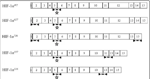
NM_001530 as a reference. Primer and probe sets were designed using the Primer Express software (Applied Biosystems, Courtaboeuf, France) and chosen based on their exon-exon junction location (Figure 1).HIF-1a736, HIF-1a557andHIF-1a516splice variant specific TaqMan assays, and an assay specific to a common region pre-sent in all known variants and located at exons 5 and 6, were designed. Specific probes and primers on adjacent exons were also designed to detect HIF-1a transcripts containing a three base pairs TAG insertion between exon 1 and exon 2 (which we designated HIF-1aTAG). For this variant, primers and probes were designed to detect intron length between exons rather than exon-exon junctions. The length of introns between these exons on DNA is long enough to avoid DNA amplification.
Optimisation of RT-qPCR
each variant in a total volume of 25 μl containing 1 × TaqMan universal PCR MasterMix (Applied Biosys-tems, Courtaboeuf, France) and both forward and reverse primers and probes. Different ranges of primer concentrations were tested and optimised for each amplicon to obtain the most specific, sensitive and effi-cient RT-PCR amplification. Selected primer and probe concentrations are shown in Table 1. Optimised RT-qPCR reaction conditions for gene amplification were as follows: 50°C for 2 min, denaturation for 10 min at 95°C followed by 50 cycles of 30 s at 95°C, 1 min annealing and extension at 60°C for all tran-scripts except for HIF-1aTAG with 1 min at 62°C. For validation of assays specificity, the primers and probes used to detect each splice variant were tested by RT-PCR with 1 μg RNA of the HEK293 cell line (DSMZ), 1 μg genomic DNA of peripheral blood mononuclear cells (PBMCs) and standards (106 copies of plasmids) specific for another splice variant. No template control (NTC) was also performed. RT-qPCR products were analysed by Agilent 2100 Electrophoresis Bioanalyzer (Agilent Technologies, Santa Clara, California). Serial dilutions of specific standard plasmids were used to validate sensitivity, linearity and detection limits of
RT-qPCR assays. Intrareproducibility and inter-reprodu-cibility assays were performed by four RT-qPCR experi-ments, each time in triplicate.
RT-qPCR assays
Amplifications were performed with 5 μl of cDNA of each sample using RT-qPCR optimised conditions as previously described. mRNA from HEK293 cells was used as positive control throughout the study. Standard (106 copies of plasmid) specifics of another splice variant were amplified as negative control throughout the study and NTC was performed. Standard curves were gener-ated by serial dilutions of HIF-1asplice variant plasmids ranging from 1 to 106copies and used for calculation of copy numbers (CN) for each transcript. Transcripts of the gene coding for the TATA box-binding protein (TPB) were also quantified as endogenous gene controls, and each sample was normalised on the basis of its TBP content as previously described [31]. All experiments were carried out with duplicates for each data point. For each experiment sample, the mRNA copy number of HIF-1a gene (CNHIF-1a) and endogenous reference TBP
gene (CNTBP) were quantified from standard curves. Final HIF-1amRNA concentrations were expressed in
Figure 1Position of primers and oligonucleotide probes designed to amplify hypoxia inducible factor 1a(HIF-1a) mRNA splice variants. The diagram illustrates the combination of primers and probes used to detect mRNA of four alternative spliced isoforms ofHIF-1a.
Boxes represent exons designated by 1 to 15. Primers are indicated by solid arrowhead and probe by thick solid lines. Wild-typeHIF-1a(
HIF-1awt) mRNA consists of 15 exons and 14 introns. Variants reported here are generated by a TAG insertion between exon 1 and exon 2 (HIF-1
a827 andHIF-1a736), exon 13 to 15 alternative splicing (HIF-1
a736), exon 11 to 13 alternative splicing (HIF-1
a557) and exon 10 to 13 alternative splicing
(HIF-1a516). Primers on exons 5 and 6 (asterisk) are suitable for quantification of allHIF-1atranscripts includingHIF-1awtas well as each isoform.
Primers pairs on exons 13 and 15, 11 and 13, 10 and 13 are suitable for specific detection ofHIF-1a736,HIF-1a557andHIF-1a516variants
normalised copy numbers (NCNHIF-1a), as follows:
NCNHIF-1a=CNHIF-1a/CNTBP.
Statistical analysis
Differences among groups of breast tissue specimens (normal/benign tissues vs carcinomas) in terms of their HIF-1amRNA levels were assessed using Kruskal-Wallis and Mann-Whitney tests with Bonferroni correction. Since levels of expression in breast cancer specimens showed non-Gaussian distribution, non-parametric tests were used for the analysis of correlation with clinico-pathological parameters. Metastasis-free survival (MFS) time, defined as the time from surgery until diagnosis of metastasis, was used as a follow-up endpoint. Regression models (univariate and multivariate) were used to com-pare survival and identify predictors of survival. All pre-operative predictors (clinicopathological and biological parameters) were included in the analysis. The predic-tors with P value < 0.20 in the univariate model were tested in the multivariate regression model. Then back-ward conditional method was used for variable selection by the Cox multivariate regression model. Finally, vari-ables with adjusted Pvalues < 0.05 were kept into the final model. All statistical analyses were performed with SPSS V.13.0.1 software (SPSS, Chicago, IL, USA).
Results
Clinicopathological parameters
Tumour characteristics are summarised in Table 2. Tumours corresponded to ductal carcinomas (n = 41)
and lobular carcinomas (n = 12). Tumours were grade 1 in 9 cases, grade 2 in 26 cases and grade 3 in 17 cases. A mean of 16.9 (SD ± 5.1) lymph nodes were found in axillary node excision, and 22 patients were node posi-tive. All patients underwent axillary node excision com-bined with wide local excision with margins clearance or mastectomy in the same department. Treatment post surgery consisted of radiotherapy, chemotherapy and hormone therapy performed by the same group of oncologists. Duration of follow-up ranged from 9 to 162 months. The 2004 records showed that among 53 patients, 22 (41.5%) patients relapsed, among whom 18 died.
Optimisation and validation of RT-PCR assays
Primers and probes sets forHIF-1avariants were tested using RT-qPCR on the HEK293 RNA and specific stan-dard plasmids. Primer concentration optimisation assays were carried out to determine the optimal concentra-tions for each primer and probe set. To test the specifi-city and crossreaction of the primers used for RT-qPCR, experiments were performed using genomic DNA, stan-dard plasmids specific of another splice variant, and nNTC. No amplification was observed with genomic DNA of PBMCs, specific standard plasmids of other splice variants and NTC. Serial dilutions ofHIF-1a var-iants specific standard plasmid were used to generate a standard curve to assess the intra-assay and interassay reproducibility and sensitivity. For each variant, excel-lent reproducibility and linearity of standard curves
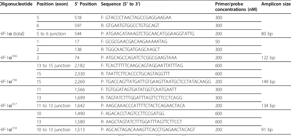
Table 1 Details of oligonucleotide sequences of primers and probes used to quantify hypoxia inducible factor 1a (HIF-1a) mRNA splice variants by real-time quantitative reverse transcription PCR assays
Oligonucleotide Position (exon) 5’Position Sequence (5’to 3’) Primer/probe concentrations (nM)
Amplicon size
5 518 F: GTACCCTAACTAGCCGAGGAAGAA 300
6 597 R: GTGAATGTGGCCTGTGCAGT 300
HIF-1a(total) 5 to 6 junction 544 P: ATGAACATAAAGTCTGCAACATGGAAGGTATTG 200 80 bp
1 17 F: GCGCGAACGACAAGAAAAATAG 50
2 138 R: TGGCAACTGATGAGCAAGCT 300
HIF-1aTAG 2 74 P: ATGCAGCCAGATCTCGGCGAAGTAAA 200 122 bp
13 to 15 junction 2,182 F: TCACTTTTTCAAGCAGTAGGAATTATTTAG 600
15 2,330 R: TAATTCTTCACCCTGCAGTAGGTTT 600
HIF-1a736 15 2,260 P: TGACCAGTTATGATTGTGAAGTTAATGCTCCTATACAAGG 200 149 bp
11 1,566 F: TGTGGATAGTGATATGGTCAATGAATT 300
13 1,699 R: TAGTATCTTTGGATTTAGTTCTTCCTCAGG 300
HIF-1a557 11 to 13 junction 1,642 P: AAGCAAACCCATTTTCTACTCAGAACTACA 200 134 bp
10 1,490 F: AGACACCTAGTCCTTCCGATGG 600
13 1,580 R: AAGCTAGTATCTTTGGATTTAGTTCTTCCT 600
HIF-1a516 10 to 13 junction 1,513 P: AGCACTAGACAAAGTTCACCTGAGAACTACAGT 200 91 bp
were found. All runs exhibited amplification slopes between -3.31 and -3.55, which correspond to efficien-cies higher than 92% and coefficients of correlation between 0.993 and 1. The assays sensitivity was found at 10 copies for all variants. We did not succeed in quanti-fying theHIF-1a417isoform.
Differential expression ofHIF-1asplice variants in normal/benign and malignant breast tissues
Expression levels of HIF-1a mRNAs were determined in each of the 82 samples of breast tissue.HIF-1a splice variants were detected in every sample of normal/benign and malignant breast tissues at varying levels. HIF-1aTAG mRNA levels were higher than the other HIF-1asplice variants in the majority of breast tissues, and low levels were only found forHIF-1a736,HIF-1a516 andHIF-1a557isoforms (Figure 2).HIF-1aTAG mRNAs were expressed at similar levels to the total HIF-1a
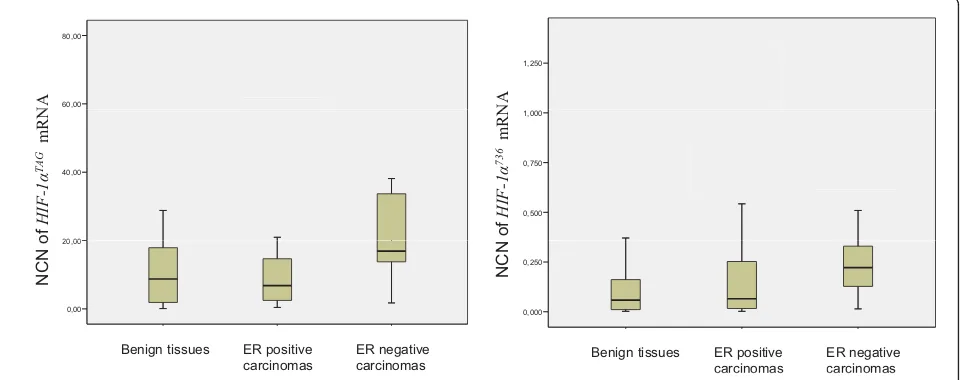
expression suggesting that unknown isoforms devoid of exons 5 and 6 may contain the three base pairs TAG insertion. Splice variants HIF-1a736 and HIF-1a516 mRNAs were expressed 100-fold lower than HIF-1aTAG and variant HIF-1a557 mRNA was expressed at levels 1,000-fold lower than HIF-1aTAG. Expression levels of HIF-1a516 andHIF-1a557 mRNA that were very low (mean normalised copy numbers < 0.1) were not included in statistical analyses. There was no significant statistical difference in the total HIF-1a expression between normal/benign and malignant breast tissues. HIF-1aTAGmRNAs were also expressed at similar level between the two categories of tissues. HIF-1a736 mRNAs showed a link with malignant phenotype but did not reach statistical significance (P= 0.053). Inter-estingly,HIF-1aTAGandHIF-1a736mRNAs were found to be expressed at higher levels in OR-negative carcino-mas compared to normal/benign tissues (P= 0.009 and P = 0.004 respectively) (Figure 3). HIF-1aTAG mRNAs were also higher in OR-negative carcinomas compared to OR-positive ones (P= 0.005).
HIF-1asplice variants expression correlates with
prognostic clinicopathological parameters of breast cancer
Levels of eachHIF-1a variant mRNA were determined in tumour samples from 53 patients with breast cancer and were compared with lymph node status, tumour size, tumour grade, peritumoural vascular invasion, OR and PgR status. They were also compared with micro-vessel density of tumours (Table 3). Lymph node status was significantly associated with HIF-1aTAG mRNA levels (P= 0.037). Statistically significant association was found between tumour grade and totalHIF-1a(P= 0.048) orHIF-1aTAG(P= 0.048). Peritumoural vascular invasion correlated with total HIF-1a (P= 0.028). Interestingly HIF-1aTAGmRNA levels inversely correlated with both OR and PgR status. No significant association was found between any of theHIF-1amRNA levels and tumour size or microvessel density.
Expression ofHIF-1aTAGsplice variant correlates with patient survival
The strength of association between clinicopathological tumour characteristics (lymph node status, tumour size, tumour grade, peritumoural invasion, OR and PgR sta-tus, tumour microvessel density) and expression levels of HIF-1a mRNAs with metastasis-free survival is shown in Table 4. Lymph node status (P= 0.003), OR status (P = 0.021), PgR status (P = 0.005) and HIF-1aTAGmRNA levels (P= 0.01) were found to be signifi-cantly predictive of metastasis free survival. HIF-1aTAG mRNAs were expressed at varying levels. In our series, we used the median value (9.95) of these levels as a cut
Table 2 Distribution of clinical and histopathological characteristics of breast cancer patients
Parameter No. of patients No. of patients with metastasis
Age, years:
≤50 21 8
> 50 30 13
Positive lymph node:
0 25 6
1 to 3 11 9
4 to 10 4 3
> 10 7 3
Pathological tumour size, mm:
≤10 3 1
11 to 20 25 8
21 to 50 23 12
> 50 2 1
Tumour grade:
1 9 1
2 26 13
3 17 8
Histological type:
Tubular 41 18
Lobular 12 4
Peritumoural vascular invasion:
Absent 12 4
Present 40 18
OR status:
OR positive 32 10
OR negative 16 10
PgR status:
PgR positive 26 6
PgR negative 24 16
off to define two groups of patients (the group with mRNA levels higher than 9.95 was considered as a high level of expression and the group with mRNA levels lower than 9.95 was considered as a low level of expres-sion) to evaluate their association with metastasis survi-val. Breast cancer patients with high expression levels of HIF-1aTAG
mRNA had a significantly higher risk of
relapse (Figure 4). Expression levels of the otherHIF-1a transcripts were not associated with prognosis (not shown). Multivariate Cox analysis identified lymph node status (P = 0.005) and PgR status (P = 0.012) as inde-pendent predictors of metastasis free survival. HIF-1aTAG
mRNA levels did not remain a significant independent prognostic variable.
Figure 2Distribution of hypoxia inducible factor 1a(HIF-1a) mRNA levels in 82 breast tissues determined by real-time quantitative reverse transcription PCR assay.HIF-1amRNA levels were expressed in normalised copy numbers (NCN) on the basis of TATA box-binding protein (TPB) gene content of the tissues as described in the Methods section. Patients grouped according to histological type of tissues (x axis).
HIF-1amRNA levels (y axis) were represented according to these different groups of patients. Results were plotted on a logarithmic scale.HIF-1a
splice variants were detectable in all samples at varying levels. Splice variantHIF-1a736andHIF-1a516mRNAs were expressed at levels 100-fold
lower thanHIF-1aTAG. VariantHIF-1a557mRNAs that were expressed at levels 1,000-fold lower thanHIF-1aTAGare not shown.
80,00
60,00
34
1,250
7
26
N
A NA
E1E2/
TB
P
40,00
20 00
57
26 8
E13E15/T
BP
1,000
0,750
0,500
30
39
of
HIF-1α
TAG
mR
N
of
HIF-1α
736
mR
N
Groupe_RE
RE2cat0 Abs RE2cat1 Prés
Bénin 20,00
0,00
Groupe_RE
RE2cat0 Abs RE2cat1 Prés
Bénin 0,250
0,000
NCN
o
NCN
Benign tissues ER positive
carcinomas ER negative carcinomas Benign tissues ER positive carcinomascarcinomas ER negative carcinomascarcinomas
Figure 3Distribution of hypoxia inducible factor 1a(HIF-1a)TAGand HIF-1a736mRNA levels in 82 breast tissues determined by real-time quantitative reverse transcription PCR assay.HIF-1amRNA levels were expressed in normalised copy numbers (NCN) on the basis of TATA box-binding protein (TPB) gene content of the tissues as described in the Methods section. Patients grouped according to histological
type of tissues (x axis).HIF-1aTAGandHIF-1a736mRNAs were expressed at higher levels in oestrogen receptor (OR)-negative carcinomas
compared to normal/benign tissues (P= 0.009 andP= 0.004 respectively).HIF-1aTAGmRNAs were also higher in OR-negative carcinomas
Discussion
Several isoforms of HIF-1a resulting from alternative splicing have been described in various human tumour cell lines or tissues. Only one isoform (HIF-1a736) has been documented in human breast cancer [24]. More-over, only a few studies have been performed to link HIF-1a mRNA expression and tumour-specific para-meters [32,33]. Specific antibodies allowing HIF-1a splice variant detection at the protein level are not cur-rently available. We used a real-time quantitative reverse transcription PCR assay to measure mRNA expression levels of 4 alternatively spliced transcripts of HIF-1a in breast specimens of 53 primary cancers and 29 normal tissues or benign lesions.
Interestingly, this work shows for the first time higher expression levels of two HIF-1a splice variants ( HIF-1aTAGandHIF-1a736) in OR-negative carcinomas com-pared to normal/benign tissues. We also investigated the prognostic value ofHIF-1a transcript expression levels in breast cancer and found a significant relationship between either clinicopathological characteristics or patient metastasis-free survival. First, we found that HIF-1aTAG
mRNAs levels were substantially higher in high grade and steroid hormone receptor-negative tumours. The second and most striking observation was
that HIF-1aTAGmRNA levels were indicative of shorter metastasis-free survival, and that this correlated with lymph node status. Contrary to the Cayre et al. report [24], we did not find significant correlation between total HIF-1amRNA expression and lymph node status but we observed significant association with tumour grade. This could be because our series was smaller than series of Cayre et al. or because the technology used in our study was more sensitive. In our series, total HIF-1amRNA expression negatively correlated with OR status. Similar to Cayreet al. we did not find any corre-lation between totalHIF-1amRNA expression and out-come. Our results showing that HIF-1a736 mRNA expression does not correlate with clinicopathological characteristics of tumours also concur with earlier find-ings of the same group [24]. In this study, effect of adju-vant (none, chemotherapy or hormone) treatment on survival and interactions with expression levels of HIF-1a splice variants were not checked because of the lim-ited number of patients in each subgroup. Further experiments in a larger series are required to answer this question.
Alternative splicing is known to play an important role in gene expression regulation by modulating the func-tional properties of transcription factors [34]. In this
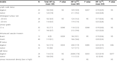
Table 3 Relationship between the hypoxia inducible factor 1a(HIF-1a) splice variant’s expression levels (normalised copy numbers) and clinicopathological factors or microvessel density in tumour tissue specimens from 53 breast cancer patients
Variable N TotalHIF-1a,
mean (SD) P
value HIF-1aTAG, mean (SD) P
value HIF-1a736, mean (SD) P
value
Lymph node status:
Negative 25 10.0 (9.0) NS 10.9 (10.9) 0.037 0.19 (0.29) NS
Positive 22 12.8 (7.8) 21.1 (19.2) 0.26 (0.28)
Pathological tumour size:
< 20 mm 28 10.2 (8.3) NS 12.4 (13.2) NS 0.17 (0.26) NS
≥20 mm 25 11.8 (8.3) 17.9 (17.4) 0.25 (0.28)
Tumour grade:
1/2 vs 35 9.2 (7.7) 0.048 11.8 (12.3) 0.048 0.19 (0.30) NS
3 17 14.6 (8.7) 21.5 (19.4) 0.23 (0.20)
Peritumoural vascular invasion:
Absent 12 8 (9) 0.028 9.6 (10.1) NS 0.19 (0.36) NS
Present 40 11.9 (8.1) 16.6 (16.6) 0.20 (0.24)
OR status:
Negative 16 14.2 (7.3) 0.024 20.8 (11.9) 0.005 0.25 (0.19) 0.06
Positive 32 10 (8.8) 12.7 (17.1) 0.20 (0.31)
PgR status:
Negative 24 11.8 (7.3) NS 20.08 (17.8) 0.033 0.23 (0.19) 0.077
Positive 26 10.8 (9.4) 11.3 (12.3) 0.2 (0.34)
Tumour microvessel density (low vs high) NS NS NS
Because of missing data, the numbers do not always add up to 53. Expression levels ofHIF-1a516andHIF-1a557mRNA were not included in statistical analyses because of their very low levels (mean normalised copy numbers < 0.1).
regard, alternative splicing can modify DNA-binding properties of transcription factors [35], introduce or eliminate activating domains or increase the in vivo sta-bility of a given isoform [36]. Moreover, the abundance of specific isoforms is likely to result from differential expression, RNA stability and selective splicing process leading to an increase of some mRNA species. Recent evidence indicates that in several cancers the ratio of splice variants is dramatically altered and that differen-tial expression of alternatively spliced isoforms in cancer patients can have severe implications for clinical out-come [37]. Remarkably, statistical association has pre-viously been reported betweenHIF-1b splicing variant expression, oestrogen receptors and breast cancer survi-val [38]. It should be noted that the primers designed on exons 1 and 2 in our study allowed quantification of the HIF-1aTAG sequence present in bothHIF-1a827and HIF-1a736splice variants [17]. The HIF-1aTAGtranscript is characterised by insertion of a three base pairs TAG
insertion that may be generated by the use of two potential splice acceptor dinucleotides (AG) of intron 1 at a splice junction site as previously described [39,1]. This splicing results in the replacement of Lys12 by Asn12 and the addition of Arg13 residue located upstream from the bHLH domain of the protein. This replacement may modify the DNA binding affinity of the protein complex as previously shown for Arg14and Arg15 residues in aryl hydrocarbon receptor (AHR)/aryl hydrocarbon receptor nuclear translocator (ARNT) het-erodimer [40]. Further biochemical and structural stu-dies are required for a better understanding of the functional properties of this variant.
Conclusions
To our knowledge, our results suggest for the first time that at least one HIF-1aspliceavariant may be a mar-ker for the advanced clinical and oestrogen-resistant
Table 4 Univariate and multivariate (backward conditional selection) Cox regression analysis of metastasis-free survival (MFS) in breast cancer patients considering clinicopathological characteristics, tumour microvessel density and hypoxia inducible factor 1a(HIF-1a)TAGmRNA levels
Characteristics Univariate analysis of MFS, Exp B (95% CI) P
value Multivariate analysis of MFS,
Exp B (95% CI) P value
Age, years:
< 50 1 0.561
≥50 1.32 (0.52 to 3.37)
Positive lymph node:
0 1 0.003 1 0.005
1 to 3 8.00 (2.56 to 24.81) 5.56 (1.75 to 17.63)
4 to 9 7.50 (1.70 to 30.00) 6.68 (1.48 to 30.14)
> 10 1.20 (0.14 to 10.00) 0.48 (0.05 to 4.25)
Pathological tumour size, mm:
< 10 1 0.39
10 to 20 1.10 (0.10 to 8.70)
21 to 49 2.40 (0.30 to 18.90)
> 50 2.70 (0.20 to 42.90)
Tumour grade:
1/2 vs 1 0.24
3 1.70 (0.70 to 4.40)
Peritumoural vascular invasion:
Absent 1 0.104
Present 3.40 (0.80 to 14.80)
OR status: Positive
Negative 0.33 (0.13 to 0.85) 0.021
PgR status: Positive
Negative 0.20 (0.07 to 0.60) 0.005 0.23 (0.07 to 0.72) 0.012
Tumour microvessel density 1.02 (0.07 to 0.60) 0.22
HIF-1aTAGmRNA levels 1.03 (1.00 to 1.05) 0.01
stage of breast cancer. Based on their correlation with survival, HIF-1aTAG mRNA levels may be a potential useful prognostic indicator whose value should be further validated in prospective studies.
Acknowledgements
This work was supported by grants from Institut National du Cancer (ACI2004, PL2006). We are grateful to Jacques Pouyssegur for his generous gifts of plasmids and discussion. Claudine Andonian from the department of Pathology is also acknowledged for her excellent work with collecting and archiving the breast tumour samples.
Author details
1Plateforme Transcriptome, CRO2, Marseille, France.2Department of
Pathology, Hôpital Nord, Marseille, France.3Biochemistry and Molecular
Biology, Hôpital Nord, Marseille, France.4Etablissement Français du Sang
Alpes Méditerranée, Marseille, France.5Department of Medical Information,
Hôpital Sainte Marguerite, Marseille, France.6Department of Gynaecologic
Oncology, Hôpital de la Conception, Assistance-Publique Hôpitaux de Marseille, Université de la Méditerranée, Marseille, France.
Authors’contributions
NB, MS and CP carried out the molecular genetic studies, participated in the sequence alignment and drafted the manuscript. CF participated in the sequence alignment. CC, SG analysed the immunohistochemistry results. VP and VP performed the statistical analysis. J-PD and JG conceived of the study, and participated in its design and coordination. All authors read and approved the final manuscript.
Competing interests
The authors declare that they have no competing interests.
Received: 26 May 2010 Accepted: 12 July 2010 Published: 12 July 2010
References
1. Iyer NV, Leung SW, Semenza GL:The human hypoxia-inducible factor 1a
gene: HIF1A structure and evolutionary conservation.Genomics1998, 52:159-165.
2. Wang GL, Jiang BH, Rue EA, Semenza GL:Hypoxia-inducible factor 1 is a
basic-helix-loop-helix-PAS heterodimer regulated by cellular O2 tension.
Proc Natl Acad Sci USA1995,92:5510-5514.
3. Pouysségur J, Dayan F, Mazure NM:Hypoxia signalling in cancer and
approaches to enforce tumour regression.Nature2006,441:437-443.
4. Ryan HE, Poloni M, McNulty W, Elson D, Gassmann M, Arbeit JM,
Johnson RS:Hypoxia-inducible factor-1ais a positive factor in solid tumor growth.Cancer Res2000,60:4010-4015.
5. Wang GL, Semenza GL:Purification and characterization of
hypoxia-inducible factor 1.J Biol Chem1995,270:1230-1237.
6. Jiang BH, Semenza GL, Bauer C, Marti HH:Hypoxia-inducible factor 1
levels vary exponentially over a physiologically relevant range of O2 tension.Am J Physiol1996,271:C1172-1180.
7. Huang LE, Arany Z, Livingston DM, Bunn HF:Activation of
hypoxia-inducible transcription factor depends primarily upon redox-sensitive stabilization of itsasubunit.J Biol Chem1996,271:32253-32259.
8. Semenza GL:HIF-1 and tumor progression: pathophysiology and
therapeutics.Trends Mol Med2002,8:S62-67.
9. Zhong H, De Marzo AM, Laughner E, Lim M, Hilton DA, Zagzag D,
Buechler P, Isaacs WB, Semenza GL, Simons JW:Overexpression of
hypoxia-inducible factor 1ain common human cancers and their metastases.Cancer Res1999,59:5830-5835.
10. Dales JP, Garcia S, Meunier-Carpentier S, Andrac-Meyer L, Haddad O, Lavaut MN, Allasia C, Bonnier P, Charpin C:Overexpression of hypoxia-inducible factor HIF-1apredicts early relapse in breast cancer: retrospective study in a series of 745 patients.Int J Cancer2005, 116:734-739.
11. Schindl M, Schoppmann SF, Samonigg H, Hausmaninger H, Kwasny W,
Gnant M, Jakesz R, Kubista E, Birner P, Oberhuber G:Overexpression of hypoxia-inducible factor 1ais associated with an unfavorable prognosis in lymph node-positive breast cancer.Clin Cancer Res2002,8:1831-1837.
12. Huang LE, Gu J, Schau M, Bunn HF:Regulation of hypoxia-inducible factor
1ais mediated by an O2-dependent degradation domain via the ubiquitin-proteasome pathway.Proc Natl Acad Sci USA1998,95:7987-7992.
13. Jiang BH, Rue E, Wang GL, Roe R, Semenza GL:Dimerization, DNA binding,
and transactivation properties of hypoxia-inducible factor 1.J Biol Chem
1996,271:17771-17778.
14. Jiang BH, Zheng JZ, Leung SW, Roe R, Semenza GL:Transactivation and
inhibitory domains of hypoxia-inducible factor 1a. Modulation of transcriptional activity by oxygen tension.J Biol Chem1997, 272:19253-19260.
15. Pagé EL, Robitaille GA, Pouysségur J, Richard DE:Induction of hypoxia-inducible factor-1aby transcriptional and translational mechanisms.J Biol Chem2002,277:48403-48409.
16. Semenza GL, Rue EA, Iyer NV, Pang MG, Kearns WG:Assignment of the
hypoxia-inducible factor 1agene to a region of conserved synteny on mouse chromosome 12 and human chromosome 14q.Genomics1996, 34:437-439.
17. Gothié E, Richard DE, Berra E, Pagès G, Pouysségur J:Identification of alternative spliced variants of human hypoxia-inducible factor-1a.J Biol Chem2000,275:6922-6927.
18. Chun YS, Choi E, Yeo EJ, Lee JH, Kim MS, Park JW:A new HIF-1avariant
induced by zinc ion suppresses HIF-1-mediated hypoxic responses.J Cell Sci2001,114:4051-4061.
19. Chun YS, Choi E, Kim TY, Kim MS, Park JW:A dominant-negative isoform
lacking exons 11 and 12 of the human hypoxia-inducible factor-1a gene.Biochem J2002,362:71-79.
20. Chun YS, Lee KH, Choi E, Bae SY, Yeo EJ, Huang LE, Kim MS, Park JW:
Phorbol ester stimulates the nonhypoxic induction of a novel hypoxia-inducible factor 1aisoform: implications for tumor promotion.Cancer Res2003,63:8700-8707.
21. Lee KH, Park JW, Chun YS:Non-hypoxic transcriptional activation of the
aryl hydrocarbon receptor nuclear translocator in concert with a novel hypoxia-inducible factor-1aisoform.Nucleic Acids Res2004,32:5499-5511.
22. Depping R, Hägele S, Wagner KF, Wiesner RJ, Camenisch G, Wenger RH,
Katschinski DM:A dominant-negative isoform of hypoxia-inducible
factor-1aspecifically expressed in human testis.Biol Reprod2004, 71:331-339.
23. Lukashev D, Sitkovsky M:Preferential expression of the novel alternative
isoform I.3 of hypoxia-inducible factor 1ain activated human T lymphocytes.Hum Immunol2008,69:421-425.
24. Cayre A, Rossignol F, Clottes E, Penault-Llorca F:aHIF but not HIF-1a transcript is a poor prognostic marker in human breast cancer.Breast Cancer Res2003,5:R223-230.
25. Bloom HJ, Richardson WW:Histological grading and prognosis in breast
cancer; a study of 1409 cases of which 359 have been followed for 15 years.Br J Cancer1957,11:359-377.
26. Elston CW, Ellis IO:Pathological prognostic factors in breast cancer. I. The value of histological grade in breast cancer: experience from a large study with long-term follow-up.Histopathology1991,19:403-410.
27. Charpin C, Martin PM, De Victor B, Lavaut MN, Habib MC, Andrac L, Toga M:
Multiparametric study (SAMBA 200) of estrogen receptor
immunocytochemical assay in 400 human breast carcinomas: analysis of estrogen receptor distribution heterogeneity in tissues and correlations with dextran coated charcoal assays and morphological data.Cancer Res
1988,48:1578-1586.
28. Charpin C, Jacquemier J, Andrac L, Vacheret H, Habib MC, Devictor B,
Lavaut MN, Toga M:Multiparametric analysis (SAMBA 200) of the
progesterone receptor immunocytochemical assay in nonmalignant and malignant breast disorders.Am J Pathol1988,132:199-211.
29. Dales JP, Garcia S, Andrac L, Carpentier S, Ramuz O, Lavaut MN, Allasia C,
Bonnier P, Charpin C:Prognostic significance of angiogenesis evaluated
by CD105 expression compared to CD31 in 905 breast carcinomas:
correlation with long-term patient outcome.Int J Oncol2004, 24:1197-1204.
30. Gabert J, Beillard E, van der Velden VH, Bi W, Grimwade D, Pallisgaard N, Barbany G, Cazzaniga G, Cayuela JM, Cavé H, Pane F, Aerts JL, De Micheli D, Thirion X, Pradel V, González M, Viehmann S, Malec M, Saglio G, van Dongen JJ:Standardization and quality control studies of‘real-time’ quantitative reverse transcriptase polymerase chain reaction of fusion gene transcripts for residual disease detection in leukemia - a Europe Against Cancer program.Leukemia2003,17:2318-2357.
31. Bièche I, Laurendeau I, Tozlu S, Olivi M, Vidaud D, Lidereau R, Vidaud M: Quantitation of MYC gene expression in sporadic breast tumors with a real-time reverse transcription-PCR assay.Cancer Res1999,59:2759-2765.
32. Nakayama K, Kanzaki A, Hata K, Katabuchi H, Okamura H, Miyazaki K,
Fukumoto M, Takebayashi Y:Hypoxia-inducible factor 1a(HIF-1a) gene
expression in human ovarian carcinoma.Cancer Lett2002,176:215-223.
33. Søndergaard KL, Hilton DA, Penney M, Ollerenshaw M, Demaine AG:
Expression of hypoxia-inducible factor 1ain tumors of patients with glioblastoma.Neuropathol Appl Neurobiol2002,28:210-217.
34. López AJ:Developmental role of transcription factor isoforms generated
by alternative splicing.Dev Biol1995,172:396-411.
35. Kozmik Z, Czerny T, Busslinger M:Alternatively spliced insertions in the paired domain restrict the DNA sequence specificity of Pax6 and Pax8.
EMBO J1997,16:6793-6803.
36. Foulkes NS, Mellström B, Benusiglio E, Sassone-Corsi P:Developmental
switch of CREM function during spermatogenesis: from antagonist to activator.Nature1992,355:80-84.
37. Venables JP:Unbalanced alternative splicing and its significance in
cancer.Bioessays2006,28:378-386.
38. Qin C, Wilson C, Blancher C, Taylor M, Safe S, Harris AL:Association of ARNT splice variants with estrogen receptor-negative breast cancer, poor induction of vascular endothelial growth factor under hypoxia, and poor prognosis.Clin Cancer Res2001,7:818-823.
39. Wenger RH, Rolfs A, Spielmann P, Zimmermann DR, Gassmann M:Mouse
hypoxia-inducible factor-1ais encoded by two different mRNA isoforms: expression from a tissue-specific and a housekeeping-type promoter.
Blood1998,91:3471-3480.
40. Wache SC, Hoagland EM, Zeigler G, Swanson HI:Role of arginine residues
14 and 15 in dictating DNA binding stability and transactivation of the aryl hydrocarbon receptor/aryl hydrocarbon receptor nuclear translocator heterodimer.Gene Expr2005,12:231-243.
Pre-publication history
The pre-publication history for this paper can be accessed here: http://www.biomedcentral.com/1741-7015/8/44/prepub
doi:10.1186/1741-7015-8-44
Cite this article as:Daleset al.:Hypoxia inducible factor 1agene (
HIF-1a) splice variants: potential prognostic biomarkers in breast cancer.
BMC Medicine20108:44.
Submit your next manuscript to BioMed Central and take full advantage of:
• Convenient online submission
• Thorough peer review
• No space constraints or color figure charges
• Immediate publication on acceptance
• Inclusion in PubMed, CAS, Scopus and Google Scholar • Research which is freely available for redistribution