R E V I E W
Open Access
The expansion of targetable biomarkers for
CAR T cell therapy
Michelle H. Townsend
1, Gajendra Shrestha
1,2, Richard A. Robison
1and Kim L. O
’
Neill
1*Abstract
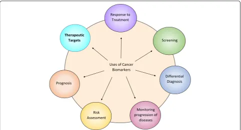
Biomarkers are an integral part of cancer management due to their use in risk assessment, screening, differential
diagnosis, prognosis, prediction of response to treatment, and monitoring progress of disease. Recently, with the
advent of Chimeric Antigen Receptor (CAR) T cell therapy, a new category of targetable biomarkers has emerged.
These biomarkers are associated with the surface of malignant cells and serve as targets for directing cytotoxic T cells.
The first biomarker target used for CAR T cell therapy was CD19, a B cell marker expressed highly on malignant B cells.
With the success of CD19, the last decade has shown an explosion of new targetable biomarkers on a range of human
malignancies. These surface targets have made it possible to provide directed, specific therapy that reduces healthy
tissue destruction and preserves the patient
’
s immune system during treatment. As of May 2018, there are over 100
clinical trials underway that target over 25 different surface biomarkers in almost every human tissue. This expansion
has led to not only promising results in terms of patient outcome, but has also led to an exponential growth in the
investigation of new biomarkers that could potentially be utilized in CAR T cell therapy for treating patients. In this
review, we discuss the biomarkers currently under investigation and point out several promising biomarkers in the
preclinical stage of development that may be useful as targets.
Keywords:
CAR T cell therapy, Biomarkers, Hematological malignancies, Solid tumors, Targets, Tumor associated
antigens (TAA), Tumor specific antigens (TSA), Combination therapy
Background
As the new paradigm shift in cancer treatment,
immunother-apy is the epitome of personalized medicine, as a patient’
s
immune system is enlisted to fight their own cancer.
Origin-ally manifest as monoclonal antibody therapy,
immunother-apy now has a broadened definition that encompasses tumor
vaccines, checkpoint blockades, bispecific antibodies, tumor
infiltrating lymphocytes (TILs), and most recently, chimeric
antigen receptor (CAR) T cell therapy. T cells are a critical
component of the adaptive immune system as they not only
orchestrate cytotoxic effects, but also provide long term
cel-lular
‘memory’
of specific antigens [
1
]. Commonly, a patient
will have TILs specific for their tumor but these cells are
often retrained by the tumor microenvironment to become
anergic and nonfunctional [
2
]. T cells endogenously require
the interaction between MHC displayed peptides and their
TCR to activate [
3
], but CAR T cells have been engineered
to activate via a tumor-associated or tumor-specific antigen
(TAA and TSA, respectively). CAR T cells are a
“living
drug”
comprised of a targeting domain (single chain
vari-able fragment (scFv), peptides, polypeptides, ligands,
muteins, etc.) fused to the signaling domain of a T cell
[
4
,
5
]. Upon recognition and binding to the scFv target, the
T cell activates and subsequent target cell killing is initiated.
CAR T cell therapy has been revolutionary in the treatment
of hematological malignancies with the targets CD19 and
CD20 but has been unable to translate effectively to solid
tumors. A major drawback for CAR therapy in solid
malig-nancies is the lack of cancer-specific tumor targets. While
hematological malignancies do not necessarily require
complete antigen target specificity towards cancer cells,
solid tumor targets are more delicate and targets ideally
cannot be expressed on normal tissue. With the struggles
facing CAR T cell therapy (on-target off-tumor cytotoxicity,
persistence in vivo, immunosuppressive tumor
microenvir-onment, cytokine release syndrome, etc.), biomarker
dis-covery and specificity is essential for further CAR T cell
development and success.
* Correspondence:kim_oneill@byu.edu
1Department of Microbiology and Molecular Biology, Brigham Young University, 3142 LSB, Provo, UT 84602, USA
Full list of author information is available at the end of the article
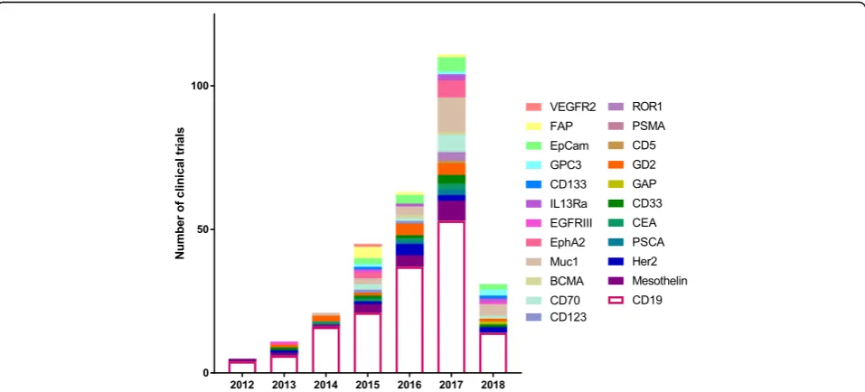
With over 300 CAR T cell therapy clinical trials ongoing
in CAR therapy as of May 2018, there has been an equally
impressive effort to identify and characterize TAA or TSA
surface biomarkers in solid tumors. Biomarkers have been
an integral component of cancer for several decades, and
with the expansion of CAR T cell therapy, a new category
of therapeutic biomarkers has arisen. These markers can be
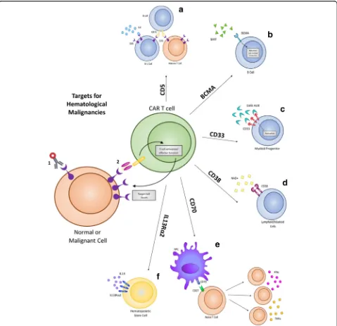
used to direct CAR T cells to malignant target cells (Fig.
1
).
The effort to identify and characterize these therapeutic
biomarkers has been substantial and has increased
expo-nentially over the last decade. As a result, 18 surface
bio-markers are currently being evaluated in clinical trials
(Fig.
2
). In addition, there is also a significant number of
pre-clinical biomarkers that have shown promise as targets
for CAR therapy due to their unique expression on cancer
cells. Here, we summarize the biomarkers currently under
investigation in clinical trials for both hematological and
solid malignancies, along with those that may prove useful
in future CAR therapies for solid tumors.
Surface biomarkers have expanded significantly
over the last decade
CAR T cell therapy was initially conceptualized in 1989 [
6
]
and was recognized as an effective therapeutic after
target-ing CD19 for the treatment of lymphomas and leukemias
[
7
–
9
]. This led to an exponential growth in CAR therapy
and as a direct consequence, in surface biomarker discovery
(Fig.
3
). In 2012, there were a total of 5 clinical trials, four
targeting CD19 and one targeting Mesothelin. This number
has continued to grow and the number of biomarkers
tested in a clinical setting has also expanded from 2 to 25.
The year 2017 saw more clinical trials than any previous
year with 111 initiated, targeting 17 different biomarkers
(Table
1
). This growth demonstrates not only the efficacy
of CAR T cell therapy, but also the huge push in
immuno-therapy to find new and better targets.
Current clinical targets for hematological
malignancies
As the most studied and researched target for CAR therapy,
CD19 has shown impressive success in clinical settings to
treat Acute Lymphoblastic Leukemia (ALL), Non-Hodgkin
Lymphoma (NHL), and Chronic Lymphocytic Leukemia
(CLL) [
10
]. Despite the high levels of complete response
rates in patients, relapse from CD19 CAR therapy can
occur via a suppressive tumor microenvironment or
antigen escape [
11
–
13
]. With this in mind, new targets are
being identified and evaluated to treat hematological
malig-nancies. Among these new targets are CD5, CD123, CD33,
CD70, CD38, and BCMA. These same targets have already
shown promise using drug-conjugated antibodies, and
several have been FDA approved for treatment (Figs.
1
,
2
,
3
and
4
). These biomarkers are now being evaluated as
tar-gets for adoptive T cell CAR therapy to treat hematological
malignancies.
CD5
CD5 is a negative regulator of TCR signaling and is
expressed on the surface of most T cells and on a
spe-cific subpopulation of B cells (B-1) found most
com-monly in fetal cells [
14
] (Fig.
4a
). CD5 has high
expression in approximately 80% of T-cell acute
lympho-blastic leukemia (T-ALL) and T cell lymphomas along
and also has significant expression on B-cell lymphomas
[
15
]. CD5 was first utilized as an immunotherapy
treat-ment via immunotoxin-conjugated antibodies [
16
–
22
]
Fig. 2Current CAR T cells in clinical trials. From the initial success of CD-19 CAR T cell therapy, several new biomarker targets have emerged and are being tested in clinical trials. This expansion of targets has expanded CAR T cell therapy to the treatment of not just hematological malignancies, but also to solid tumors as wellTable 1
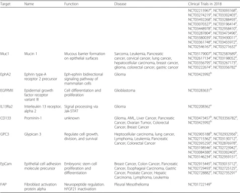
Current Clinical Trials (as of April 2018)
Target Name Function Disease Clinical Trials in 2018
CD19 Cluster of Differentiation 19
Dominant signaling component on mature B cells
ALL, B cell lymphoma, leukemia, Non-Hodgkin lymphoma,
NCT03366350b, NCT03366324b, NCT02546739b, NCT03448393b, NCT03467256b, NCT03488160b, NCT03016377b, NCT03468153b, NCT03483688b, NCT03398967b, NCT03229876b, NCT03455972b, NCT03423706b, NCT03497533b
Mesothelin exact function of
mesothelin in these normal mesothelial cells is unclear.
Pancreatic cancer, Cervical Cancer, Ovarian Cancer, Lung Cancer, Peritoneal carcinoma, Fallopian tube cancer, Colorectal Cancer, Breast Cancer
NCT02930993a, NCT03182803a, NCT03030001a, NCT02706782a, NCT01583686a, NCT03356795a, NCT03054298a, NCT03267173a, NCT02792114a, NCT02959151a, NCT02580747a, NCT02414269a, NCT02465983a, NCT03323944a,
Her2 Human Epidermal Growth Factor Receptor 2
Activate intracellular signaling pathways in response to extracellular signals.
CNS tumor, Breast Cancer, Ovarian Cancer, Lung Cancer, Gastric Cancer, Colorectal Cancer, Glioma, Pancreatic Cancer, Glioblastoma
NCT03500991b, NCT03423992b, NCT02713984a, NCT03267173a, NCT02792114a, NCT02442297a, NCT00889954a, NCT03423992a, NCT01109095a, NCT02706392a, NCT00902044a, NCT03389230a, NCT01818323a
PSCA Prostate Stem Cell Antigen
Not well understood Pancreatic cancer, lung cancer CT03198052a, NCT02744287a, NCT03267173a
CEA Carcinoembryonic antigen
Cell adhesion Liver metastases, lung cancer, colorectal cancer, gastric cancer, breast cancer, pancreatic cancer,
NCT02850536a, NCT02349724a, NCT03267173a, NCT02959151, NCT01212887a
CD33 Siglec-3 Transmembrane
receptor on myeloid lineage
Myeloid leukemia, NCT03473457b, NCT02958397a, NCT03126864a, NCT03222674a,
GAP GTPase-activating protein
Terminating G protein signaling
Solid tumors NCT02932956b
GD2 Ganglioside G2 Glioma, Cervical cancer, sarcoma,
neuroblastoma,
NCT03423992b, NCT03356795a, NCT02992210a, NCT01953900a, NCT02761915, NCT03373097a, NCT02765243a, NCT03423992a, NCT03294954a, NCT03356782a, NCT02919046a,
CD5 Cluster of
differentiation 5
TCR inhibitory molecule T cell acute lymphoblastic lymphoma, T-non-Hodgkin lymphoma,
NCT03081910a,
PSMA (PSMA/TGFβ)
Prostate specific membrane antigen
Transmembrane protein Cervical cancer, Prostate cancer, Bladder cancer
NCT03356795a, NCT03089203a (-TGFβ), NCT03185468a, NCT01140373a
ROR1 Receptor Tyrosine Kinase like Orphan Receptor 1
Modulates neurite growth in the CNS
Breast cancer, lung cancer, lymphoblastic leukemia,
NCT02706392a,
CD123 IL-3RA Involved in hematopoietic
progenitor cell differentiation and proliferation
AML, Leukemia, NCT03473457b, NCT03125577a,
NCT02937103a, NCT03114670a, NCT02159495a, NCT03098355a, NCT03222674a, NCT03203369a, NCT03190278a,
CD70 Cluster of differentiation 70
Induces proliferation of costimulated T cells
B cell malignancies, pancreatic cancer, renal cell cancer, breast cancer, melanoma, ovarian cancer
NCT03125577a, NCT02830724a,
CD38 Cluster of differentiation 38
Cell adhesion, signal transduction, and calcium signaling
Myeloma, NCT03464916b, NCT03473496b,
NCT03473457b, NCT03125577a, NCT03222674a, NCT03271632a,
BCMA B cell maturation antigen
Mediates the survival of plasma cells
Myeloma NCT03448978b, NCT03473496b,
that aided in the depletion of malignant T cell populations
in treated patients. More recently, CD5 has been utilized
as a CAR target to treat T cell malignancies directly. As
CD5 is not cancer specific, this treatment results in T cell
aplasia [
23
,
24
]. While this therapy is effective in
eliminat-ing malignant T cells, sustained T cell aplasia is a
poten-tially undesirable outcome of treatment.
IL3R
α
Interleukin-3 receptor alpha chain (IL3R
α
or CD123) is
a surface receptor found overexpressed in several
hematological malignancies including blastic
plasmacy-toid dendritic cell neoplasm (BPDCN) [
25
], hairy cell
leukemia [
26
,
27
], B-cell acute lymphocytic leukemia
(B-ALL) [
26
,
28
], and Acute myeloblastic leukemia
(AML) [
29
,
30
]. As the receptor expression is limited on
hematopoietic stem cells, the receptor has promising use
as a targetable biomarker for CAR therapy [
30
,
31
]
(Fig.
4f
). Initial targeting of IL3R
α
was conducted
utiliz-ing the natural ligand, IL-3, but CAR T cell approaches
are now being utilized to further target this receptor to
treat primarily AML patients. Initial trials with CD123
CAR cells showed potent cytotoxicity against AML cells
within mice [
32
–
35
] and in human patients [
36
]. This
preliminary success has led to its further testing in
clinical trials, evaluating this therapy for both safety and
efficacy against AML. IL3R
α
, like CD5, is not cancer
specific, and the consequence of CD5 CAR T cells is
severe myeloablation [
37
,
38
].
CD33
CD33 is a transmembrane receptor that binds sialic acid
and causes inhibition of activation. The protein is
expressed on AML blasts and normal myeloid
progeni-tors [
39
–
43
] (Fig.
4c
). Because CD33 is absent in adult
pluripotent hematopoietic stem cells and has elevated
Table 1
Current Clinical Trials (as of April 2018)
(Continued)
Target Name Function Disease Clinical Trials in 2018
NCT02215967a, NCT03093168a, NCT03274219a, NCT03302403a, NCT03492268a, NCT03288493a, NCT03070327a, NCT03196414a, NCT03448978a, NCT02958410a, NCT03287804a, NCT03473496a, NCT03380039a, NCT03430011a, NCT03361748a, NCT03455972a, NCT02546167a, NCT03271632a
Muc1 Mucin 1 Mucous barrier formation
on epithelial surfaces
Sarcoma, Leukemia, Pancreatic cancer, cervical cancer, lung cancer, hepatocellular carcinoma, breast cancer, glioma, colorectal cancer, gastric cancer
NCT03179007a, NCT02587689a, NCT02617134a, NCT03198052a, NCT03356795a, NCT03267173a, NCT03222674a, NCT03356782a
EphA2 Ephrin type-A receptor 2 precursor
Eph-ephrin bidirectional signaling pathway of mammalian cells
Glioma NCT03423992b
EGFRVIII Epidermal growth factor receptor variant III
Cell differentiation and proliferation
Glioblastoma NCT03283631b
IL13Ra2 Interleukin 13 receptor, alpha 2
Signal processing via Jak-STAT
Glioma NCT02208362a
CD133 Prominin-1 unknown Glioma, AML, Liver Cancer, Pancreatic
Cancer, Ovarian Tumor, Colorectal Cancer, Breast Cancer
NCT03473457b, NCT03356782a, NCT03423992b
GPC3 Glypican 3 Regulate cell growth,
division, and survival
Heptocellular carcinoma, lung cancer, Lymphoma, Leukemia, Pancreatic Cancer, Colorectal Cancer
NCT02905188b, NCT02932956b, NCT02715362a, NCT03130712a, NCT02395250a, NCT02876978a, NCT03198546a, NCT02723942a, NCT03084380a, NCT03302403a, NCT03146234a, NCT02959151a,
EpCam Epithelial cell adhesion molecule precursor
Embryonic stem cell proliferation and differentiation
Breast Cancer, Colon Cancer, Pancreatic Cancer, Esophageal Carcinoma, Gastric Cancer, Prostate Cancer, Hepatic Carcinoma, Lymphoma, Leukemia
NCT02915445a, NCT03013712a, NCT02729493a, NCT02725125a, NCT02728882a, NCT02735291a
FAP Fibroblast activation protein alpha
Neuropeptide regulation. hFGF21 inactivation
Pleural Mesothelioma NCT01722149a
Note.a
; indicate trials ongoing/active,b
expression on approximately 85–90% of AML patients,
the antigen has gained clinical significance as a TAA
[
44
–
46
]. In initial trials testing the efficacy of CD33
CAR T cells, patients showed signs of an inflammatory
reaction in response to infused CAR T cells: chills, fever,
and elevated cytokine levels. This resulted in reduced
blasts within the bone marrow following two weeks of
therapy [
47
]. Following these preliminary tests, clinical
trials are ongoing to determine if CD33 is a safe and
ef-fective treatment for myeloid leukemia.
CD70
CD70 is a target that is being utilized to treat both
hematological malignancies as well as solid tumors (Table
[
51
,
62
–
64
], ovarian cancer [
65
–
67
], and pancreatic cancer
[
65
,
68
]. Targeting this antigen is feasible as CD70/CD27
signaling is not essential for the development of a
func-tional immune system as CD27
−/−mice recover from
infec-tion in a similar time frame as CD27
WTmice [
69
,
70
].
Targeting was first performed using monoclonal antibodies
against CD70, and this showed promise in animal models
[
51
,
71
,
72
]. CD70 CAR T cells contain the human CD27,
the natural binding partner of CD70, fused to the CAR
signaling domain [
48
].
CD38
CD38 is a glycoprotein associated within lipid rafts and
is specific to cell surface receptors that function to
regu-late calcium flux and mediate signal transduction in both
lymphoid and myeloid cells [
73
–
75
]. While CD38 is
expressed consistently on myeloma cells [
73
,
76
], it’s
ex-pression is limited on normal lymphoid and myeloid
cells [
77
] (Fig.
4d
). As a TAA, CD38 has been used as a
target via monoclonal antibody treatment
(Daratumu-mab) [
73
], which was approved by the FDA in 2015 for
patients with multiple myeloma [
78
]. Daratumumab
showed an overall response rate of 31%, which
demon-strates the success of utilizing CD38 as a target. CD38
CAR T cells have shown similar efficacy against
double-hit lymphoma cells (MYC rearrangement along
with BCL2 or BCL6 rearrangement) [
79
]. With
promis-ing data, CD38 CAR T cells are currently in phase I
trials against myeloma to test safety and dosing.
BCMA
B cell maturation antigen (BCMA) is a TNF receptor
that binds B-cell activating factor (BAFF) and is
univer-sally expressed on myeloma cells but has insignificant
expression on major adult organs [
80
] (Fig.
4b
). BCMA
is exclusively expressed in B-cell lineage cells, and is
expressed during plasma cell differentiation [
81
]. In
pre-clinical models, anti-BCMA CAR T cells have shown
ef-fective killing of myeloma cells both in vitro and in vivo
[
82
,
83
]. Following Phase I safety studies, some patients
experienced neurotoxicity and cytokine release
syn-drome, which are common side effects of CAR T cell
treatment [
84
]. Other side effects of targeting BCMA are
similar to those of other hematological malignancies, as
patients suffer from partial or complete B cell aplasia.
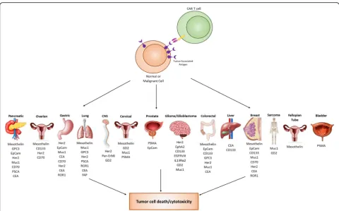
Current clinical targets for solid tumors
While CAR T cell therapy has been very successful against
hematological malignancies, it has been challenging to
apply this technology to solid tumors. This challenge has
resulted in a strong effort to discover biomarkers for solid
malignancies. As such, there are 17 biomarkers currently
in clinical trials for solid tumors (Fig.
5
).
Mesothelin
Mesothelin (MSLN), the second most frequently
targeted biomarker after CD19, has emerged as an
attractive target for cancer immunotherapy. MSLN is a
cell-surface glycoprotein with presence in the sera of
cancer patients as soluble MSLN-related peptide
(SMRP). Within normal tissue, the expression of MSLN
is restricted to mesothelial cells lining the pericardium,
peritoneum, and pleura. Yet, in cancer cells, MSLN is
overexpressed on nearly a third of human malignancies
[
85
]. Elevated levels of MSLN have been reported on
ovarian cancers [
86
,
87
], non-small-cell lung cancers
[
88
,
89
], breast cancers [
90
,
91
], esophageal cancers
[
92
], colon and gastric cancers [
93
], and pancreatic
can-cers [
94
]. In addition, Lamberts et al. reported MSLN
expression in other solid tumors such as thyroid cancer,
renal cancer, and synovial sarcoma [
95
]. The biological
function of MSLN is nonessential given that MSLN
−/−mice do not show any phenotypic abnormalities [
96
].
However, the overexpression of MSLN has been
associ-ated with cancer cell proliferation, increased local
inva-sion and metastasis, and resistance to apoptosis
induced by cytotoxic agents [
91
,
97
–
99
]. MSLN-CAR T
cells have been created and tested against ovarian
can-cer, and lung cancer [
97
]. These CAR T cells have
shown significant increases in T cell proliferation, T cell
redistribution to metastatic sites, reduction in tumor
burden, and increased overall survival. This promising
pre-clinical data has led to several Phase I clinical trials
to test the safety and efficacy of MSLN CAR T cell
ther-apy against several tumors. Initial Phase I clinical trials
have shown transient expression of the MSLN-CAR T
cells and minimal cytokine release syndrome or
on-target, off-tumor effects (NCT01355965, NCTO
02159716 & NCTO01897415). A single infusion of
MSLN-CAR T cells resulted in decreased tumor burden
and patients had no signs of long-term toxicities
1–2 months post infusion [
100
].
Her2
preclinical and early clinical studies [
106
,
107
].
Trastu-zumab, a humanized monoclonal antibody developed to
target overexpressed HER2 receptor, has also shown
suc-cess as an immunotherapy treatment. Trastuzumab,
along with chemotherapy, has increased overall survival
and risk of recurrence compared to chemotherapy alone
in HER2 overexpressing breast cancer patients [
108
].
Several groups have reported the anti-tumor activity,
persistence, and application feasibility of HER2 CAR T
cells preclinically in HER2 overexpressing cancer as an
alternative targeted therapy [
109
–
111
]. The success of
preclinical experiments of HER2 CAR T cell has led to
the initiation of several clinical trials for the treatment of
various cancers [
112
–
114
]. Additionally, Her2 is also
used as a target in combinatorial therapy engaging
mul-tiple targets as well as modified receptors that enhance
T cell signaling. T1E28z CAR T cells engage multiple
ErbB dimers, including Her2-containing heterodimers.
The CAR is co-expressed with a chimeric cytokine
re-ceptor called 4
αβ
that amplifies mitogenic stimulus
de-livered by IL-4, providing a convenient tool to enrich
CAR T cells ex vivo [
115
]. Initial trials using these
com-binatorial CARs have shown safe intra-tumoral
adminis-tration in patients with advanced head and neck
squamous cancer [
116
].
GD2
taken to avoid neurotoxicity as GD2 has expression in
normal neural cells. GD2, as of May 2018, has 10 ongoing
clinical CAR T cell trials targeting primarily
neuroblast-oma. A majority of these clinical trials are in phase I status
to determine the safety of the treatment. One of the
clin-ical trials (NCT02765243) is testing the incorporation of a
kill switch, which is an engineered suicide gene (iCasp9)
to help avoid neurotoxicity.
MUC1
MUC1 is a large transmembrane glycoprotein that is
tran-scriptionally upregulated in breast and ovarian tumors
[
130
,
131
]. MUC1 expression is confined to normal
luminal epithelium, and the expression is lost upon
trans-formation [
132
–
136
]. MUC1 has recently become an
interesting target in cancer immunotherapy because of the
overexpression of aberrantly glycosylated MUC1 in most
solid tumors and several hematological malignancies. This
is in addition to the role of MUC1 in cancer progression,
invasion, metastasis, angiogenesis, and chemoresistance.
Although expressed significantly on malignant cells,
MUC1 targeting presents some complications as MUC1 is
shed and may inhibit tumor antibody binding/recognition
[
137
]. MUC1 also has the ability to inhibit T cell function
and thereby promotes an anti-inflammatory TME [
138
].
CAR T-cell therapy targeting MUC1 has been beset with
several
challenges
such
as
steric
hindrance
and
glycosylation-related epitope heterogeneity [
139
].
Follow-ing CAR optimization with tripartite endodomains and
high affinity screening for effective ScFv fragments,
MUC1-CAR T cells showed significant delays in tumor
growth in mouse xenograft models [
139
]. MUC1-CAR T
cells also show enhanced proliferation, increased IFN-
ϒ
secretion, and enhanced anti-tumor efficacy when
com-pared to control CAR T cells in vitro [
140
]. Based on the
success of these preclinical MUC1-CAR T cells, several
clinical trials targeting MUC1 in several cancer types have
begun. Early phase 1 clinical trials revealed no initial
adverse side-effects and patient cytokine levels increased,
indicating a positive response as tumor necrosis was
observed [
141
].
GPC3
Glypican-3 (GPC3) is a GPI bound sulfate proteoglycan
involved in cellular growth, differentiation, and
migra-tion [
142
,
143
]. GPC3 shows elevated expression in
approximately 75% of hepatocellular carcinoma samples,
but had no expression in corresponding normal tissue
[
144
,
145
]. GPC3 is also elevated within breast cancer
[
146
], melanoma [
147
], and pancreatic cancer [
148
,
149
]
demonstrating its use across a wide variety of cancer
types. GPC3 CAR T cells showed promising preclinical
results targeting tumors in mouse xenograft models
[
150
]. In human trials there was minimal toxicity and all
patients tolerated the treatment (NCT02395250) [
151
].
Further clinical trials targeting lung cancer, pancreatic
cancer, and colorectal cancer are ongoing.
IL13R
α
2
There are currently two clinical trials, one initiated in
2015 and one in 2018, testing the efficacy and safety of
IL13R
α
2 directed CAR T cells against glioma patients.
IL-13 is a T helper 2 (TH2) derived cytokine involved in
immune regulation. IL13R
α
2 is an IL-13 receptor that
acts as a decoy by directly competing with the IL13R
α
1
receptor to elicit downstream STAT signaling [
152
,
153
].
IL13R
α
2 receptors are upregulated in approximately
50% of glioma patients and have a strong correlation
with poor survival [
154
]. As a gene that is highly
expressed in tumor infiltrating macrophages (TIM) and
tumor-associated macrophages (TAM), but shows
min-imal expression in normal brain tissue, IL13R
α
2 has
been previously studied as a cancer vaccine, and more
recently as a direct target for CAR therapy. Initially,
IL13R
α
2 CAR T cells were developed utilizing a
mem-brane-tethered IL13 ligand mutated at residue 13 (E
➔
Y)
[
154
] as the antigen recognition domain. Unfortunately, it
was determined that these domains also recognized
IL13R
α
1 receptors as well, which raised significant safety
concerns. New CAR T cell constructs targeting IL13R
α
2
therapy rely on scFv-based targeting. With this
modifica-tion in antigen specificity, scFv-based IL13R
α
2 CARs
in-duce tumor regression in mouse xenograft models of
glioma and show insignificant recognition of IL13R
α
1
re-ceptors [
155
]. In 2016, a patient who received Il13R
α
2
CAR T cells through two intracranial delivery routes
followed by infusions into the ventricular system over
220 days showed regression of all intracranial and spinal
tumors which continued 7.5 months after the initiation of
the therapy [
156
]. This remarkable sustained response by
this patient demonstrates the promise of targeting
IL13R
α
2.
PSCA
targeting PSCA induced significant antitumor activity in
pancreatic cancer [
168
]. Although initial results have
been promising, preclinical reports have shown that
tu-mors can escape PSCA-CAR T cells and while treatment
does prolong survival, it does not necessarily eradicate
PSCA-expressing tumors [
169
,
170
].
VEGFR2
Vascular endothelial growth factor receptor 2 (VEGFR2) is
an important mediator of tumor angiogenesis [
171
,
172
].
VEGFR2 is involved in microvascular permeability,
endo-thelial cell proliferation, invasion, migration, and survival
[
173
]. Overexpression of VEGFR2 has been associated with
increased metastasis in several malignancies [
174
,
175
],
and VEGFR2 expression has also been shown on
squa-mous cell carcinomas of the head and neck [
176
],
colorec-tal cancer [
177
,
178
], breast cancer [
179
,
180
], and NSCLC
[
181
–
183
]. While overexpressed in cancer, the expression
of VEGFR2 in normal tissue is restricted to endothelia and
mesothelial [
184
]. Initial targeting of VEGFR2 with
mono-clonal antibodies has resulted in growth inhibition and
de-creased micro vessel density while simultaneously inducing
tumor cell apoptosis and necrosis [
185
,
186
]. These
pre-clinical results have been shown in NSCLC, renal
carcin-oma, hepatocellular carcincarcin-oma, melancarcin-oma, ovarian cancer,
and colorectal cancer [
174
,
187
–
191
]. To date, only one
clinical trial has been enrolled utilizing CAR T cells against
VEGFR2 (NCT01218867) [
192
].
CEA
Carcinoembryonic antigen (CEA) is a glycoprotein on
the surface of several carcinomas [
193
]. The most
stud-ied use for CEA as a surface biomarker has been in liver
metastasis, especially originating from colorectal cancer
[
194
–
196
]. CEA is also significantly expressed on the
surface of gastric cancer, pancreatic cancer, ovarian
can-cer, and lung cancers [
197
]. While CEA is expressed on
the surface of some normal cells, including epithelial
cells in the pulmonary tract and in the gastrointestinal
tract, these normal sites of expression are invisible to
immune detection as CEA is restricted to the apical
sur-face of the epithelial cells that sur-face the lumen in normal
adults [
198
,
199
]. As the cells are
‘invisible’
to immune
detection it renders CEA an attractive target with
lim-ited bystander cytotoxicity. Following cancer
develop-ment,
epithelial
cells
lose
apical
polarity,
which
subsequently results in CEA gaining access to the blood
stream and into the serum of the patient [
200
]. This
ren-ders CEA a useful diagnostic biomarker, as serum
detec-tion can serve to identify cancer development for several
cancer types including breast [
201
–
203
], skin cancer
[
204
], NSCLC [
205
–
207
], gastric [
202
,
208
–
211
], and
pancreatic cancer [
202
,
212
–
215
]. Preclinical testing
with CEA-CAR T cells has shown that lymphodepletion
or myeloablation prior to infusion is required to induce
a response in mice with CEA+ tumors [
198
]. Initially,
CEA was targeted utilizing engineered TCRs, but trials
were halted as patients developed severe colitis as a
re-sult of off target killing of normal epithelial cells [
216
].
These same results have yet to be observed with CAR T
cell therapy targeting CEA, but patients are treated with
caution to avoid on-target, off-tumor cytotoxicity.
PSMA
Prostate specific membrane antigen (PSMA), or Glutamate
carboxypeptidase II (GCPII) [
158
], is a glycoprotein [
217
]
with three known activities including folate hydrolase
[
218
], NAALADase [
219
], and dipeptidyl peptidase [
217
].
While PSMA is expressed in normal prostate epithelium
[
217
], it has been shown in 90% of human prostate tumors
including their respective metastatic sites [
158
,
220
,
221
].
PSMA has also been expressed in low levels in salivary
glands, brain, and kidneys [
222
–
224
]. In initial pre-clinical
models, anti-PSMA CAR T cells were able to effectively
target and eliminate 60% of tumors in treated animals
while significantly improving overall survival
in viv o
[
225
].
Following Phase I clinical trials, no anti-PSMA toxicities
were noted and 40% of patients achieved clinical partial
re-sponses (PR) [
226
]. More recently, PSMA CAR T cells have
been designed to resist TGF
β
suppression, which is
com-monly found in prostate cancers, via a negative TGF
β
receptor II [
7
]. In patients with castrate metastatic prostate
cancer, PSMA-CAR T cell therapy is not only safe, but
patients experience cytokine production suggestive of
persistence of T cells in the blood for up to 2 weeks
(NCT01140373) [
227
].
ROR1
Receptor tyrosine kinase like orphan receptor 1 (ROR1) is
a Wnt5a surface receptor expressed during embryonic
de-velopment, but generally absent from adult tissue with the
exception of adipocytes, gut, pancreas, and parathyroid
glands [
228
–
230
]. In the case of cancer, ROR1 has shown
high levels in several solid malignancies: pancreatic [
231
,
FAP
Fibroblast activation protein (FAP) is a transmembrane
serine protease with high expression on cancer-associated
stromal cells (CASC) in epithelial cancers [
247
–
249
]. In
pancreatic tumors, FAP shows significant elevation and is
correlated with worse clinical outcome [
250
]. In colorectal
cancer, patients with high levels of FAP were more likely
to develop metastasis, recurrence, and aggressive disease
progression [
251
]. FAP does not have this same expression
within normal cells, as most stromal cells have
insignifi-cant levels of the protein [
252
–
254
]. As a therapeutic
target, FAP has been utilized as a useful cancer vaccine in
inhibiting tumor growth and increasing cytotoxicity [
247
,
255
,
256
]. As the biomarker has shown success as a
target-ing agent, CAR T cells targettarget-ing FAP have been developed.
These FAP CAR T cells show conflicting results as some
groups report limited antitumor efficacy [
257
], while
others report significant tumor cytotoxicity with minimal
off-tumor killing [
258
] along with prolonged survival
[
259
]. While the use of FAP CAR T cells may extend to
many different organ sites, current clinical trials are
designed to treat pleural mesothelioma.
EpCAM
Epithelial cell adhesion molecule (EpCAM or CD326) is a
transmembrane glycoprotein that functions to abrogate
E-cadherin-mediated cell adhesion, and functions within
transcriptional complexes inducing c-myc and cyclin A &
E expression [
260
,
261
]. EpCAM has shown
overexpres-sion in a range of tumors including colon adenocarcinoma,
stomach adenocarcinoma, pancreatic adenocarcinoma,
lung adenocarcinoma, ovarian adenocarcinoma, breast
adenocarcinoma, and AML [
262
–
265
]. The protein is
found at the basolateral cell membrane of normal adult
tis-sue [
266
]. EpCAM has shown significance as a biomarker
for early cancer development [
267
]. Like several other
bio-marker targets described, antibody therapy targeting
EpCAM (Catumaxomab) has been used in patients to treat
peritoneal carcinomatosis (PC) which resulted in a slight
increase in survival [
268
]. Further clinical trials with
Catu-maxomab have been used to target bladder cancer [
269
],
head and neck cancer [
270
], ovarian cancer [
271
], and
metastatic disease [
272
]. These trials resulted in an increase
in overall patient survival. EpCAM specific CAR T cells
have been developed to treat prostate, breast, and
periton-eal cancers and have shown suppressed tumor
progres-sion/delayed disease as well as CAR T cell trafficking into
the tumor site [
273
–
276
].
EGFRvIII
Epidermal growth factor receptor variant III (EGFRvIII)
is a gain of function mutated EGFR that arises from the
genomic deletion of exons 2–7. The deletion of these
exons leads to a ligand-independent receptor that
endows cells with a significant growth advantage over
normal cells [
277
]. EGFRVIII is commonly found within
glioblastoma patients, especially in CD133+ glioblastoma
cancer stem cells [
278
]. As a tumor-specific antigen,
EGFRvIII has been targeted utilizing FDA approved
can-cer vaccines (Rindopepimut), which result in significant
improved survival [
279
]. Due to its success as a cancer
vaccine, CAR T cells have been developed to directly
target malignant cells expressing EGFRvIII. These CAR
T cell therapies have shown delayed tumor growth,
elim-ination of EGFRVIII+ tumor cells, and increased
pro-inflammatory
cytokine
release
in
an
antigen
dependent manner [
280
–
283
]. A first-in-human study of
intravenous delivery of a single dose of autologous
EGFRvIII-CAR T cells (NCT02209376) had reported
that the infusion of cells was feasible and safe, with no
off-tumor toxicity or cytokine release syndrome. In this
study, 10 patients with recurrent glioblastoma (GBM)
were treated with EGFRvIII-CAR T cells. At least one
patient achieved stable disease for over 18 months with
a single infusion of CAR T cells. The median overall
sur-vival was about 8 months in all patients. The study,
however, found that tumor microenvironment increased
the expression of inhibitory molecules and infiltration by
regulatory T cells which suppressed effector CAR T cell
functions [
284
]. While there are promising results using
this target, there may be suppressive factors that limit its
efficacy in patients. There are nine clinical trials ongoing
(as of May 2018) targeting a variety of tumor types.
EphA2
Ephrin type A receptor (EphA2) is a receptor tyrosine
kinase that plays a key role in the development of cancer
disease. EphA2 enhances tumorigenesis and progression
via interactions with other cell-surface receptors such as
EGFR and HER2/ErbB2, which in turn amplify MAPK,
Akt, and Rho family GTPase activities [
285
–
287
]. EphA2
has shown expression in normal brain, skin, bone marrow,
lung, thymus, spleen, liver, small intestine, colon, bladder,
kidney, uterus, testis and prostate at low levels [
288
,
289
].
Overexpression of EphA2 has been observed in malignant
tissue which has been linked to poor clinical prognosis
[
290
–
292
]. EphA2 has been targeted through a variety of
avenues including viral vectors, RNA interference,
small molecule inhibitors, recombinant proteins, and
immunotherapy.
Small
molecule
inhibitors
(FDA
approved-Dasatinib)
of
EphA2
have
significantly
reduced tumor growth in several cancer types, and have
shown anti-tumor efficacy via the reduction of EphA2
expression and kinase activity upon treatment [
293
,
[
297
] which have all demonstrated cytotoxic effects
both in vitro and in vivo [
298
].
Combination therapy with multiple biomarker
targets
To aid in providing both specificity and longevitiy of
CAR T cells, efforts have been made to combine
differ-ent biomarker targets to elicit T cell responses. Initially
designed as enhancers of co-stimulation [
299
], these
CARs are termed
“tandem CARs”
and are designed to
express two antigen binding domains. Following binding
of both scFv fragments, CAR T cells are able to send an
activation signal and elicit target cell death, but are
un-able to do this if only one scFv binds [
300
]. BCMA CAR
T cells have been linked to CS1-CAR T cells and
de-signed to express both CAR molecules on the cell
sur-face. They found that this combination elicited potent
and specific anti-tumor activity through both antigens in
vitro and in vivo [
301
]. HER2/IL-13RA2 CAR T cells
have been designed and showed additive T cell activation
when both receptors were engaged, resulting in superior
sustained activity [
302
]. ErbB2/MUC1 CAR T cells have
been shown to kill ErbB2 expressing cells efficiently and
proliferate in a MUC1 dependent manner [
303
].
Mean-while, pan-ErbB CARs are designed to target 8 distinct
homo- and hetero-dimers formed by the ErbB network
[
115
]. These tandem CARs avoided antigen escape,
which is the primary drawback from CAR therapy as
cancer evolves to sequester target antigen expression.
CD20/CD19 tandem CARs have also been developed,
but showed no difference between tandem CAR killing
and single antigen specificity CARs in this context [
304
].
This demonstrates that only certain combinations of
biomarker targets are effective in a tandem CAR design.
CD19 has also been combined with Her2 and showed
the engineered cells could preserve the cytolytic activity
of T cells [
305
]. This is an ongoing worthwhile pursuit
to develop CARs that have specific killing with minimal
cytotoxic effects to healthy tissue. By activating upon
two ScFv signals, bystander organ killing could be
reduced as different antigen combinations can decrease
on-target, off-tumor killing. In addition, as another
mechanism to enhance CAR efficacy in vivo, CAR T
cells are also being constructed to induce transcriptional
activation of synthetic notch receptors upon antigen
binding. By combining this form of activation with a
standard CAR target, cytokine secretion profiles, T cell
differentiation, and local delivery of therapeutics can be
controlled [
306
].
In an effort to increase CAR
–
tumor specificity and
reduce off-tumor toxicity inhibitory chimeric antigen
receptors (iCARs) have been developed to ensure healthy
tissue is not targeted by CAR T cells. iCAR cells are
de-signed with an ingrained override signal. When in contact
with only the tumor antigen, CAR T cells elicit a cytotoxic
response to the target cell, but when in contact with
nor-mal tissue antigens, the T cells are effectively turned
‘
off
’
via anti-inflammatory co-stimulation. This new technique
may provide a way for biomarkers to be used in
combin-ation to elicit extremely specific effects within cancer and
avoid healthy tissue toxicity [
307
,
308
].
Up and coming biomarkers
As CAR therapy expands, so does the need for discovering
new cancer-specific biomarkers that can serve as targets.
We show some biomarkers with preliminary preclinical
data that may be useful as future CAR targets.
CT antigens
Cancer/testis (CT) antigens have normal expression
lim-ited to adult testicular germ cells, but have shown
expression in various tumor cells such as ovarian cancer,
lung cancer, melanoma, breast cancer, glioma, and colon
cancer [
309
–
316
]. Because male germ cells are unable to
present antigens to T cells, CT antigens can be targeted
with minimal cytotoxicity to normal tissue. While
current efforts to target CT antigens are primarily
focused on modified high specific TCR regions [
317
],
there is an opportunity to target these antigens using
CAR T cells as well.
GUCY2C
Guanylyl cyclase C (GUCY2C) is a membrane-bound
pro-tein found on the apical surfaces of intestinal epithelial
cells, but is also a cancer mucosa antigen that is
overex-pressed in both primary and metastatic colorectal cancers
as well as esophageal and gastric cancers [
318
–
323
]. It has
been determined that CD8+ T cell responses are expanded
when cells are vaccinated against GUCY2C. These cells
are effective at eliminating metastatic colorectal tumors
[
324
,
325
]. Initial GUCY2C targeting with CAR T cells has
shown promising specificity and demonstrated reduced
tumor number and increased survival in mice with
GUCY2C+ tumors. This target shows potential for the
possible CAR T cell treatment of colorectal tumors in
human patients.
TAG-72
expression in normal patient tissue during treatment
[
334
]. As such, TAG-72 may have potential as a possible
biomarker for the treatment of some cancer types.
HPRT1/TK1
Salvage enzymes Thymidine Kinase 1 (TK1) and
Hypo-xanthine guanine phosphoribosyltransferase (HPRT1)
have recently shown potential as surface antigens for
CAR T cell therapy. HPRT1 is a salvage pathway enzyme
that synthesizes guanine and inosine throughout the cell
cycle [
335
]. The protein is a housekeeping protein that is
found within all normal somatic cells in low levels [
336
].
There is an upregulation of HPRT1 in certain cancer
types, making it a promising biomarker for the
treat-ment of these cancers [
337
,
338
]. In addition, the protein
has also been shown to have significant surface
localization on certain malignancies such as lung and
colorectal cancer [
339
,
340
]. As HPRT1 expression is
limited to the cytosol within normal cells, the unique
surface localization of the protein makes it promising as a
targetable biomarker. TK1 is another salvage enzyme
responsible for the synthesis of thymidine in the cell cycle
and has been used as a serum biomarker for cancer
detec-tion and recurrence [
341
–
344
]. Recently, there has been
evidence that shows that TK1 may also be upregulated
within some malignancies and displayed on the surface of
the cell [
345
]. As proteins normally restricted
intracellu-larly, TK1 and HPRT could be used as surface antigens for
CAR therapy with minimal bystander cytotoxicity.
Conclusions
As CAR T cell therapy expands, so does the search for
new biomarker targets for both hematological and solid
malignancies. We have provided an analysis of the
biomarker targets currently under investigation in clinical
trials, in addition to those that may show clinical
signifi-cance in the future upon further development.
Immuno-therapy is becoming the new standard in patient care and
has experienced huge growth and expansion over the last
decade. As CAR T cells become more sophisticated and as
new biomarkers are discovered to expand treatment to
numerous cancer types, the field of immunotherapy will
reach more patients and aid in the improvement of care.
Abbreviations
BCMA:B cell maturation antigen; CD133: Prominin-1; CD19: Cluster of Differentiation 19; CD33: Siglec-3; CD38: Cluster of differentiation 38; CD5: Cluster of differentiation 5; CD70: Cluster of differentiation 70; CEA: Carcinoembryonic antigen; CT antigens: Cancer/testis;
EGFRvIII: Epidermal growth factor receptor variant III; EpCam: Epithelial cell adhesion molecule precursor; EphA2: Ephrin type-A receptor 2 precursor; FAP: Fibroblast activation protein alpha; GAP: Ganglioside G2; GPC3: Glypican 3; GUCY2C: Guanylyl cyclase C; Her2: Human Epidermal Growth Factor Receptor 2; HPRT1: Hypoxanthine guanine phosphoribosyltransferase; IL13Rα2: Interleukin 13 receptor, alpha 2; K1: Thymidine Kinase I; MUC1: Mucin 1; PSCA: Prostate stem cell antigen; PSMA: Prostate specific
membrane antigen; ROR1: Receptor tyrosine kinase like orphan receptor 1; TAG-72: Tumor associated glycoprotein-72; TME: Tumor microenvironment
Funding
This research was supported by the Department of Microbiology and Molecular Biology, the College of Life Sciences, and the Simmons Center for Cancer Research.
Authors’contributions
MT created the general outline and drafted the majority of the manuscript. GS contributed to the writing of clinically established biomarkers, RR was involved in the revising of the manuscript, KO participated in the design of the manuscript and the revision. All authors read and approved the final manuscript.
Ethics approval and consent to participate Not applicable.
Consent for publication Not applicable.
Competing interests
The authors declare that they have no competing interests.
Publisher
’
s Note
Springer Nature remains neutral with regard to jurisdictional claims in published maps and institutional affiliations.
Author details
1Department of Microbiology and Molecular Biology, Brigham Young University, 3142 LSB, Provo, UT 84602, USA.2Thunder Biotech, Highland, UT, USA.
Received: 7 June 2018 Accepted: 28 June 2018
References
1. Pennock ND, White JT, Cross EW, Cheney EE, Tamburini BA, Kedl RM. T cell responses: naive to memory and everything in between. Adv Physiol Educ. 2013;37:273–83. American Physiological Society. Cited 22 May 2018. Available from:http://www.ncbi.nlm.nih.gov/pubmed/24292902. 2. Cohen IJ, Blasberg R. Impact of the tumor microenvironment on
tumor-infiltrating lymphocytes: focus on breast Cancer. Breast Cancer (Auckl). 2017;11:1178223417731565.https://doi.org/10.1177/1178223417731565. 3. Riberdy JM, Mostaghel E, Doyle C. Disruption of the CD4-major
histocompatibility complex class II interaction blocks the development of CD4(+) T cells in vivo. Proc Natl Acad Sci U S A. 1998;95:4493–8. National Academy of Sciences. Cited 22 May 2018. Available from:http://www.ncbi. nlm.nih.gov/pubmed/9539765.
4. Finney HM, Akbar AN, Lawson ADG. Activation of resting human primary T cells with chimeric receptors: costimulation from CD28, inducible costimulator, CD134, and CD137 in series with signals from the TCR zeta chain. J Immunol. 2004;172:104–13. Cited 25 Jun 2018. Available from:
http://www.ncbi.nlm.nih.gov/pubmed/14688315.
5. Finney HM, Lawson AD, Bebbington CR, Weir AN. Chimeric receptors providing both primary and costimulatory signaling in T cells from a single gene product. J Immunol. 1998;161:2791–7. Available from:http://www.ncbi. nlm.nih.gov/pubmed/9743337.
6. Gross G, Waks T, Eshhar Z. Expression of immunoglobulin-T-cell receptor chimeric molecules as functional receptors with antibody-type specificity. Proc Natl Acad Sci. 1989;86:10024–8. Available from:http://www.pnas.org/ cgi/doi/10.1073/pnas.86.24.10024.
7. Kloss C, Lee J, June C. 638. TGFBeta signaling blockade within PSMA targeted CAR human T cells for the eradication of metastatic prostate Cancer. Mol Ther. 2016;24:S252–3. Elsevier. Cited 22 May 2018. Available from:http://linkinghub.elsevier.com/retrieve/pii/S1525001616334463. 8. Kochenderfer JN, Dudley ME, Feldman SA, Wilson WH, Spaner DE, Maric I, et
transduced T cells. Blood. 2012;119(12):2709–20.https://doi.org/10.1182/ blood-2011-10-384388.
9. Kochenderfer JN, Dudley ME, Kassim SH, Somerville RPT, Carpenter RO, Maryalice SS, et al. Chemotherapy-refractory diffuse large cell lymphoma and indolent B-cell malignancies can be effectively treated with autologous T B-cells expressing an anti-CD19 chimeric antigen receptor. J Clin Oncol. 2015;33:540–9.
10. Hay KA, Turtle CJ. Chimeric antigen receptor (CAR) T cells: lessons learned from targeting of CD19 in B-cell malignancies. Drugs. 2017;77:237–45. Springer International Publishing. Cited 22 May 2018. Available from:http:// link.springer.com/10.1007/s40265-017-0690-8.
11. Ruella M, Maus M V. Catch me if you can: leukemia escape after CD19-directed T cell immunotherapies. Comput Struct Biotechnol J. 2016;14:357– 362. Natrix Separations. Available from: doi:https://doi.org/10.1016/j.csbj. 2016.09.003.
12. Jackson H, Brentjens R. Overcoming antigen escape with CART-cell therapy. Cancer Discov. 2016;27:138–44.
13. Lai X, Liu J-Q, Dong L, Ou-Yang H-M, Dian Z-J, Song J-X, et al. CD19 epitope escape after 4SCAR19 T cell therapy resulted in re-establishment of chemo-sensitivity in adult B-cell acute lymphocytic leukemia patients. Blood. 2016; 128 Cited 22 May 2018. Available from:http://www.bloodjournal.org/ content/128/22/1633?sso-checked=true.
14. Su W, Yeong KF, Spencer J, Su W, Yeong KF, Spencer J.
Immunohistochemical analysis of human CD5 positive B cells : mantle cells and mantle cell lymphoma are not equivalent in terms of CD5 expression short reports Immunohistochemical analysis of human CD5 positive B cells : mantle cells and mantle cell lympho. J Clin Pathol. 2000;53(5):395–7. 15. Doronin II, Vishnyakova PA, Kholodenko IV, Ponomarev ED, Ryazantsev DY,
Molotkovskaya IM, et al. T-cell modulatory properties of CD5 and its role in antitumor immune responses. Leukemia. 2007;9:865–77. BioMed Central Ltd. Cited 10 Jan 2018. Available from:https://www.ncbi.nlm.nih.gov/pmc/ articles/PMC3583937/.
16. Filipovich AH, Vallera D, McGlave P, Polich D, Gajl-Peczalska K, Haake R, et al. T cell depletion with anti-CD5 immunotoxin in histocompatible bone marrow transplantation. The correlation between residual CD5 negative T cells and subsequent graft-versus-host disease. Transplantation. 1990;50: 410–5. Lippincott Williams and Wilkins. Cited 15 May 2018. Available from:
http://www.ncbi.nlm.nih.gov/pubmed/1698319.
17. Gasanov SE, Rael ED, Gasanov NE, Vernon LP. In vitro evaluation of Pyrularia thionin-anti-CD5 immunotoxin. Cancer Immunol Immunother. 1995;41:122–8. 18. Antin JH, Bierer BE, Smith BR, Ferrara J, Guinan EC, Sieff C, et al. Selective
depletion of bone marrow T lymphocytes with anti-CD5 monoclonal antibodies: effective prophylaxis for graft-versus-host disease in patients with hematologic malignancies. Blood. 1991;78:2139–49. Available from:
http://www.ncbi.nlm.nih.gov/pubmed/1717080.
19. Hertler AA, Schlossman DM, Borowitz MJ, Blythman HE, Casellas P, Frankel AE. An anti-CD5 immunotoxin for chronic lymphocytic leukemia: enhancement of cytotoxicity with human serum albumin-monensin. Int J Cancer. 1989;43:215–9. Wiley-Blackwell. Cited 15 May 2018. Available from:
http://doi.wiley.com/10.1002/ijc.2910430207.
20. Ravel S, Colombatti M, Casellas P. Internalization and intracellular fate of anti-CD5 monoclonal antibody and anti-CD5 ricin A-chain immunotoxin in human leukemic T cells. Blood. 1992;79:1511–7.
21. Manske JM, Buchsbaum DJ, Vallera DA. The role of ricin B chain in the intracellular trafficking of anti-CD5 immunotoxins. J Immunol. 1989;142: 1755–66. Available from:http://www.ncbi.nlm.nih.gov/entrez/query. fcgi?cmd=Retrieve&db=PubMed&dopt=Citation&list_uids=2465347. 22. Vallera DA, Manske JM, Buchsbaum J, Azemove SM, Hanna DE. Antigenic
modulation by anti-CD5 information about subscribing to the journal of immunology is online at : ANTIGENIC MODULATION BY ANTI-CD5 IMMUNOTOXINS’. 2018;
23. Mamonkin M, Rouce RH, Tashiro H, Brenner MK. IMMUNOBIOLOGY a T-cell– directed chimeric antigen receptor for the selective treatment of T-cell malignancies. Blood. 2015;126:983–93. Available from:http://www. bloodjournal.org/content/126/8/983.abstract.
24. Chen KH, Wada M, Pinz KG, Liu H, Lin KW, Jares A, Firor AE, Shuai X, Salman H, Golightly M, Lan F, Senzel L, Leung EL, Jiang X, Ma Y. Preclinical targeting of aggressive T-cell malignancies using anti-CD5 chimeric antigen receptor. Leukemia. 2017;31(10):2151–60.
25. MacDonald KP, Munster DJ, Clark GJ, Dzionek A, Schmitz J, Hart DNJ. Characterization of human blood dendritic cell subsets. Cell. 2002;100:4512– 20. Available from:http://www.ncbi.nlm.nih.gov/pubmed/12393628.
26. Muñoz L, Nomdedéu JF, López O, Carnicer MJ, Bellido M, Aventín A, et al. Interleukin-3 receptor alpha chain (CD123) is widely expressed in hematologic malignancies. Haematologica. 2001;86:1261–9. Available from:
http://www.ncbi.nlm.nih.gov/pubmed/11726317.
27. Shao H, Calvo KR, Grönborg M, Tembhare PR, Kreitman RJ, Stetler-stevenson M, et al. Development and validation of diagnostic criteria. Leuk Res. 2013; 37:1–9. Available from: doi:https://doi.org/10.1016/j.leukres.2012.11.021. 28. Testa U, Riccioni R, Militi S, Coccia E, Stellacci E, Samoggia P, et al.
Associated with enhanced blast proliferation, increased cellularity, and elevated expression of IL-3R␣in acute myelogenous leukemia is associated with enhanced blast proliferation, increased cellularity, and poor prognosis. 2011;100:2980–8.
29. Jordan CT, Upchurch D, Szilvassy SJ, Guzman ML, Howard DS, Pettigrew AL, et al. The interleukin-3 receptor alpha chain is a unique marker for human acute myelogenous leukemia stem cells. Leukemia. 2000;14:1777–84. Cited 16 May 2018.Available from:http://www.ncbi.nlm.nih.gov/pubmed/11021753. 30. Testa U, Pelosi E, Frankel A. CD 123 is a membrane biomarker and a therapeutic
target in hematologic malignancies. Biomark Res. 2014;2:4. Available from:http:// biomarkerres.biomedcentral.com/articles/10.1186/2050-7771-2-4.
31. Testa U, Fossati C, Samoggia P, Masciulli R, Mariani G, Hassan HJ, et al. Expression of growth factor receptors in unilineage differentiation culture of purified hematopoietic progenitors. Blood. 1996;88:3391–406. Available from:http://www.ncbi.nlm.nih.gov/pubmed/8896404.
32. Mardiros A, Dos SC. T cells expressing CD123-specific chimeric antigen receptors exhibit specific cytolytic effector functions and antitumor effects against human acute myeloid leukemia. Blood. 2013;122:3138–48. 33. Kim E, Ilagan JO, Liang Y, Daubner GM, Lee SC, Ramakrishnan A, Li Y, Chung
YR, Micol JB, Murphy ME, Cho H, Kim MK, Zebari AS, Aumann S, Park CY, Buonamici S, Smith PG, Deeg HJ, Lobry C, Aifantis I, Modis Y, Allain FH, Halene S, Bradley RK, Abdel-Wahab O. Mutations Contribute to Myelodysplasia by Mutant-Specific Effects on Exon Recognition. Cancer Cell. 2015;11;27(5):617–30. 34. Fan M, Li M, Gao L, Geng S, Wang J, Wang Y, et al. Chimeric antigen
receptors for adoptive T cell therapy in acute myeloid leukemia. J Hematol Oncol. 2017;10:151. Available from:http://jhoonline.biomedcentral.com/ articles/10.1186/s13045-017-0519-7.
35. Tettamanti S, Biondi A, Biagi E, Bonnet D. CD123 AML targeting by chimeric antigen receptors: a novel magic bullet for AML therapeutics?
Oncoimmunology. 2014;3:e28835.
36. Luo Y, Chang L-J, Hu Y, Dong L, Wei G, Huang H. First-in-man CD123-specific chimeric antigen receptor-modified T cells for the treatment of refractory acute myeloid leukemia. Blood. 2015;126 Cited 16 May 2018. Available from:http://www.bloodjournal.org/content/126/23/3778. 37. Pizzitola I, Anjos-Afonso F, Rouault-Pierre K, Lassailly F, Tettamanti S, Spinelli
O, et al. Chimeric antigen receptors against CD33/CD123 antigens efficiently target primary acute myeloid leukemia cells in vivo. Leukemia. 2014;28: 1596–605. Cited 25 Jun 2018. Available from:http://www.ncbi.nlm.nih.gov/ pubmed/24504024.
38. Gill S, Tasian SK, Ruella M, Shestova O, Li Y, Porter DL, et al. Preclinical targeting of human acute myeloid leukemia and myeloablation using chimeric antigen receptor–modi fi ed T cells. Blood. 2014;123:2343–54. 39. Walter RB, Gooley TA, Van Der Velden VHJ, Loken MR, Van DJJM, Flowers
DA, et al. Brief report CD33 expression and P-glycoprotein–mediated drug efflux inversely correlate and predict clinical outcome in patients with acute myeloid leukemia treated with gemtuzumab ozogamicin monotherapy. Response. 2007;109:4168–70.
40. Griffin JD, Linch D, Sabbath K, Larcom P, Schlossman SF. A monoclonal antibody reactive with normal and leukemic human myeloid progenitor cells. Leuk Res. 1984;8:521–34. Cited 16 May 2018. Available from:http:// www.ncbi.nlm.nih.gov/pubmed/6590930.
41. Dinndorf P, Andrews R, Denis B, Derry R, Wolff L, Bernstein I. Expression of normal myeloid-associated antigens by acute leukemia. Cell. 2016;67:1048–53. 42. Schwonzen M, Diehl V, Dellanna M, Staib P. Immunophenotyping of surface
antigens in acute myeloid leukemia by flow cytometry after red blood cell lysis. Leuk Res. 2007;31:113–6.
43. Hoyer JD, Grogg KL, Hanson CA, Gamez JD, Dogan A. CD33 detection by immunohistochemistry in paraffin-embedded tissues: a new antibody shows excellent specificity and sensitivity for cells of myelomonocytic lineage. Am J Clin Pathol. 2008;129:316–23.