Open Access
Research
Overexpression of thioredoxin in islets transduced by a lentiviral
vector prolongs graft survival in autoimmune diabetic NOD mice
Feng-Cheng Chou
1and Huey-Kang Sytwu*
1,2Address: 1Graduate Institute of Life Sciences, National Defense Medical Center, Taipei, Taiwan, Republic of China and 2Graduate Institute of
Medical Sciences, National Defense Medical Center, Taipei, Taiwan, Republic of China
Email: Feng-Cheng Chou - slow@mail2000.com.tw; Huey-Kang Sytwu* - sytwu@ndmctsgh.edu.tw * Corresponding author
Abstract
Pancreatic islet transplantation is considered an appropriate treatment to achieve insulin independence in type I diabetic patients. However, islet isolation and transplantation-induced oxidative stress and autoimmune-mediated destruction are still the major obstacles to the long-term survival of graft islets in this potential therapy. To protect islet grafts from inflammatory damage and prolong their survival, we transduced islets with an antioxidative gene thioredoxin (TRX) using a lentiviral vector before transplantation. We hypothesized that the overexpression of TRX in islets would prolong islet graft survival when transplanted into diabetic non-obese diabetic (NOD) mice.
Methods
Islets were isolated from NOD mice and transduced with lentivirus carrying TRX (Lt-TRX) or enhanced green fluorescence protein (Lt-eGFP), respectively. Transduced islets were transplanted under the left kidney capsule of female diabetic NOD mice, and blood glucose concentration was monitored daily after transplantation. The histology of the islet graft was assessed at the end of the study. The protective effect of TRX on islets was investigated.
Results
The lentiviral vector effectively transduced islets without altering the glucose-stimulating insulin-secretory function of islets. Overexpression of TRX in islets reduced hydrogen peroxide-induced cytotoxicity in vitro. After transplantation into diabetic NOD mice, euglycemia was maintained for significantly longer in Lt-TRX-transduced islets than in Lt-eGFP-transduced islets; the mean graft survival was 18 vs. 6.5 days (n = 9 and 10, respectively, p < 0.05).
Conclusion
We successfully transduced the TRX gene into islets and demonstrated that these genetically modified grafts are resistant to inflammatory insult and survived longer in diabetic recipients. Our results further support the concept that the reactive oxygen species (ROS) scavenger and antiapoptotic functions of TRX are critical to islet survival after transplantation.
Published: 12 August 2009
Journal of Biomedical Science 2009, 16:71 doi:10.1186/1423-0127-16-71
Received: 19 March 2009 Accepted: 12 August 2009
This article is available from: http://www.jbiomedsci.com/content/16/1/71
© 2009 Chou and Sytwu; licensee BioMed Central Ltd.
Background
Autoimmune diabetes is an inflammatory disease that causes the loss of insulin-secreting β-cells and hyperglyc-emia. Islet transplantation can provide near perfect, moment-by-moment control of the homeostasis of blood glucose concentration and is much more effective than insulin injection, which cannot prevent nephropathy, retinopathy, vascular, and heart disease. However, inflam-mation, allorejection, and recurrent autoimmune damage can contribute to early graft loss and are major obstacles to successful islet transplantation [1]. The process of islet isolation also triggers a cascade of stressful events in the islets involving the induction of apoptosis or necrosis and production of proinflammatory molecules that negatively influence islet viability and function. Proinflammatory cytokines such as IL-1β and TNF-α produced by islet-resi-dent macrophages are toxic to islets and can induce ROS formation in islet cells [2,3]. Inflammatory cytokines and free oxygen radicals released in situ can cause apoptosis and loss of islets after implantation, and eventually graft failure [4,5].
Previous studies have demonstrated that by using differ-ent strategies to protect islet from those detrimdiffer-ental immune response result in greatly improving graft func-tion and prolonging graft survival. These strategies include modulating immune response by CTLA-4-Ig or TGF-β [6,7], inflammatory blockade by chemicals [2,8], overexpressing antiapoptotic gene Bcl-2 [9], and reducing oxidative stress by overexpression of antioxidative genes [10]. However, different animal models used made it dif-ficult to compare with each work.
Because islets produce very low antioxidative enzymes and are very sensitive to oxidative stress, clearance of ROS is crucial in the viability of islet graft. The concept of anti-oxidative treatment in islets has been proven in many models, including the transgenic mouse model with spe-cific expression of antioxidative genes in islets or islet grafts transduced by viral vector carrying those genes. Transgenic expression of antioxidant genes in islets, such as glutathione peroxidase, different isoforms of superox-ide dismutase, metallothionein, or catalase, significantly increases the islet viability and reduces ROS formation after challenge with free radical donor or hypoxia expo-sure in vitro [11-13]. The role of ROS scavengers in islet transplantation has also been investigated in these trans-genic mouse models using syngeneic or allogeneic islet transplantation. However, only islets from metal-lothionein transgenic mice showed prolonged islet graft survival in an allogeneic transplantation model [13]. In summary, most results regarding that overexpression of antioxidative genes in islets protects them from oxidative injury were obtained from in vitro experiments, the in vivo
function and survival of these genetically-modified islets in diabetic recipients was not clear.
Thioredoxin (TRX) is a small, ubiquitously expressed pro-tein in the cell. TRX has many biological functions includ-ing the regulation of the cellular reduction-oxidation balance, promotion of cell growth, inhibition of apopto-sis, and regulation of gene expression [14-16]. Previous studies demonstrated that administration of recombinant TRX protein reduces brain damage induced by focal cere-bral ischemia in mice [17]. In other studies, transgenic overexpression of TRX by the β-actin promoter protected from the disease or reduced the disease severity in differ-ent disease models such as acute hepatitis [18] and ischemic brain injury [19] which are caused mainly by oxidative stress, indicating that TRX has strong cytoprotec-tive properties. TRX with antioxidacytoprotec-tive and antiapoptotic functions has been demonstrated to prevent β cells from autoimmune destruction in a β cell-specific TRX trans-genic NOD model, strongly suggesting that oxidative stress plays an essential role in the destruction of β cells by infiltrating inflammatory cells in pancreas [20]. These results suggested that TRX is a better candidate gene than other antioxidative genes which may have therapeutic application in prevention of islet grafts from inflamma-tory insults. To improve islet grafts viability and maintain euglycemia after islet transplantation in autoimmune dia-betes, we used a lentiviral vector delivery system to carry the TRX gene into islets before transplantation. In the present study, islet cells overexpressing TRX were more resistant to hydrogen peroxide (H2O2)-induced cell toxic-ity and had prolonged islet survival after transplant into diabetic recipients. Our results show that controlling the inflammation-mediated and ROS-mediated islet graft damage is critical for successful islet transplantation.
Methods
Animals
NOD/Sytwu (Kd, Db, Ld, I-Ag7) mice were purchased orig-inally from Jackson Laboratory (Bar Harbor, ME, USA) and were subsequently bred and maintained under spe-cific pathogen-free conditions at the Animal Center of the National Defense Medical Center (Taipei, Taiwan), which was accredited by AAALAC. Male mice aged 5–8 weeks were used as islet donor and female mice with blood glu-cose concentration 300–500 mg/dl were selected as recip-ients.
Construction of the plasmid and generation of the lentivirus
HIV-based virus was produced by three-plasmid cotrans-fection of TE671 cells with the packaging helper construct, pHP, the envelope expression construct, pHEF-VSV-G, and the transducing self-inactivating vector carrying the target gene under control of the elongation factor-1α pro-moter. The viruses were concentrated by ultracentrifuga-tion and titered by transducultracentrifuga-tion of confluent TE671 cells as described before [21].
Islet isolation and viral transduction
Islets were purified from 6-week-old male NOD mice using the collagenase-digesting method as described pre-viously [22]. Briefly, the common bile duct was clamped at its entrance to the duodenum, and 2.5 ml of cold Hank's balanced salt solution containing 1.5 mg/ml of collagenase XI (Sigma-Aldrich, St Louis, MO, USA) was injected into the common bile duct. The islets were incu-bated in a 37°C water bath for 20 min and then separated by a density gradient using Histopaque 1077–1 (Sigma-Aldrich). Finally, islets with a diameter between 75 μm and 250 μm were handpicked under a dissecting micro-scope and confirmed by dithizone (Sigma-Aldrich) stain-ing. Purified islets were suspended in 0.5 ml of culture medium containing 8 μg/ml polybrene (Sigma-Aldrich) and infected with lentivirus at a multiplicity of infection (MOI) of 10. Islet is a 3-dimensional architecture which is composed by α, β, δ, ε and PP cells and contains on aver-age 1000 cells. The MOI was calculated according to the assumption that islets contain on average 1000 cells. Islets were incubated at 37°C for 3 h with lentivirus in about 0.5 ml of medium and then cultured in F12K (Invitrogen, Carlsbad, CA, USA) medium supplemented with 10% fetal bovine serum, 1% penicillin-streptomycin (10,000 units/ml) (Invitrogen), and 1% L-glutamine (2 mmol/l) (Invitrogen) at 37°C in 5% CO2 before use for transplan-tation or in vitro analysis. When using the replication-defective lentiviral vectors to infect islets, only part of cells surrounded of the islet could be efficiently infected and the transduction efficiency is around 5% of total islet cells [23].
In vitro studies of islet cell function
After incubation with lentivirus for 24 h, islets were washed with RPMI-1640, and a glucose-stimulated insu-lin-secretion test was performed using Millicell® Cell Cul-ture Inserts (Corning Inc., Corning, NY, USA). Twenty-five islets were cultured in the Millicell inserts, washed with glucose-free 1640, and then preincubated in RPMI-1640 containing 2.8 mM glucose for 30 min. Islets were stimulated first with RPMI-1640 containing 2.8 mM glu-cose for 1 h and then moved to a second well containing 16.7 mM glucose for an additional 1 h. Insulin released into the culture medium was measured by ELISA (Merco-dia, Uppsala, Sweden).
MTT cell viability assay
Forty-eight hours after virus transduction, islets were chal-lenged with H2O2 at different concentrations (50, 75, and 100 μM) for 18 h. Islet cell viability was measured by the MTT assay (Sigma-Aldrich) as described previously [24].
RNA extraction and quantitative PCR analysis
Total RNA was extracted using TRIzol reagent (Invitrogen) and reverse transcribed to cDNA using SuperScript™ III Reverse transcriptase kit (Invitrogen). PCR for TRX was run at 35 cycles (45 s of denaturation at 94°C, 45 s of annealing at 55°C, and 45 s of extension at 72°C), and the PCR products were separated on a 1.2% agarose gel. The cDNA was used as a template in the subsequent PCR analyses. Transcript levels were determined by real-time PCR using Bio-Rad iCycler and iQ SYBR Green Supermix (Bio-Rad, Hercules, CA, USA). PCR primers for TRX were forward 5'-GGAAT TCTTTCCATCGGTCCTTACAGC-3' and reverse GGAATTCGCAGATGGCAACTGGGTTTA-3'. Primers for real-time PCR were list below: forward 5'-GCCACCAAGGAGGTACACAT-3' and reverse 5'-GCTT-GTTGCGCTCTATCTCC-3' for HO-1, forward ATCCTT-GGAGCCAGTCAAGA-3' and reverse ATGATGCCGGAAACAAGAAG-3' for c-fos, forward 5'-TCCCCTATCGACATGGAGTC-3' and reverse 5'-TTTT-GCGCTTTCAAGGTTTT-3' for c-jun, forward ACG-GTCTGATCCGCAAATAC-3' and reverse 5'-AGCATGATCGGTTCCACTTG-3' for Rps29 (housekeep-ing gene).
Transient transfection of reporter gene system
Lt-eGFP- or Lt-TRX-transduced NIT-1 cells (1 × 10⁵/well) were seeded and maintained in 24-well plate for 24 hours in F-12K medium and were then transfected with pCMV-luciferase (Promega, Milan, Italy) and pAP1-SEAP (Clon-tech, San Jose, CA) at a ratio of 1:50 for 4 hours using the Lipofectamine 2000 (Invitrogen, Carlsbad, CA, USA) fol-lowing the manufacturer's recommendations. Transfected cells were maintained for 20 hours in the complete medium and then cultured in serum free F-12K medium for an additional 24 hours. The SEAP activity was deter-mined in culture supernatants, and luciferase activity was measured in cell lysates to normalize the transfection effi-ciency.
Immunoblot analysis of human TRX expression
Islet transplantation
Twenty-four hours after virus transduction, marginal islets were collected and washed, and a total of around 700 islets were implanted into the left renal capsule of newly diabetic NOD female mice whose blood glucose concen-tration was 300–500 mg/dl. Blood glucose concenconcen-tration was monitored daily after islet transplantation. Loss of graft function was defined as a blood glucose concentra-tion > 300 mg/dl on two consecutive days.
Histological analysis of the grafts
Graft-bearing kidneys were removed on day 7 after trans-plantation and embedded in OCT for frozen sectioning. The sections were stained with hematoxylin and eosin (Sigma-Aldrich). For immunohistochemical analysis, tis-sue sections were stained with either an insulin (Abcam), TRX (R&D systems, Minneapolis, MN) or HO-1 (Stress-gen, Ann Arbor, MI) primary antibody and followed by an HRP-conjugated secondary antibody. Chromogen sub-strate 3-3' diaminobenzidine (DAKO, Carpinteria, CA) was added for enzymatic stain development which resulted in a brown colored precipitate at the antigen sites. Mayer's hematoxylin was applied as a counterstain.
Statistical analysis
Insulin secretion and islet viability data were analyzed using Student's t test and islet graft survival was analyzed using the Kaplan-Meier method. A p value < 0.05 was defined as significant.
Results
TRX transgene expression in lentiviral vector-transduced islets
The lentiviral vector is an excellent tool for experimental gene transfer because it infects both dividing and nondi-viding cells such as pancreatic islets. The transgene carried by this vector can be stably expressed over the long term in host cells after virus transduction [25]. To investigate the transduction efficiency of the lentivirus in the freshly isolated islets from NOD mice, we transduced islets with lentivirus at various MOIs and checked the mRNA and protein levels of the transgene expression after virus trans-duction. Islets were isolated from 6-week-old male NOD mice and morphologically intact islets were selected for the experiment. Islet cells were transduced with the lenti-virus carrying eGFP or TRX. Transgene expression was confirmed by fluorescent microscopic imaging for eGFP protein expression (Fig. 1a) and RT-PCR analysis for human TRX mRNA expression (Fig. 1b) in lentivirus-transduced islets. The TRX protein expression in Lt-TRX-transduced islets was confirmed by an immunoblot anal-ysis (Fig. 1c). At an MOI of 10, transgenes were expressed successfully in the islets, and we used an MOI of 10 to transduce islets in further experiments. Our data demon-strate that the lentivirus delivered the target genes into
Lentiviral vector-transduced islets
Figure 1
islets effectively and that it has potential as a tool for genetic manipulation in these almost nondividing islets.
Insulin-secreting function of islets after lentivirus transduction
The insulin-secreting function of islets is tightly regulated in response to environmental glucose concentration, and this control is important for glucose homeostasis. To eval-uate whether the insulin-secreting function was affected in lentivirus-transduced islets, a glucose-induced insulin-secretion assay was performed. One day after virus trans-duction, islets of similar size and with intact morphology were selected for the glucose-induced insulin-secretion assay. The concentration of insulin released into the medium was measured by ELISA. The concentration of insulin released into the medium did not differ between control islets and lentivirus-transduced islets (Fig. 2). Our data demonstrate that overexpression of eGFP or TRX in islets did not alter the glucose-stimulated insulin-secret-ing function of islets, suggestinsulin-secret-ing that in vitro manipulation of islets by lentivirus did not affect their health or physio-logical function
H2O2-induced cytotoxicity in Lt-eGFP- or Lt-TRX-transduced islets
TRX regulates the cellular redox status through its two cysteine residues and counteracts the toxic effect of ROS. H2O2 is toxic to cells at low concentrations, and this toxic-ity can be neutralized by TRX. To evaluate the protective effect of transgenic TRX on the islet damage mediated by oxidative stress, we used H2O2 as a ROS donor to induce
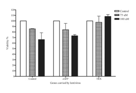
islet cell death and investigated the protective effect of TRX on islets under H2O2 stimulation. eGFP- and Lt-TRX-transduced islets were incubated with various con-centrations of H2O2 for 18 h, and the MTT assay was per-formed to evaluate the islet cell viability. Islet cells overexpressing TRX were more resistant to H2O2-induced cytotoxicity at a concentration of 100 μM (Fig. 3). These data indicate that overexpression of TRX in islets by the lentiviral vector did not affect the insulin-secreting func-tion of islets and further protected the islets from exoge-nous stress stimulation, suggesting that TRX is a potential candidate for genetic manipulation of islet grafts in trans-plantation.
Survival of TRX-transduced islet grafts after transplantation
Oxidative stress induced by inflammation and islet isola-tion and transplantaisola-tion are major obstacles to islet replacement therapy because they may cause primary non-function of islets and early graft loss. Considering the advantages of the antioxidative and antiapoptotic func-tions of TRX, we hypothesized that overexpression of this gene would protect islets from the stress induced by the transplantation procedure and subsequent autoimmune attack. Freshly isolated islets were transduced with Lt-TRX or Lt-eGFP and cultured for 1 day. Around 700 marginal islets were transplanted under the left kidney capsule of newly diabetic female NOD mice. Islet grafts with TRX transduction showed better glycemic control (Fig. 4a) and prolonged islet graft survival in diabetic recipients (mean graft survival days, 18 vs. 6.5, n = 9 and 10, respectively, p < 0.05) (Fig. 4b). We also performed nephrectomy of the
Insulin-secreting function of islets
Figure 2
Insulin-secreting function of islets. Freshly isolated islets were transduced with Lt-eGFP or Lt-TRX. After 24 h cul-ture, the glucose-stimulated insulin-secretion test was per-formed. The concentration of insulin released into the medium was measured by ELISA. The concentration of insu-lin release is expressed as mean ± SD of three independent experiments.
Overexpression of TRX reduces H2O2-induced cytotoxicity
Figure 3
Overexpression of TRX reduces H2O2-induced cyto-toxicity. Freshly isolated islets were transduced with Lt-eGFP or Lt-TRX. After 48 h culture, islets were treated with H2O2 at different concentrations for 18 h. Islet cell viability
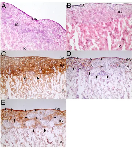
left kidney implanted with grafted islets to confirm further the glucose-regulatory function of the Lt-TRX-transduced islet grafts. The NOD recipients with normal glycemia became hyperglycemic after the islet-implanted kidney was removed, indicating that the islet grafts were still functional and maintained the normal glycemia in trans-planted recipients (data not shown). Histological analysis of the graft-bearing kidneys revealed that although Lt-TRX-transduced islets showed much less leukocytic infil-tration than the Lt-eGFP-transduced islets, they were not completely free from lymphocyte infiltration (Fig. 5a and 5b). Immunohistochemical staining showed that Lt-TRX transduced islet grafts were insulin-producing (Fig. 5c) and TRX-expressing (Fig. 5d), suggesting that these islets with transgene expression were still functional. Since
heme oxygenase-1 (HO-1) mediates a strong cytoprotec-tion in cells under a variety of stresses, the induccytoprotec-tion of HO-1 in TRX-transduced islets may have a synergistic effect with TRX to protect them from apoptosis and improve islet survival and function after transplantation. Results from immunohistochemical staining revealed that the distribution of HO-1-expressing cells was quite close to that of the TRX-expressing cells (Fig. 5e), consistent with the previous reports that TRX facilitates the induction of HO-1 [26]. These results indicate that overexpression of TRX in islet grafts protected them from inflammation-induced stress and prolonged graft survival after trans-plantation, although it could not inhibit leukocytic infil-tration completely and prevent loss of the grafts.
Cellular responses to the TRX overexpression
Previous reports have demonstrated that TRX selectively activates a number transcription factors and facilitates the induction of cytoprotective genes [14], suggesting that TRX-mediated islet graft protection may not only acts through free radical scavenger. To investigate the protec-tive mechanisms mediated by TRX in islet grafts, we fur-ther analyzed the expression of genes such as c-fos, c-jun, and HO-1 that were regulated by TRX. We also analyzed AP-1 activity in β cell-derived NIT-1 cells after Lt-TRX virus transduction. Quantitative PCR analysis reveals that expression of HO-1 and c-fos was up-regulated after Lt-TRX transduction (Fig. 6a).
To further determine the AP-1 activity in Lt-TRX-trans-duced cells, we transfected cells with a SEAP reporter plas-mid which containing AP-1 binding sites on the upstream promoter region. The AP-1 activity was around 3-fold increase in Lt-TRX-transduced cells than in Lt-eGFP-trans-duced cells, suggesting that TRX could regulate gene tran-scription through the up-regulation of AP-1 activity (Fig. 6b). These results are consistent with previous reports that TRX regulates cellular redox status and promotes cell sur-vival through inducing cell protective genes expression.
Discussion
Clinical trials of islet transplantation are showing remark-able success since the Edmonton protocol [27] was devel-oped, and this glucocorticoid-free immunosuppressive protocol was replicated successfully [28]. However, inad-equate islet donors and recurrent autoimmunity are major obstacles in the treatment of type I diabetes. The trans-plantation procedure such as collagenase-based islet isola-tion triggers proinflammatory cytokine and chemokine production by islets, which contributes to early graft loss or primary loss of function [3,29]. In this study, we over-expressed TRX in islet grafts to determine whether this would protect them from oxidative stress-induced cell damage. We found that TRX did not alter the glucose-induced insulin-secreting function of the islets but
pro-Function of marginal islets and diabetic-free frequency of dia-betic recipients
Figure 4
Histology of islet grafts (IG) under the capsule (CA) of the kidney (K)
Figure 5
tected them from ROS challenge. In the islet transplanta-tion experiment, TRX-overexpressed islets showed better glycemic control and longer graft survival, indicating that TRX has a strong cytoprotective effect on islets.
Many approaches have been shown to have protective effects in islets, including the regulation of the immune response by overexpressing anti-inflammatory cytokines
or cytokine inhibitors [6,30,31]; and by reducing cellular stress by overexpression of antiapoptotic and antioxida-tive genes in islets [9,10,12]. These approaches might pro-tect islets from apoptosis or promote islet function in vitro or in vivo, although the choice of target genes and the methods to overexpress the target genes can affect the results significantly. In general, therapeutic targets that have a paracrine action have a more marked biological effect than do genes that target intracellular molecules. In addition, the selected vectors for carrying the target genes to the islets should have low immunogenicity, and long-term and stable gene expression in islets is required. In this work, we used a lentivirus to carry the TRX gene into the islets, which do not divide or have a low rate of divi-sion. This approach has some advantages. First, lentivirus can infect both dividing and nondividing or quiescent cells efficiently and provide for the stable integration into the host cell genome. Second, this method does not elicit an immune response in vivo. Third, TRX is a strong ROS scavenger in the cytoplasm and can clear H2O2 directly and repair the oxidized proteins.
TRX also regulates the expression or activity of other pro-teins. In some circumstances, TRX translocates to the nucleus to regulate transcription factor activity [32] or can be secreted and act as a growth factor [33]. Under stimu-lation by inflammatory mediators, TRX regulates AP-1 activity and enhances heme oxygenase-1 (HO-1) expres-sion [26]. The AP-1 activity was around 3-fold increase in Lt-TRX-transduced cells than in Lt-GFP-transduced cells, suggesting that TRX could regulate gene transcription through the up-regulation of AP-1 activity in our model. HO-1 expression was observed in Lt-TRX-transduced islets and induction of HO-1 in islet grafts have been demon-strated to protect them from apoptosis and improve their function after transplantation [34]. Thus, TRX and HO-1 may play coordinated roles in protecting cells from inflammatory stress. The reduced form of TRX also binds to a variety of cellular proteins. Apoptosis signal-regulat-ing kinase 1 (ASK1) is one TRX-bindsignal-regulat-ing protein that medi-ates stress- and cytokine-mediated apoptosis [35]. TRX binds to the N-terminus of ASK1, and activation of ASK1 requires dissociation of the TRX [16,36]. Taken together, the antioxidative and antiapoptotic functions of TRX may contribute to protecting islets from inflammation-induced cell injury.
Conclusion
It is unlikely that any single biological agent will be suffi-cient to stop such complicated autoimmune processes such as cytokine imbalance, free radical formation, and cellular apoptosis [37]. Using newly diabetic NOD mice that had developed strong autoimmunity to pancreatic islets as the recipients, we showed that TRX can prolong graft survival significantly. This effect may reflect the
mul-Cellular responses to TRX overexpressing
Figure 6
tiple biological functions of TRX, which has both antioxi-dative and antiapoptotic activities. These are important functions for preventing islets from immune attack, and this idea has been demonstrated in the transgenic model [20]. However, despite its beneficial effects, TRX could not inhibit the leukocytic infiltration into the islet grafts com-pletely, and future studies using combinations of immu-noregulatory genes may help prolong graft survival and maintain long-term glucose homeostasis in diabetic recip-ients.
Competing interests
The authors declare that they have no competing interests.
Authors' contributions
FCC ried out the molecular genetic studies, participated in the sequence alignment and drafted the manuscript, par-ticipated in the design of the study and performed the sta-tistical analysis, conceived of the study, and participated in its design and coordination and helped to draft the manuscript. HKS carried out the molecular genetic stud-ies, participated in the sequence alignment and drafted the manuscript, participated in the design of the study and performed the statistical analysis, conceived of the study, and participated in its design and coordination and helped to draft the manuscript
Acknowledgements
This work was supported by the National Science Council, Taiwan, ROC (NSC96-2628-B-016-002-MY3 to H.-K. Sytwu). We thank Shing-Jia Shieh for technical assistance.
References
1. Gysemans CA, Waer M, Valckx D, Laureys JM, Mihkalsky D, Bouillon R, Mathieu C: Early graft failure of xenogeneic islets in NOD mice is accompanied by high levels of interleukin-1 and low levels of transforming growth factor-beta mRNA in the grafts. Diabetes 2000, 49:1992-1997.
2. Matsuda T, Omori K, Vuong T, Pascual M, Valiente L, Ferreri K, Todorov I, Kuroda Y, Smith CV, Kandeel F, Mullen Y: Inhibition of p38 pathway suppresses human islet production of pro-inflammatory cytokines and improves islet graft function.
Am J Transplant 2005, 5:484-493.
3. Bottino R, Balamurugan AN, Tse H, Thirunavukkarasu C, Ge X, Pro-fozich J, Milton M, Ziegenfuss A, Trucco M, Piganelli JD: Response of human islets to isolation stress and the effect of antioxidant treatment. Diabetes 2004, 53:2559-2568.
4. Corbett JA, Sweetland MA, Wang JL, Lancaster JR Jr, McDaniel ML: Nitric oxide mediates cytokine-induced inhibition of insulin secretion by human islets of Langerhans. Proc Natl Acad Sci USA
1993, 90:1731-1735.
5. Barshes NR, Wyllie S, Goss JA: Inflammation-mediated dysfunc-tion and apoptosis in pancreatic islet transplantadysfunc-tion: impli-cations for intrahepatic grafts. J Leukoc Biol 2005, 77:587-597. 6. Suarez-Pinzon WL, Marcoux Y, Ghahary A, Rabinovitch A: Gene
transfection and expression of transforming growth factor-beta1 in nonobese diabetic mouse islets protects beta-cells in syngeneic islet grafts from autoimmune destruction. Cell Transplant 2002, 11:519-528.
7. Fernandes JR, Duvivier-Kali VF, Keegan M, Hollister-Lock J, Omer A, Su S, Bonner-Weir S, Feng S, Lee JS, Mulligan RC, Weir GC: Trans-plantation of islets transduced with CTLA4-Ig and TGFbeta using adenovirus and lentivirus vectors. Transpl Immunol 2004, 13:191-200.
8. Yang Z, Chen M, Ellett JD, Fialkow LB, Carter JD, Nadler JL: The novel anti-inflammatory agent lisofylline prevents autoim-mune diabetic recurrence after islet transplantation. Trans-plantation 2004, 77:55-60.
9. Contreras JL, Bilbao G, Smyth CA, Jiang XL, Eckhoff DE, Jenkins SM, Thomas FT, Curiel DT, Thomas JM: Cytoprotection of pancreatic islets before and soon after transplantation by gene transfer of the anti-apoptotic Bcl-2 gene. Transplantation 2001, 71:1015-1023.
10. Bertera S, Crawford ML, Alexander AM, Papworth GD, Watkins SC, Robbins PD, Trucco M: Gene transfer of manganese superoxide dismutase extends islet graft function in a mouse model of autoimmune diabetes. Diabetes 2003, 52:387-393.
11. Chen H, Li X, Epstein PN: MnSOD and catalase transgenes demonstrate that protection of islets from oxidative stress does not alter cytokine toxicity. Diabetes 2005, 54:1437-1446. 12. Mysore TB, Shinkel TA, Collins J, Salvaris EJ, Fisicaro N, Murray-Segal
LJ, Johnson LE, Lepore DA, Walters SN, Stokes R, et al.: Overex-pression of glutathione peroxidase with two isoforms of superoxide dismutase protects mouse islets from oxidative injury and improves islet graft function. Diabetes 2005, 54:2109-2116.
13. Li X, Chen H, Epstein PN: Metallothionein protects islets from hypoxia and extends islet graft survival by scavenging most kinds of reactive oxygen species. J Biol Chem 2004, 279:765-771. 14. Powis G, Montfort WR: Properties and biological activities of
thioredoxins. Annu Rev Biophys Biomol Struct 2001, 30:421-455. 15. Burke-Gaffney A, Callister ME, Nakamura H: Thioredoxin: friend
or foe in human disease? Trends Pharmacol Sci 2005, 26:398-404. 16. Saitoh M, Nishitoh H, Fujii M, Takeda K, Tobiume K, Sawada Y, Kawa-bata M, Miyazono K, Ichijo H: Mammalian thioredoxin is a direct inhibitor of apoptosis signal-regulating kinase (ASK) 1. Embo J 1998, 17:2596-2606.
17. Hattori I, Takagi Y, Nakamura H, Nozaki K, Bai J, Kondo N, Sugino T, Nishimura M, Hashimoto N, Yodoi J: Intravenous administration of thioredoxin decreases brain damage following transient focal cerebral ischemia in mice. Antioxid Redox Signal 2004, 6:81-87.
18. Okuyama H, Nakamura H, Shimahara Y, Araya S, Kawada N, Yamaoka Y, Yodoi J: Overexpression of thioredoxin prevents acute hepatitis caused by thioacetamide or lipopolysaccha-ride in mice. Hepatology 2003, 37:1015-1025.
19. Takagi Y, Mitsui A, Nishiyama A, Nozaki K, Sono H, Gon Y, Hashim-oto N, Yodoi J: Overexpression of thioredoxin in transgenic mice attenuates focal ischemic brain damage. Proc Natl Acad Sci USA 1999, 96:4131-4136.
20. Hotta M, Tashiro F, Ikegami H, Niwa H, Ogihara T, Yodoi J, Miyazaki J: Pancreatic beta cell-specific expression of thioredoxin, an antioxidative and antiapoptotic protein, prevents autoim-mune and streptozotocin-induced diabetes. J Exp Med 1998, 188:1445-1451.
21. Chang LJ, Urlacher V, Iwakuma T, Cui Y, Zucali J: Efficacy and safety analyses of a recombinant human immunodeficiency virus type 1 derived vector system. Gene Ther 1999, 6:715-728. 22. Juang JH, Kuo CH, Hsu BR: Effects of multiple site implantation
on islet transplantation. Transplant Proc 2002, 34:2698-2699. 23. Curran MA, Ochoa MS, Molano RD, Pileggi A, Inverardi L, Kenyon
NS, Nolan GP, Ricordi C, Fenjves ES: Efficient transduction of pancreatic islets by feline immunodeficiency virus vectors1.
Transplantation 2002, 74:299-306.
24. Janjic D, Wollheim CB: Islet cell metabolism is reflected by the MTT (tetrazolium) colorimetric assay. Diabetologia 1992, 35:482-485.
25. Bottino R, Lemarchand P, Trucco M, Giannoukakis N: Gene- and cell-based therapeutics for type I diabetes mellitus. Gene Ther
2003, 10:875-889.
26. Wiesel P, Foster LC, Pellacani A, Layne MD, Hsieh CM, Huggins GS, Strauss P, Yet SF, Perrella MA: Thioredoxin facilitates the induc-tion of heme oxygenase-1 in response to inflammatory medi-ators. J Biol Chem 2000, 275:24840-24846.
Publish with BioMed Central and every scientist can read your work free of charge "BioMed Central will be the most significant development for disseminating the results of biomedical researc h in our lifetime."
Sir Paul Nurse, Cancer Research UK
Your research papers will be:
available free of charge to the entire biomedical community
peer reviewed and published immediately upon acceptance
cited in PubMed and archived on PubMed Central
yours — you keep the copyright
Submit your manuscript here: BioMedcentral
28. Shapiro AM, Ricordi C, Hering BJ, Auchincloss H, Lindblad R, Robert-son RP, Secchi A, Brendel MD, Berney T, Brennan DC, et al.: Inter-national trial of the Edmonton protocol for islet transplantation. The New England journal of medicine 2006, 355:1318-1330.
29. Abdelli S, Ansite J, Roduit R, Borsello T, Matsumoto I, Sawada T, Alla-man-Pillet N, Henry H, Beckmann JS, Hering BJ, Bonny C: Intracel-lular stress signaling pathways activated during human islet preparation and following acute cytokine exposure. Diabetes
2004, 53:2815-2823.
30. Gallichan WS, Kafri T, Krahl T, Verma IM, Sarvetnick N: Lentivirus-mediated transduction of islet grafts with interleukin 4 Results in sustained gene expression and protection from insulitis. Human gene therapy 1998, 9:2717-2726.
31. Deng S, Ketchum RJ, Yang ZD, Kucher T, Weber M, Shaked A, Naji A, Brayman KL: IL-10 and TGF-beta gene transfer to rodent islets: effect on xenogeneic islet graft survival in naive and B-cell-deficient mice. Transplant Proc 1997, 29:2207-2208. 32. Hayashi T, Ueno Y, Okamoto T: Oxidoreductive regulation of
nuclear factor kappa B. Involvement of a cellular reducing catalyst thioredoxin. J Biol Chem 1993, 268:11380-11388. 33. Hori K, Katayama M, Sato N, Ishii K, Waga S, Yodoi J:
Neuroprotec-tion by glial cells through adult T cell leukemia-derived fac-tor/human thioredoxin (ADF/TRX). Brain research 1994, 652:304-310.
34. Pileggi A, Molano RD, Berney T, Cattan P, Vizzardelli C, Oliver R, Fraker C, Ricordi C, Pastori RL, Bach FH, Inverardi L: Heme oxyge-nase-1 induction in islet cells Results in protection from apoptosis and improved in vivo function after transplanta-tion. Diabetes 2001, 50:1983-1991.
35. Ichijo H, Nishida E, Irie K, ten Dijke P, Saitoh M, Moriguchi T, Takagi M, Matsumoto K, Miyazono K, Gotoh Y: Induction of apoptosis by ASK1, a mammalian MAPKKK that activates SAPK/JNK and p38 signaling pathways. Science 1997, 275:90-94.
36. Liu H, Nishitoh H, Ichijo H, Kyriakis JM: Activation of apoptosis signal-regulating kinase 1 (ASK1) by tumor necrosis factor receptor-associated factor 2 requires prior dissociation of the ASK1 inhibitor thioredoxin. Mol Cell Biol 2000, 20:2198-2208.


