R E S E A R C H
Open Access
A novel method to establish glucocorticoid
resistant acute lymphoblastic leukemia cell
lines
Ling Gu
1,2*, Ge Zhang
3and Yanle Zhang
1Abstract
Background:Drug-resistant cell lines, established from drug-sensitive cell lines by drug exposure in vitro, are the most useful cancer models in studies on the mechanism of chemoresistance. However, the success rate of the traditional approaches to construct such cell lines is low because a long time is required for the addition of drugs.
Methods:A cell culture technique was used to establish the drug-resistant cell lines from their parental cells. Molecular and cellular biological techniques including flow cytometry, MTT assay, western blotting, and DNA fingerprinting analysis were used to characterize the drug-resistant cell lines. Nude mice were used for xenograft studies.
Results:We established novel glucocorticoid (GC)-resistant cell lines from 3 GC-sensitive acute lymphoblastic leukemia (ALL) cell lines. First, we established a novel GC-resistant T-ALL cell line, CEM-C7/HDR, by mimicking the
microenvironment of the bone marrow and culturing GC-sensitive CEM-C7–14 cells under hypoxia for 5 weeks with a single dexamethasone (Dex) treatment. The CEM-C7/HDR cells had been cultured continuously in drug-free medium under normoxia for 1 year. The IC50and resistance index (RI) to Dex were maintained at 60~70μM and 1500~1800, respectively, which is in consistent with the IC50and RI of GC-resistant CEM-C1–15 cells. To clarify the reliability of the method, we subcloned CEM-C7–14 cells, and obtained Dex-resistant cell lines, CEM-C7-SC2/HDR and CEM-C7-SC14/HDR, from 2 monoclonal cells of CEM-C7–14 by the same method. Moreover, we obtained two additional Dex-resistant B-ALL cell lines, NALM-6/HDR and HXEX-ALL1/HDR, from NALM-6 and HXEX-ALL1 cells with the same approach.
Conclusions:CEM-C7/HDR, NALM-6/HDR and HXEX-ALL1/HDR cell lines may serve as useful GC-resistant ALL models for both in vitro and in vivo studies. Culturing under hypoxic condition with a single Dex treatment is a novel and
convenient approach for generating stable GC resistant cell lines.
Keywords:Method, Glucocorticoid resistance, Acute lymphoblastic leukemia, Hypoxia, Cell line
Background
Drug resistance remains a major challenge in cancer therapy. Although contemporary chemotherapy regi-mens have improved the long-term survival rate of childhood acute lymphoblastic leukemia (ALL) to nearly 90%, relapsed/refractory ALL is still a leading cause of
tumor-related death in children [1]. The principal cause of treatment failure in relapsed/refractory ALL is che-moresistance, especially resistance to glucocorticoids (GCs) [2,3]. Drug-resistant cell lines that are established from drug-sensitive cell lines are the most useful models in tumor research [4,5]. The first in vitro-derived, drug-resistant cell line was reported by Biedler and Riehm in 1970 [6]. Drug-resistant cell lines have been used to investigate the mechanisms of drug resistance for nearly 50 years, but the methods used to construct such cell lines have remained essentially unchanged [4]. Two methods are available to acquire drug resistance in cell lines. One is stepwise dose-escalation continuous expos-ure, and the other is high-concentration pulsatile
© The Author(s). 2019Open AccessThis article is distributed under the terms of the Creative Commons Attribution 4.0 International License (http://creativecommons.org/licenses/by/4.0/), which permits unrestricted use, distribution, and reproduction in any medium, provided you give appropriate credit to the original author(s) and the source, provide a link to the Creative Commons license, and indicate if changes were made. The Creative Commons Public Domain Dedication waiver (http://creativecommons.org/publicdomain/zero/1.0/) applies to the data made available in this article, unless otherwise stated.
* Correspondence:guling@scu.edu.cn
1
Laboratory of Hematology/Oncology, Department of Pediatric Hematology/ Oncology, Key Laboratory of Birth Defects and Related Diseases of Women and Children (Sichuan University), Ministry of Education, West China Second University Hospital, Sichuan University, No.20, Section 3, Renmin South Road, Chengdu 610041, People’s Republic of China
2Joint laboratory of West China Second University Hospital, Sichuan
University and School of Life Science, Fudan University for Pulmonary Development and Disease, Chengdu, China
exposure. The stepwise escalation method has been used more commonly because it has a higher success rate [5]. Approaches that combine these two methods have also been successful at constructing drug-resistant cell lines. However, all such approaches took a long time (6~16 months, or more) [5]. Furthermore, the success rate for such approaches is low because a long time is required for the addition of drugs and cell recovery [4].
Drugs at a high concentration can kill all parental cells, which makes construction of a highly resistant cell line difficult. The resistance index (RI), defined as the ratio of the IC50(drug concentration that inhibits 50% of
cell growth) of the resistant cell line to that of its paren-tal cell line, is approximately 2~80 in most cases [5]. The resistant phenotype may remain for several weeks or months after discontinuation of drug exposure; after that time, the cells have to be re-exposed to the drugs to regain resistance [4]. The time required to develop a resistant cell line and the phenotype obtained vary depending on the type of cell line and the selection agent being used [4]. The most important features of drug-resistant cell lines are those that conform to the clinical setting.
In a previous study, we observed the glycolytic pheno-type in most drug-resistant leukemia cells, which is in concordance with the results of other studies [7]. Enhancement of the glycolytic phenotype may induce drug resistance [8]. ALL cells cultured under a hypoxic condition were more tolerant to chemotherapeutic drugs, including GCs [9]. Bone marrow (BM), which exists in a hypoxic microenvironment [10], is the most common site of relapse in ALL. Therefore, we attempted to simulate the clinical setting by establishing resistant cell lines under a hypoxic condition.
Methods
Cell lines and culture conditions
CEM-C7–14(GC sensitive) and CEM-C1–15 (GC resist-ant) cell lines were subcloned from the CCRF-CEM cell line [11], which was established from a patient with T-ALL [12]. The two cell lines were kindly provided by Dr. Thompson (The University of Texas Medical Branch). The GC-sensitive B-ALL cell line, NALM-6 was pur-chased from Shanghai Institute Cell Resources Bank; HXEX-ALL1 was established from a B-ALL patient by our laboratory. All cell lines were maintained in RPMI 1640 (HyClone, Logan, UT, USA) supplemented with 10% fetal bovine serum (FBS; HyClone) at 37 °C under a humidified atmosphere with 5% carbon dioxide (CO2)
and 21% oxygen (O2; normoxic condition).
Reagents and antibodies
Dexamethasone (Dex; Sigma-Aldrich, St. Louis, MO, USA) was dissolved in ethanol and used at a concentration of
0.1~ 0.5μM. The final concentration of ethanol in the medium was 0.001%~ 0.005%, at which cell growth was not obviously altered. Propidium iodide (PI), trypan blue and 3-(4,5-dimethylthiazol-2-yl)-2,5-diphenyltetrazolium bromide (MTT) were purchased from Sigma. The Annexin V-PI Kit was purchased from Roche (Mannheim, Germany). Anti-bodies to Glut-1, HKII, LDH, LDH (Tyr10), 4E-BP1, p-4E-BP1 (Thr37/46), p70S6K, p-p70S6K (Thr389), AMPK, AMPK (Thr172), glucocorticoid receptor (GR), and p-GR (Ser211) were purchased from Cell Signaling Technol-ogy (Beverly, MA, USA). Horseradish peroxidase (HRP)– conjugated donkey anti-rabbit antibody and HRP-conjugated sheep anti-mouse antibodies were obtained from Santa Cruz Biotech (Santa Cruz, CA, USA). The β -Actin antibody was obtained from Kangchen Bio-Tech (Shanghai, China).
Establishment of Dex-resistant ALL cell lines
Logarithmically growing cells were harvested and seeded in 6-well sterile plastic culture plates (Corning Inc., Corning, NY, USA) at a density of 1~3 × 10⁴/ml in RPMI-1640 medium supplemented with 10% FBS at 37 °C, and cultured in a tri-gas CO2incubator (Thermo
Fisher, Carlsbad, CA, USA) with a 5% CO2and 1% O2
atmosphere (hypoxic condition). When the cell density reached 1~3 × 10⁵/ml, various concentrations of Dex (0.10μM, 0.25μM and 0.50μM) were added to the cul-ture plates. After 10~14 days in the lag-phase, the cells began to grow. The medium was replaced every 3~4 days to maintain the cells at a density of 5~10 × 105/ml in the next 2~3 weeks. After 5~6 weeks of culture under the hypoxic condition, the cells were transferred to the normoxic condition and cultured in drug-free medium continuously for 1 year.
Subcloning of ALL cells
Logarithmically growing cells were harvested and seeded in 6-well sterile plastic culture plates at a density of 5 × 10²/ml in methylcellulose RPMI-1640 medium contain-ing 0.9% methylcellulose (MethoCult GFH4434; Sigma, St. Louis, MO, USA) and 10% FBS at 37 °C under a hu-midified atmosphere with 5% CO2and 21% O2. Random
aspiration of individual colonies growing in methylcellu-lose was undertaken on day 8 of the culture. Next, each colony was cultured in RPMI-1640 complete medium.
Cell growth and viability assay
Cells were cultured in a 6-well sterile plastic culture plates at 1 × 10⁵/ml in RPMI-1640 medium with 10% FBS and grown for 7 days. Viable cells were counted using trypan blue staining every day. Doubling time (Td) was calculated for cells in exponential growth with the following equation: Td (h) = t × lg2/lg(Nt/N0), where t is
cells, and N0is the initial number of cells. Cell viability
was evaluated by MTT assay. Briefly, cells were seeded in 96-well plates. Next, 0.5 mM MTT (final concentra-tion) was added to each well for 4 h at 37 °C. Then, solubilization buffer (10% SDS in 0.01 M HCl) was added to each well, and the plates were further incu-bated for 24 h at 37 °C. The spectrophotometric absorb-ance was measured at 570 nm (reference 690 nm) using a multiplate reader (Multiskan Spectrum, Thermo Elec-tron Co., Waltham, MA, USA). Values were obtained by comparing the experimental cells with their respective controls. Mean values were calculated from triplicate cultures.
Chemosensitivity assays
The chemotherapeutic drugs, Dex, daunorubicin, vin-cristine, arabinoside, methotrexate and asparaginase, were purchased from Sigma. All cells were treated with increasing concentrations of different drugs for 48 h, followed by assessment of cell viability by MTT assay. The IC50was calculated by linear interpolation.
Cell cycle analysis
For each analysis, 106 cells were harvested 48 h after treatment and fixed overnight in 70% ethanol at 4 °C. The cells were then washed and stained with 5μg/ml PI in the presence of DNAse-free RNAse (Sigma). After 30 min at room temperature, the cells were analyzed via flow cytometry (Cytomics FC 500 and CXP & Multicycle software, Beckman Coulter Inc., Miami, FL, USA), acquiring 30,000 events.
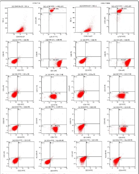
Immunophenotype analysis
For the detection of the immunophenotype of the CEM-C7/HDR and CEM-C7–14 cells, we used antibodies against the following targets: CD33, CD34, HLA-DR, cyTdT, cCD3, CD3, CD4, CD5, CD7, CD8, CD117, CD1α, CD2, CD10, CD19, CD20, CD13 and CD45 (Bec-ton Dickinson Inc., Franklin Lakes, NJ, USA). Positivity for the antigens was determined using a FACSCalibur flow cytometer (Becton Dickinson Inc.).
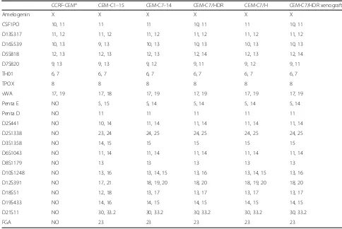
DNA fingerprinting analysis
The identity of the CEM-C1–15, CEM-C7–14, CEM-C7/HDR and CEM-C7/H cell lines was checked using DNA fingerprinting. DNA was prepared from these cells using the Qiagen DNeasy Blood Kit (Qiagen), according to the instructions provided by the manufacturer. The following 22 highly polymorphic short tandem repeat (STR) loci were tested by a multiplex PCR:Amelogenin, CSF1PO, D13S317, D16S539, D5S818, D7S820, TH01, TPOX, vWA, Penta E, Penta D, D2S441, D2S1338, D3S1358, D6S1043, D8S1179, D10S1248, D12S391, D18S51,D19S433,D21S11andFGA.
Western blotting analysis
Briefly, cells (106) were washed twice in cold PBS and then lysed by Laemmli sample buffer (Bio-Rad). Samples were boiled for 5 min at 100 °C. Proteins were separated by 8%~ 15% SDS–polyacrylamide gel electrophoresis and transferred onto nitrocellulose membranes (0.22μm or 0.45μm, Millipore). Non-specific binding sites were blocked with 5% non-fat dry milk dissolved in TBS (10 mM Tris-HCl, pH 7.6, 137 mM NaCl) with 0.1% Tween 20 (TTBS) for 2 h at room temperature, followed by incubation with primary antibody for 2 h at room temperature or at 4 °C overnight. The membranes were then washed 3 times in TTBS and incubated for 2 h at room temperature with secondary HRP–conjugated donkey anti-rabbit antibody or HRP-conjugated sheep anti-mouse antibody diluted 1:5000 in TTBS with 5% non-fat milk. Proteins were visualized by incubation with ECL plus (Millipore). All experiments were conducted independently at least 3 times. The level of the β-Actin protein was used as a control for the amount of protein loaded into each lane.
Animal experiments
Cultured 5 × 10⁶cells were subcutaneously injected into the right flanks of 6-week-old female BALB/c (nu/nu) nude mice, and 0.1 ml of PBS was injected into the left flanks as the control. Once palpable tumors were estab-lished, animals were randomized into 2 groups, each containing 4 mice. Mice were injected intraperitoneally daily with 15 mg/kg/d Dex (Dex group), or PBS (Control group) for 14 days. Tumor size was measured by calipers every 2 days. The approximate tumor volume was calculated using the eq. V = (length×width×depth)/2. All animals were ear-tagged and monitored individually throughout the experiment. All animal care was in com-pliance with the guidelines established by the internal Institutional Animal Care and Use Committee and Ethics Committee of Sichuan University. After the mice were euthanized, the tumor mass was excised, fixed in 10% formalin, and routinely processed for paraffin embed-ding. Five-millimeter-thick sections were obtained and prepared for standard histopathological examination.
Statistical analysis
All assays were performed in triplicate, and data are expressed as mean values ± SD. One-way ANOVA was used to compare two groups. A p-value < 0.05 was considered to be significant.
Results
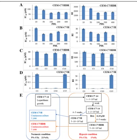
Establishment of Dex-resistant ALL cell lines
were transferred to the normoxic condition for 1 week. At that time, an MTT assay revealed that the IC50values
of the Dex groups, with a single treatment of 0.10μM, 0.25μM or 0.50μM Dex respectively, were 50~150μM at 48 h. We chose the 0.25μM Dex group to be cultured continuously and constructed a stable resistant cell line, which was designated CEM-C7/HDR. The IC50 of Dex
in the CEM-C7/HDR cells at a 10 cell population doub-ling level (PDL) was 128.1 ± 4.73μM. Next, the IC50and
the RI of those cells decreased slowly, resulting in the PDL increasing from 10 to 100, where it stabilized after 100 PDL (Fig. 1a). After 1 year of Dex-free continuously culturing, the IC50 and RI were 60~70μM and
1500~1800, respectively (Fig.1a). Unexpectedly, the con-trol group, which was cultured under the hypoxic condi-tion with no Dex treatment, also gained Dex resistance. We named the control cell line CEM-C7/H, and its RI was 10~20 after 100 PDL (Fig. 1b). All CEM-C7/HDR subclones were resistant to Dex with RIs ranging from 1200 to 2000 (Fig.1c). However, we obtained both resist-ant and sensitive subclones of CEM-C7/H, with RIs ran-ging from 0.3 to 33.4 (Fig.1d). Interestingly, CEM-C7/H cells obtained cross-resistance to daunorubicin and vincristine, with RIs of 2.02 ± 0.23 and 1.71 ± 0.19, re-spectively. However, CEM-C7/HDR cells did not obtain cross-resistance to daunorubicin, vincristine, arabino-side, methotrexate or asparaginase. Figure1e showed the procedure for constructing above cell lines. To clarify the mechanisms of resistance development, we selected two GC-sensitive CEM-C7–14 mono-clonal cells, named CEM-C7-SC2 and CEM-C7-SC14, with IC50 values of
0.04μM and 0.06μM, respectively. This method resulted in the development of two resistant cell lines named CEM-C7-SC2/HDR and CEM-C7-SC14/HDR, with IC50
values of 100~150μM at 10PDL. The control groups, CEM-C7-SC2/H and CEM-C7-SC14/H, which were cul-tured under hypoxic condition with no Dex treatment, did not express the resistant phenotype. Additional file1: Figure S1 showed the origin and name of the aforemen-tioned cell lines.
To evaluate the repeatability of the method, we con-structed stable Dex-resistant cell lines, NALM-6/HDR and ALL1/HDR, from the NALM-6 and HXEX-ALL1 cell lines, using the same method. NALM-6/HDR and HXEX-ALL1/HDR were 28,000- and 4- fold more resistant to Dex than their parental cells (Additional file2: Figure S2).
Morphological and biological characteristics of CEM-C7/ HDR cells
The CEM-C7/HDR and CEM-C7/HDR-C2 (its mono-clonal cell line) cells were grown in suspension as single cells, and Wright-Giemsa staining showed no obvious differences in morphology between the two resistant
cell lines and the parental cell line, CEM-C7–14 (Fig. 2a and b). All three cells lines showed similar percentages of G0/G1 and S phase cells, with no
sta-tistically significant differences observed (P> 0.05) (Fig. 2c). They also exhibited similar growth curves (Fig. 2d). The three cell lines all stably proliferated in RPMI-1640 medium containing 10% FBS with popula-tion doubling times of 16~22 h. CEM-C7/HDR and CEM-C7–14 displayed identical immunophenotype with cCD3, cyTDT, CD4, CD5 and CD7 positive (Fig. 3). STR analysis confirmed that CEM-C7/HDR, CEM-C7/H, CEM-C7–14 and CEM-C1–15 were all derived from CCRF-CEM (Table 1).
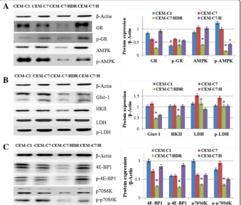
Expression of proteins associated with GC resistance and hypoxia in CEM-C7/HDR
The tumorigenicity of CEM-C7/HDR cells
In an in vivo transplantation test, subcutaneous injection of CEM-C7–14 and CEM-C7/HDR resulted in the devel-opment of tumors in all 12 mice (100%). After 20 days, the mean volume of the tumors generated by subcutaneous injection with CEM-C7–14 and CEM-C7/HDR were 717.8 ± 309.6 mm3 (n= 6) and 803.0 ± 294.9 mm3 (n= 6),
respectively. Hematoxylin and eosin (HE) staining indicated that the tumor masses were composed of leukemia cells (Fig. 5a). Immunohistochemistry (IHC) staining showed that the expression locations of GR, Glut-1, HKII, and 4E-BP1 proteins were consistent in the xenografts of CEM-C7–14 and CEM-C7/HDR (Fig. 5b-e). The results from STR analysis indicated
that subcutaneous tumors derived from the corre-sponding CCRF-CEM cells (Table 1).
Having shown the GC-resistant phenotype of CEM-C7/HDR in vitro, we examined the in vivo efficacy of Dex given intraperitoneally in C7/HDR and CEM-C7–14 xenografts in nude mice. As anticipated, 15 mg/ kg/d Dex used alone showed almost no antitumor effect (p> 0.05) in CEM-C7/HDR xenografts mice, whereas inhibited tumor growth obviously in CEM-C7–14 xeno-grafts mice (p< 0.05, Fig.5f ).
Discussion
Preclinical studies on cancer cell lines have played an important role in our understanding of cancer biology and high-throughput screening for drug development. Molecular and cellular changes that are observed in a resistant cell line, when compared to its drug-sensitive counterpart, can suggest molecular mecha-nisms underlying drug resistance, and locus of drug action is inferred by the presence of those particular
alterations. There is a controversy regarding construc-tion of resistant cell lines by the tradiconstruc-tional drug-exposure method and whether or not they can reflect the clinical setting [4]. We should establish a method that matches the manner in which chemotherapy is administered to patients.
GCs specifically induce apoptosis in malignant lymphoblasts and are thus pivotal in the treatment of lymphoid malignancies, especially ALL [18]. However, GC resistance is a therapeutic problem that accounts for most of the treatment failures [2,3]. The exact molecu-lar mechanism remains poorly understood. The hypoxic microenvironment of BM promoted ALL cells survival and conferred resistance to chemotherapy [9, 19, 20]. Hence, we aimed to construct Dex-resistant cell lines under conditions that mimic ALL cells growing under the hypoxic BM microenvironment. First, GC sensitive ALL cells were cultured under a hypoxic environment for approximately 1 week to accommodate the hypoxic condition. Second, we added a therapeutic dose of Dex
according to the clinical chemotherapy protocol to the parental cells and cultured the cells under the hypoxic condition for 4~5 weeks. Finally, the cells were trans-ferred to the normoxic condition to mimic the clinical setting, similar to how ALL cells are released from BM into the peripheral blood. To our delight, this strategy works. Using this method, we obtained Dex-resistant ALL cell lines from GC sensitive parental cell lines, CEM-C7–14, NALM-6, HXEX-ALL1 and their sub-cloned cell lines. Furthermore, the resistant phenotype acquired by this approach was stable after 1 year of continuous culture in Dex-free medium.
The established CEM-C7/HDR cells were almost iden-tical with its parental cells, CEM-C7–14, with respect to morphology, cell proliferation, STR and immunopheno-type characteristics. In addition, CEM-C7/HDR cells showed a high tumorigenic capacity similar to CEM-C7–14, and the expression location of proteins associ-ated with GC resistance and hypoxia, GR, Glut-1, HKII, and 4E-BP1, were consistent in the xenografts of CEM-C7–14 and CEM-C7/HDR. The resistance char-acteristics of CEM-C7/HDR cells were similar to that of CEM-C1–15 cells. The IC50 of Dex in CEM-C1–15
cells was 61.71 ± 4.25μM, and the RI for these cells
was 1620 ± 128. The IC50 values of Dex in other
GC-resistant leukemia-lymphoma cell lines, such as Jurkat, Raji, Molt-4 and MV4–11, ranged from 30 to 90μM. Therefore, the GC-resistant cell line CEM-C7/ HDR, which was constructed by mimicking the in vivo microenvironment of BM, does resemble the clinic setting to some extent.
It is generally accepted that GCs induce apoptosis in lymphoid blast cells through activation of the GR, and low expression of GR may contribute to GC resistance in leukemia cells [13, 14]. As anticipated, our results showed that CEM-C7/HDR exhibited an obviously lower expression of GR and p-GR (Ser211) compared with its parental cells CEM-C7–14, and p-GR (Ser211) was low expressed in both two GC-resistant cell line, CEM-C7/ HDR and CEM-C1–15. Therefore, low expression of p-GR (Ser211) might be the main cause for GC resistance in T-ALL, which needs further investigation in T-ALL patients.
Tumor cells respond to hypoxic stress by upregulating various genes involved in glucose uptake, glycolysis, and angiogenesis, all of which are essential in maintaining nutrient availability and intracellular ATP levels [21,22]. AMPK is an energy sensor that is pivotal in maintaining
Table 1STR analysis of CEM-C1–15, CEM-C7–14, CEM-C7/HDR, CEM-C7/H and cells from the CEM-C7/HDR xenografts in nude mice
CCRF-CEMa CEM-C1–15 CEM-C7–14 CEM-C7/HDR CEM-C7/H CEM-C7/HDR xenograft
Amelogenin X X X X X X
CSF1PO 10, 11 11 11 10, 11 11 10, 11 D13S317 11, 12 11, 12 11, 12 11, 12 11, 12 11, 12 D16S539 10, 13 9, 13 10, 13 10, 13 10, 13 10, 13 D5S818 12, 13 12, 13 12, 13 12, 14 12, 13 12, 14 D7S820 9, 13 9, 13 9, 12 9, 11 9, 12 9, 11 TH01 6, 7 6, 7 6, 7 6, 7 6, 7 6, 7
TPOX 8 8 8 8 8 8
vWA 17, 19 17, 18 17, 19 17, 19 17, 19 17, 19 Penta E NO 5, 15 5, 14 5, 14 5, 14 5, 14
Penta D NO 11 11 11 11 11
D2S441 NO 10, 14 11, 14 11, 14 11, 14 11, 14 D2S1338 NO 23, 24 24, 25 24, 25 24, 25 24, 25 D3S1358 NO 14, 15 15 15 15 15 D6S1043 NO 11, 14 11, 14 11, 14 11, 14 11, 14
D8S1179 NO 13 13 13 13 13
D10S1248 NO 13, 16 13, 14, 15 13, 16 13, 14, 15 13, 16 D12S391 NO 17, 21 18, 19, 20 18, 20 18, 19, 20 18, 20 D18S51 NO 12, 18 13, 17 13, 17 13, 17 13, 17 D19S433 NO 14, 16 14, 15 14, 15 14, 15 14, 15 D21S11 NO 30, 33.2 30, 33.2 30, 33.2 30, 33.2 30, 33.2
FGA NO 23 23 23 23 23
a
cell metabolic homeostasis [23, 24]. Metabolic stresses, such as glucose starvation and hypoxia, activate AMPK via threonine 172 phosphorylation by AMPK upstream kinases [23, 24]. AMPK is essential for T-ALL cell survival and disease progression [25]. Surprisingly, the expression of p-AMPK was lower in CEM-C7/HDR and CEM-C7/H than in CEM-C7–14 and CEM-C1–15. Moreover, the expression of AMPK decreased also in CEM-C7/HDR cells. It is reported that AMPK plays a role in regulating growth and survival of multiple cancer
cells including leukemia cells [26, 27]. Conversely, other studies showed that activation of AMPK may suppress the growth of some tumors including T-ALL [28–30]. A recent paper reported that levels of p-AMPK and AMPKα1 mRNA are dramatically decreased in clinical leukemia cells, which might play a role in drug resist-ance in leukemia [31]. Although AMPK plays a paradox-ical dual role in ALL survival and progression [25, 32], decreased expression of AMPK and p-AMPK might contribute to GC resistance in CEM-C7/HDR.
Hypoxic stress can induce glycolysis and the mTOR signaling pathway, which would contribute to chemore-sistance [9, 20, 21, 33]. Moreover, loss of AMPK can induce glycolysis and mTOR pathway also [24, 29, 32]. Unexpectedly, in CEM-C7/HDR, key enzymes of glycolysis, Glut-1, HKII, LDH and p-LDH, were all decreased compared to that in its parental cell line CEM-C7–14. Similarly, the mTOR signaling pathway was surprisingly depressed in CEM-C7/HDR. It is intri-guing that while AMPK functions as an antagonist of mTOR, both AMPK and mTOR signaling pathways were depressed in CEM-C7/HDR cells; this result sug-gested additional unclear regulation mechanisms, which deserves further investigation. A recent study compared overall survival in 2 subsets of patients with high and low PI3K-Akt-mTOR activation in 77 AML patients and showed no obvious difference [34]. An in vitro test showed that an AMPK activator and mTOR inhibitor, such as metformin and rapamycin, can effectively inhibit the growth of ALL cells [29, 35]. However, those drugs showed a clinically relevant response only in a minority of patients [34, 36]. Some ALL cells displayed poor response to mTOR inhibition [37]. In addition, other studies showed that long-lasting (chronic) hypoxia may inhibits mTOR pathway through multiple pathways, in-cluding BNIP3, PML, AMPK, and REDD1, to promote hypoxic tolerance [38]. Therefore, the relative low expression of AMPK and mTOR pathway might be another key point to understand the mechanisms of GC resistance, and CEM-C7/HDR may provide a novel model that represents a subset of GC-resistant ALL cells.
Resistant cell lines can be established by drug exposure and induction of extrinsic mutations or by selection of clones based on the strength of an intrinsic mutation [5]. CEM-C7/H attained a resistant phenotype by cultur-ing under hypoxia condition for 5 weeks without Dex treatment. However, it is noteworthy that only part of subclones of CEM-C7/H attained Dex resistance. More-over, NALM-6, HXEX-ALL1 and subclones of the three cell lines did not attained a resistant phenotype by only hypoxic culture. Therefore, hypoxia may act as a com-pressive stress that causes resistant clones to become dominant in the culture. In other words, hypoxia stress could not select out resistant clones from cells without intrinsic resistance-related mutations. Interestingly, CEM-C7/H cells obtained cross-resistance to daunorubi-cin and vincristine. However, CEM-C7/HDR cells did not obtain cross-resistance to daunorubicin, vincristine, arabinoside, methotrexate or asparaginase. In general, culturing under hypoxia condition with a single Dex treatment is a novel and convenient approach for gener-ating stable GC resistant cell lines. The protocol pre-sented here should be modified for use in establishing cell lines that are resistant to other drugs and for
identifying molecular inhibitors that can target cancer cells living in a hypoxic environment. Moreover, in leukemia cells, a hypoxic condition may act as a selec-tion pressure to develop resistant clones.
Conclusions
Here, we constructed a new method to establish GC-resistant leukemia cell lines. These newly established resistant cell lines may serve as valuable in vitro and in vivo tools for further investigation on the potential mechanisms of GC resistance, especially the role of the hypoxic microenvironment in GC resistance.
Additional files
Additional file 1:Figure S1.Cell lines originated from CCRF-CEM. (A) CEM-C7-14 and CEM-C1-15 cell lines were subcloned from CCRF-CEM. (B) CEM-C7-SC2 and CEM-C7-SC14 were subcloned from CEM-C7-14. After culturing under hypoxia for 5 weeks with or without Dex, CEM-C7-SC2/ HDR, CEM-C7-SC14/HDR, CEM-C7-SC2/H, and CEM-C7-SC14/H were constructed. (C) After culturing CEM-C7-14 under hypoxia for 5 weeks with or without Dex, CEM-C7/HDR and CEM-C7/H were constructed. CEM-C7/HDR were subcloned into CEM-C7/HDR-C2, CEM-C7/HDR-C3, CEM-C7/HDR-C9, and CEM-C7/HDR-C10. CEM-C7/H were subcloned into GC-resistant C7/H-C3 and C7/HDR-C15, and GC-sensitive CEM-C7/H-C8 and CEM-C7/H-C10. (TIF 294 kb)
Additional file 2:Figure S2.Resistance characteristics of NALM-6/HDR and HXEX-ALL1/HDR cell lines. (A) IC50and RI of NALM-6/HDR cells at 10~400 PDLs. (B) IC50and RI of HXEX-ALL1/HDR cells at 10~100 PDLs. Cells were cultured with increasing concentrations of Dex for 48 h. Cell viability was evaluated by MTT assays. The IC50values were calculated by linear interpolation. Experiments were performed in triplicate. (TIF 165 kb)
Abbreviations
ALL:Acute lymphoblastic leukemia; BM: Bone marrow; Dex: Dexamethasone; GC: Glucocorticoid; PDL: Population doubling level; RI: Resistance index; Td: Doubling time
Acknowledgements
We are grateful to Dr. Ji Zhang and Dr. Yufang Wang for helping us complete the experiments.
Authors’contributions
LG designed the research, performed a part of the research, analyzed the data and wrote the paper. GZ and YLZ performed a part of the research. All authors read and approved the final manuscript.
Funding
This work was supported by National Natural Science Foundation of China (Grant No.81270602), Sichuan Science and Technology Program of China (Grant No. 2018JY0044), and the Chengdu Science and Technology Huimin Project of China (Grant No. 2015-HM01–00307-SF).
Availability of data and materials
The datasets used and analyzed during the current study are available from the corresponding author on a reasonable request.
Ethics approval and consent to participate
This study was approved by the Research Institute Animal Ethics Committee of West China Second University Hospital (Chengdu, China). All animal care was in compliance with the guidelines established by the internal Institutional Animal Care and Use Committee and Ethics Committee guidelines of Sichuan University.
Consent for publication Not applicable.
Competing interests
The authors declare that they have no competing interests.
Author details 1
Laboratory of Hematology/Oncology, Department of Pediatric Hematology/ Oncology, Key Laboratory of Birth Defects and Related Diseases of Women and Children (Sichuan University), Ministry of Education, West China Second University Hospital, Sichuan University, No.20, Section 3, Renmin South Road, Chengdu 610041, People’s Republic of China.2Joint laboratory of West China Second University Hospital, Sichuan University and School of Life Science, Fudan University for Pulmonary Development and Disease, Chengdu, China.
3Department of Laboratory Medicine, West China Second University Hospital,
Sichuan University, Chengdu, China.
Received: 5 February 2019 Accepted: 14 June 2019
References
1. Pui CH, Yang JJ, Hunger SP, Pieters R, Schrappe M, Biondi A, et al. Childhood acute lymphoblastic leukemia: Progress through collaboration. J Clin Oncol. 2015;33:2938–48.
2. Inaba H, Pui CH. Glucocorticoid use in acute lymphoblastic leukaemia. Lancet Oncol. 2010;11:1096–106.
3. Ceppi F, Cazzaniga G, Colombini A, Biondi A, Conter V. Risk factors for relapse in childhood acute lymphoblastic leukemia: prediction and prevention. Expert Rev Hematol. 2015;8:57–70.
4. Coley HM. Development of drug-resistant models. Methods Mol Med. 2004; 88:267–73.
5. Watson MB, Lind MJ, Cawkwell L. Establishment of in-vitro models of chemotherapy resistance. Anti-Cancer Drugs. 2007;18:749–54.
6. Biedler JL, Riehm H. Cellular resistance to actinomycin D in Chinese hamster cells in vitro: cross-resistance, radioautographic, and cytogenetic studies. Cancer Res. 1970;30:1174–84.
7. Starkova J, Hermanova I, Hlozkova K, Hararova A, Trka J. Altered metabolism of leukemic cells: new therapeutic opportunity. Int Rev Cell Mol Biol. 2018; 336:93–147.
8. Bhattacharya B, Mohd OMF, Soong R. The Warburg effect and drug resistance. Br J Pharmacol. 2016;173:970–9.
9. Petit C, Gouel F, Dubus I, Heuclin C, Roget K, Vannier JP. Hypoxia promotes chemoresistance in acute lymphoblastic leukemia cell lines by modulating death signaling pathways. BMC Cancer. 2016;16:746.
10. Spencer JA, Ferraro F, Roussakis E, Klein A, Wu J, Runnels JM, et al. Direct measurement of local oxygen concentration in the bone marrow of live animals. Nature. 2014;508:269–73.
11. Zawydiwski R, Harmon JM, Thompson EB. Glucocorticoid-resistant human acute lymphoblastic leukemic cell line with functional receptor. Cancer Res. 1983;43:3865–73.
12. Foley GE, Lazarus H, Faraber S, Uzman BG, Boone BA, Mccarthy RE. Continous culture of human lymphoblasts from peripheral blood of a child with acute leukemia. Cancer. 1965;18:522–9.
13. Bedewy AM, El-Maghraby SM, Kandil NS, El-Bendary WR. The prognostic value of glucocorticoid receptors for adult acute lymphoblastic leukemia. Blood Res. 2015;50:235–41.
14. Bloomfield CD, Smith KA, Peterson BA, Munck A. Glucocorticoid receptors in adult acute lymphoblastic leukemia. Cancer Res. 1981;41:4857–60. 15. Yoshida GJ. Metabolic reprogramming: the emerging concept and
associated therapeutic strategies. J Exp Clin Cancer Res. 2015;34:111. 16. Martelli AM, Evangelisti C, Chappell W, Abrams SL, Bäsecke J, Stivala F, et al.
Targeting the translational apparatus to improve leukemia therapy: roles of the PI3K/PTEN/Akt/mTOR pathway. Leukemia. 2011;25:1064–79.
17. Simioni C, Martelli AM, Zauli G, Vitale M, McCubrey JA, Capitani S, et al. Targeting the phosphatidylinositol 3-kinase/Akt/mechanistic target of rapamycin signaling pathway in B-lineage acute lymphoblastic leukemia: an update. J Cell Physiol. 2018;233:6440–54.
18. Greenstein S, Ghias K, Krett NL, Rosen ST. Mechanisms of
19. Chen Y, Jacamo R, Shi YX, Wang RY, Battula VL, Konoplev S, et al. Human extramedullary bone marrow in mice: a novel in vivo model of genetically controlled hematopoietic microenvironment. Blood. 2012;119:4971–80. 20. Deynoux M, Sunter N, Hérault O, Mazurier F. Hypoxia and hypoxia-inducible
factors in leukemias. Front Oncol. 2016;6:41.
21. Wilson WR, Hay MP. Targeting hypoxia in cancer therapy. Nat Rev Cancer. 2011;11:393–410.
22. Paolicchi E, Gemignani F, Krstic-Demonacos M, Dedhar S, Mutti L, Landi S. Targeting hypoxic response for cancer therapy. Oncotarget. 2016;7:13464–78. 23. Hardie DG, Ross FA, Hawley SA. AMPK: a nutrient and energy sensor that
maintains energy homeostasis. Nat Rev Mol Cell Biol. 2012;13:251–62. 24. Inoki K, Kim J, Guan KL. AMPK and mTOR in cellular energy homeostasis
and drug targets. Annu Rev Pharmacol Toxicol. 2012;52:381–400.
25. Kishton RJ, Barnes CE, Nichols AG, Cohen S, Gerriets VA, Siska PJ, et al. AMPK is essential to balance glycolysis and mitochondrial metabolism to control T-ALL cell stress and survival. Cell Metab. 2016;23:649–62.
26. Jeon SM, Chandel NS, Hay N. AMPK regulates NADPH homeostasis to promote tumour cell survival during energy stress. Nature. 2012;485:661–5. 27. Saito Y, Chapple RH, Lin A, Kitano A, Nakada D. AMPK protects
leukemia-initiating cells in myeloid leukemias from metabolic stress in the bone marrow. Cell Stem Cell. 2015;17:585–96.
28. Faubert B, Boily G, Izreig S, Griss T, Samborska B, Dong Z, et al. AMPK is a negative regulator of the Warburg effect and suppresses tumor growth in vivo. Cell Metab. 2013;17:113–24.
29. Grimaldi C, Chiarini F, Tabellini G, Ricci F, Tazzari PL, Battistelli M, et al. AMP-dependent kinase/mammalian target of rapamycin complex 1 signaling in T-cell acute lymphoblastic leukemia: therapeutic implications. Leukemia. 2012;26:91–100.
30. Hirsch HA, Iliopoulos D, Tsichlis PN, Struhl K. Metformin selectively targets cancer stem cells, and acts together with chemotherapy to block tumor growth and prolong remission. Cancer Res. 2009;69:7507–11. 31. Yi Y, Gao L, Wu M, Ao J, Zhang C, Wang X, et al. Metformin sensitizes
leukemia cells to vincristine via activation of AMP-activated protein kinase. J Cancer. 2017;8:2636–42.
32. Herzig S, Shaw RJ. AMPK: guardian of metabolism and mitochondrial homeostasis. Nat Rev Mol Cell Biol. 2018;19:121–35.
33. Sormendi S, Wielockx B. Hypoxia pathway proteins as central mediators of metabolism in the mumor cells and their microenvironment. Front Immunol. 2018;9:40.
34. Nepstad I, Hatfield KJ, Aasebø E, Hernandez-Valladares M, Brenner AK, Bartaula-Brevik S, et al. Two acute myeloid leukemia patient subsets are identified based on the constitutive PI3K-Akt-mTOR signaling of their leukemic cells; a functional, proteomic, and transcriptomic comparison. Expert Opin Ther Targets. 2018;22:639–53.
35. Janes MR, Limon JJ, So L, Chen J, Lim RJ, Chavez MA, et al. Effective and selective targeting of leukemia cells using a TORC1/2 kinase inhibitor. Nat Med. 2010;16:205–13.
36. Fransecky L, Mochmann LH, Baldus CD. Outlook on PI3K/AKT/mTOR inhibition in acute leukemia. Mol Cell Ther. 2015;3:2.
37. Shah K, Moharram SA, Kazi JU. Acute leukemia cells resistant to PI3K/ mTOR inhibition display upregulation of P2RY14 expression. Clin Epigenetics. 2018;10:83.
38. Wouters BG, Koritzinsky M. Hypoxia signalling through mTOR and the unfolded protein response in cancer. Nat Rev Cancer. 2008;8:851–64.
Publisher’s Note