R E S E A R C H
Open Access
Elevated c-Src and c-Yes expression in malignant
skin cancers
Jang Hyun Lee
1, Jae-Kyung Pyon
2, Dong Wook Kim
3, Sang Han Lee
3, Hae Seon Nam
3, Chul Han Kim
3,
Sang Gue Kang
3, Yoon Jin Lee
3, Mi Youn Park
4, Dong Jun Jeong
3, Moon Kyun Cho
3,5*Abstracts
Background:Src family kinases (SFKs) play an important role in cancer proliferation, survival, motility, invasiveness, metastasis, and angiogenesis. Among the SFKs, c-Src and c-Yes are particularly over-expressed or hyper-activated in many human epithelial cancers. However, only a few studies have attempted to define the expression and role of c-Src and c-Yes in cutaneous carcinomas.
Objectives:To investigate the expression of c-Src and c-Yes in cutaneous carcinomas to include malignant melanoma (MM), squamous cell carcinoma (SCC) and basal cell carcinoma (BCC).
Methods:We examined 6 normal skin tissues and 18 malignant skin tumor tissues using western blotting for the expression of c-Src and c-Yes. In another set, 16 specimens of MM, 16 SCCs and 16 BCCs were analyzed for the expression of c-Src and c-Yes using immunohistochemical staining.
Results:Western blotting showed that c-Src was expressed in all malignant skin tumors, but not in normal skin, while c-Yes was expressed in MM and SCC, but not in BCC and normal skin. Immunohistochemical staining results of c-Src and c-Yes in MM, SCC, and BCC mirrored those of the western blot analysis.
Conclusions:c-Src, rather than c-Yes, plays a key role in the proliferation and progression of malignant skin cancers.
Introduction
Skin tumors have become one of the most common cancers in many countries, with rapid increasing inci-dence during the last half century. Nonmelanoma skin cancers including basal cell carcinoma (BCC) and squa-mous cell carcinoma (SCC) now make up more than one third of all cancers in the United States [1]. A large number of studies regarding the role of oncogenes and hormones, as well as environmental and predisposing factors, have been reported.
Oncogenes, especially Src family kinases (SFKs), which are activated in colon and breast cancers, are drawing attention for their involvement in malignant melanoma (MM) [2]. SFKs are non-receptor tyrosine kinases that participate in variable cellular signal transduction path-ways, with the capacity to trigger cancer with its
continuous activation. SFKs are composed of 9 mem-bers, c-Src, c-Yes, Fyn, Lyn, Lck, Hck, Blk, Rgr, and Yrk. SFKs play integral roles in cancer development to include proliferation, survival, motility, invasiveness, metastasis, and angiogenesis. Most SFKs are primarily expressed from a hematopoietic cell origin, but Src, c-Yes, and Fyn are expressed at high levels by platelets, neurons, and some epithelial tissues [3]. c-Src and c-Yes in particular are over-expressed or hyper-activated in many human epithelial cancers. The role and process of these two oncogenes in colon and breast cancer are well studied, but not in other human cancers. The role of SFKs in melanoma have been investigated with conflict-ing reports, but their overall role in nonmelanoma skin tumors has yet to be elucidated. Tyrosine kinases are known to be activated in many human MM, SCC, and BCC epithelial cancers. Therefore, we studied the expression of c-Src and c-Yes to uncover its involve-ment in malignant skin cancer developinvolve-ment.
* Correspondence: mkcho2001@hanmail.net 3
Molecular Cancer Research Center, College of Medicine, Soonchunhyang University, Chunan, Korea
Full list of author information is available at the end of the article
Materials and methods Tissue samples
A total of 8 normal skin tissues and 24 malignant skin tumor tissues were obtained from patients who under-went surgery between July 2009 and September 2009 in the Departments of Plastic and Reconstructive Surgery at Hanyang University Guri Hospital and Soonchun-hyang University Hospital in South Korea. Informed consent was obtained from the patients before surgery. The malignant skin tumor tissues, including 8 MM, 8 SCC, and 8 BCC, were obtained from patients who were treated with excisional surgery. All tumor tissues were examined using both conventional histopathological confirmation and immunohistochemical studies to con-firm the diagnosis. Clinical and histopathological data are shown in Table 1. A portion of the specimens were frozen in liquid nitrogen immediately after resection and stored at -80°C degrees for subsequent western blot ana-lysis. The human malignant melanoma cell line G361, obtained from the American Type Culture Collection (CRL 1424; Rockville, MD, USA), served as a positive control for c-Src and c-Yes expression.
Western blot analysis
Tissue samples were homogenized in WCE buffer
[25 mM HEPES (pH 7.7), 0.3 M NaCl, 1.5 mM MgCl2,
0.2 mM ethylenediamine tetraacetic acid (EDTA), 0.1% Triton X-100, 0.5 mM dithiothreitol (DTT), 20 mM-gly-cerolphosphate, 0.1 mM Na3VO4, 2 g per mL leupeptin,
2 g per mL aprotinin, 1 mM phenylmethylsulfonyl fluor-ide (PMSF), and a protease inhibitor cocktail tablet (Boehringer Mannheim)]. The tissue suspension was rotated at 4°C for 10 minutes. Supernatants were col-lected and then kept at -70°C and used for western blot-ting. Proteins from the tissue were separated by SDS-PAGE using NuSDS-PAGE 4-12% bis-Tris gels (Invitrogen, NP0335Box) and then transferred to Immobilon-P mem-branes. The membrane was blocked using 5% BSA in TBS-T (20 mM Tris, pH 7.6, 130 mM NaCl, and 0.1% Tween 20) solution. 6 MM, 6 SCC, 6 BCC and 6 normal skin tissues were then reacted with the primary antibody, Src (36D10) rabbit mAb (Cell Signaling technology®, #2109) and Yes antibody (Cell Signaling technology®, #2734) diluted to 1:1,000 concentration, at 4°C for 16 hours. This was then followed by washing buffer and TBST buffer washes (10 mM Tris-Cl, pH 8.0, 150 mM NaCl, 0.05% Tween 20), 4 times for 10 minutes, 10 min-utes, 15 minutes and 15 minutes and then reacted with anti-rabbit IgG (Cell Signaling technology®, #7074) -horseradish peroxidase-linked species-specific whole antibody dilutes to 1:2,000 for 1 hour. After the reaction with the secondary antibody, it was washed 4 times for 10 minutes, 10 minutes, 15 minutes and 15 minutes. Pro-teins on the membrane were detected using an enhanced chemiluminescence solution kit (Amersham, UK). The membranes were stripped and reblotted with anti-actin antibody (catalog number sigma A5441). Western blot-ting analysis with mouse monoclonal antibody specific for phospho-Src (Calbiochem-Novabiochem, San Diego, CA) and mouse monoclonal antibody specific for phos-pho-Yes (WAKO, Osaka, Japan), were carried out on the 2 MM, 2 SCC, 2 BCC and 2 normal skin as described.
Immunohistochemical staining
For immunohistochemical studies, the stored formalin-fixed, paraffin-embedded samples that included, 16 MM, 16 SCC, and 16 BCC were used. Parraffin sections (4μm) were deparaffinized in xylene, rehydrated in 10 mM citrate buffer (pH 6.0), and then heated in a microwave oven for 15 minutes to restore antigens. To suppress endogenous peroxidase within the tissues, the samples were treated with 3% peroxide for 5 minutes, then with blocking solution for 30 minutes. Slides were incubated with primary Src (36D10) rabbit mAb and Yes antibody in a humid chamber for 60 minutes. Tissue staining was visualized with 3,3’-Diaminobenzidine (DAB) (ScyTek, USA) substrate chromogen solution.
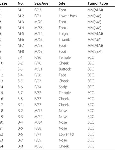
Table 1 Clinicopathological features of 24 malignant skin tumors
Case No. Sex/Age Site Tumor type
1 M-1 F/53 Foot MM(ALM)
2 M-2 F/51 Lower back MM(NM)
3 M-3 M/70 Foot MM(NM)
4 M-4 M/66 Foot MM(NM)
5 M-5 M/54 Thigh MM(ALM)
6 M-6 M/65 Thumb MM(NM)
7 M-7 M/58 Foot MM(ALM)
8 M-8 M/63 Foot MM(SSM)
9 S-1 F/86 Temple SCC
10 S-2 F/76 Cheek SCC
11 S-3 M/51 Buttock SCC
12 S-4 F/86 Face SCC
13 S-5 F/87 Cheek SCC
14 S-6 F/74 Scalp SCC
15 S-7 F/82 Temple SCC
16 S-8 F/77 Cheek SCC
17 B-1 F/67 Cheek BCC
18 B-2 M/75 Nose BCC
19 B-3 M/52 Nose BCC
20 B-4 M/64 Nose BCC
21 B-5 F/68 Nose BCC
22 B-6 F/71 Lower lid BCC
23 B-7 F/65 Nose BCC
24 B-8 M/56 Cheek BCC
Abbreviations: MM, malignant melanoma; ALM, acral lentiginous melanoma; NM, nodular melanoma; SSM, superficial spreading melanoma; SCC, squamous cell carcinoma; BCC, basal cell carcinoma.
Assessment of western blot analysis
The amount of expression in western blotting was mea-sured with TINA software (Version 2.10e). Meamea-sured amount of expression of malignant skin tumors was compared to that of normal skin.
Statistical analysis
The data from the TINA score were analyzed using the nonparametric Mann-Whitney test. A p < 0.05 was con-sidered statistically significant.
Results
Western blot analysis
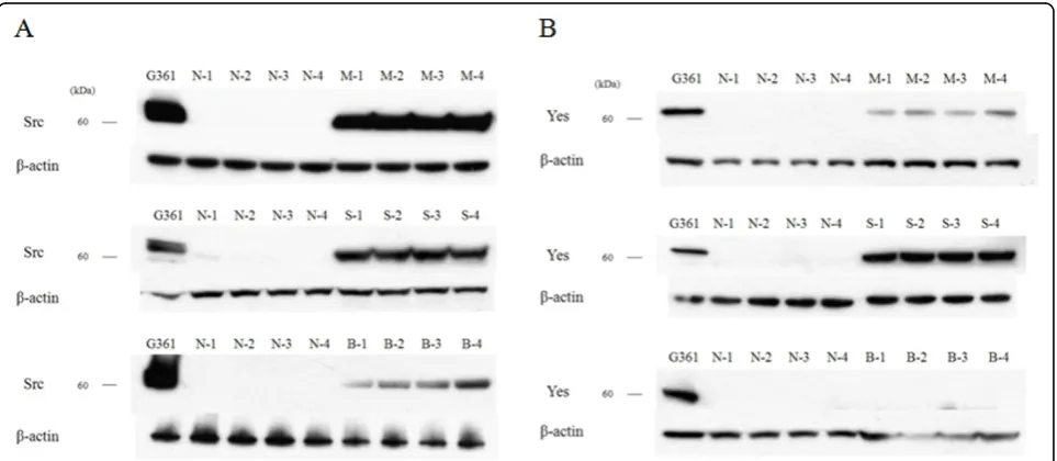
Western blot analysis was performed to determine the expression of c-Src and c-Yes in 18 malignant skin tumors and 6 normal skin tissues. c-Src was expressed in all malignant skin tumors but not expressed in nor-mal skin tissues, while c-Yes was expressed in MM and SCC and not in BCC and normal skin (Fig. 1) (data not shown for M-5, M-6, S-5, S-6, B-5, B-6, N-5, N-6). The expression amount score in western blotting was mea-sured by TINA software (Version 2.10e), and the aver-age TINA score of c-Src was 0.006 in normal skin, 1.143 in MM, 1.027 in SCC, and 0.590 in BCC. The average TINA score of c-Yes was 0.011 in normal skin, 0.374 in MM, 1.054 in SCC, and 0.012 in BCC. There were significant differences in the TINA scores of c-Src between malignant skin tumors and normal skin (p = 0.002). Of the tumor tissues, significant differences in c-Src score among the tumors (p = 0.002) were noted. With regard to c-Yes expression, there were significant
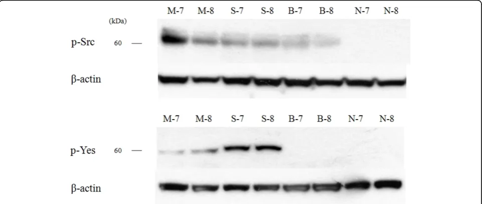
differences in the TINA scores between two skin tumors (MM and SCC) and normal skin (p = 0.002), and there was no significant difference between BCC and normal skin (p = 0.818). The expression amount score based on western blotting is graphed in Fig. 2. To confirm the expression in phosphate form, western blot analysis with phospho-Src and phospho-Yes was also performed in 2 MM, 2 SCC, 2 BCC and 2 normal skin tissues. Phos-pho-Src was expressed in all malignant skin tumors and not expressed in normal skin tissues and phospho-Yes was expressed in MM and SCC but not in BCC and normal skin (Fig. 3).
Immunohistochemical examination
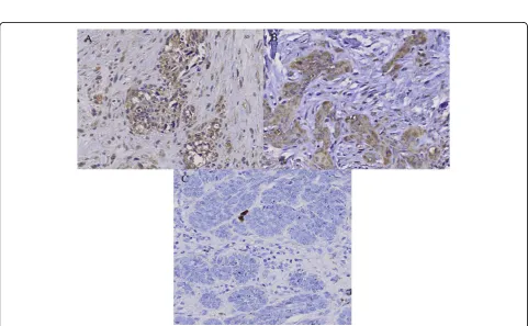
Immunohistochemical study showed that the staining pattern of c-Src and c-Yes in MM, SCC and BCC corre-lated with western blot analysis. c-Src protein was expressed in MM and SCC with moderate positivity, and BCC with mild positivity (Fig. 4). c-Yes was expressed in MM with moderate positivity and SCC with strong positivity, but not in BCC (Fig. 5).
Discussion
The activation and functions of SFKs have been more investigated and better characterized in colon cancer and breast cancer compared to skin cancers. In colon cancer studies, c-Src protein level and kinase activity in the early-stages of colon cancer were found to be greater than in normal colonic mucosa [4,5]. The activ-ity was highest in moderately to well-differentiated colo-nic lesions, while poorly differentiated carcinomas and
normal colonic mucosa showed lower c-Src kinase activ-ity [6]. Therefore, c-Src activactiv-ity is directly related to the malignant potential of the cells, providing evidence that its activation contributes to the progression of colon cancer in the early and developing stages. SFKs also play an important role in the progression of breast cancers. SFK expression, as measured by immunoblotting with
an antibody specifically recognizing Src, Fyn, and Yes, were elevated in 25 of 52 breast tumors. c-Src kinase and STAT3 activated hepatocyte growth factor expres-sion in breast carcinoma cells [7,8]. Enhanced c-Src activity is also one potential mechanism leading to tamoxifen-resistant growth in breast cancer, and activa-tion of c-Src and Fak has a close relaactiva-tionship with
Figure 2The score of expression amount using western blotting. (A) c-Src, (B) c-Yes.
Figure 4Immunohistochemical staining of c-Src in (A) malignant melanoma (MM), (B) squamous cell carcinoma (SCC) and (C) basal cell carcinoma (BCC). c-Src protein is expressed in MM and SCC with moderate positivity, and BCC with mild positivity.
distant recurrence in hormone-treated, ER-positive breast cancer [9]. In recent studies, elevated c-Src activ-ity was directly involved in the disruption of cell-cell adhesions in tamoxifen-resistant breast cancer cell lines, indicating that activated c-Src plays a role in the mislo-calization of adhesion proteins [10]. Therefore, c-Src and c-Yes play important roles in colon cancer and breast cancer.
However, a very small number of studies have been conducted on SFK expression in skin cancer, and there is some controversy as to whether c-Src or c-Yes affects melanoma. By measuring tyrosine-specific kinase activity for c-Src expression in human melanoma tissues kinase activity in melanoma was found to be greater than that in normal skin regardless of the type of melanoma or the metastatic site [11]. In one study, Src kinase inhibi-tor dasatinib inhibited melanoma cell migration and invasion by inducing cell cycle arrest and apoptosis [12]. STAT3, which has been shown to play an important role in tumor cell proliferation and survival, and c-Src tyrosine kinase are activated in melanoma cell lines. Melanoma cells undergo apoptosis when either Src kinase activity or STAT3 signaling is inhibited [13]. This supports the fact that Src activated STAT3 signaling has a key role in the survival and growth of melanoma tumor cells. c-Src activation also affects epidermal growth factor of STAT in head and neck SCCs and pro-motes the invasion and progression of SCC [14-16].
On the contrary, it has been reported that c-Yes expression and kinase activity in human melanoma cell lines are greater than that in normal melanocyte cell lines, and that c-Src expression and activity are not dif-ferent in human melanoma cell lines compared to nor-mal melanocyte cell lines [17]. Similarly, it was demonstrated in another study that c-Yes tyrosine kinase was activated more in human brain-metastatic melanoma cell lines by stimulation of neurotropin and nerve growth factor, whereas c-Src was not affected [18]. These results show that c-Yes is more important than c-Src in melanoma progression and metastasis. Therefore, we studied the expression of both c-Src and c-Yes in overall human skin cancer tissues including MM, SCC, and BCC using western blotting and immu-nochemistry. Our study results show that c-Src was expressed in all skin cancer tissues, but not in normal skin tissues. c-Yes was expressed in MM and SCC, but not in normal skin tissues or BCC. c-Src was found to be significantly more expressed in MM, followed by SCC and BCC by both western blotting and immunohis-tochemistry, and c-Yes was intensely expressed in SCC. In general, MM is the most invasive of the skin tumors, followed by SCC and BCC. Given these facts, our results suggest that c-Src is expressed more in highly aggressive skin tumors, while c-Yes is expressed more in SCC
compared to other skin cancers. We also confirmed that the expression pattern for phosphate Src and Yes forms in skin cancers were similar to the total forms. There-fore, we believe that c-Src, rather than c-Yes, plays a key role in the proliferation and progression of malig-nant skin cancers.
Abbreviations
MM: Malignant melanoma; SCC: Squamous cell carcinoma; BCC: Basal cell carcinoma
Author details
1Department of Plastic and Reconstructive surgery, Hanyang University Guri
Hospital, Guri, Korea.2Department of Plastic and Reconstructive surgery, School of medicine, Sungkyunkwan University, Seoul, Korea.3Molecular Cancer Research Center, College of Medicine, Soonchunhyang University, Chunan, Korea.4Department of Dermatology, National Medical Center, Seoul, Korea.5Department of Dermatology, College of Medicine, Soonchunhyang University, Seoul, Korea.
Authors’contributions
JHL, MKC designed the study and SHN, SHL, YJL carried out western blotting. DWK, MYP and DJJ carried out immunohistochemistry and JHL, MKC drafted the manuscript. J-KP, CHK, SGK participated in the manuscript drafting and performed the statistical analysis. All authors read and approved the final manuscript.
Competing interests
The authors declare that they have no competing interests.
Received: 13 June 2010 Accepted: 27 August 2010 Published: 27 August 2010
References
1. Gloster HM, Brodland DG:The epidemiology of skin cancer.Dermatol Surg 1996,22:217-226.
2. O’Connor TJ, Neufeld E, Bechberger J, Fujita DJ:pp60c-srcin human
Melanocytes and melanoma cells exhibits elevated specific activity and reduced tyrosine 530 phosphorylation compared to human fibroblast pp60c-src 1.Cell Growth & Differentiation1992,3:435-442.
3. Thomas SM, Brugge JS:Cellular functions regulated by Src family kinases.
Annu Rev Cell Dev Biol1997,13:513-609.
4. Rosen N, Bolen JB, Schwartz AM, Cohen P, DeSeau V, Israel MA:Analysis of pp60c-srcprotein kinase activity in human tumor cell lines and tissues.J
Biol Chem1986,261:13754-13759.
5. Bolen JB, Veillette A, Schwartz AM, Deseau V, Rosen N:Analysis of pp60c-src
in human colon carcinoma and normal human colon mucosal cells.
Oncogene Res1987,1:149-168.
6. Weber TK, Steele G, Summerhayes IC:Differential pp60c-srcactivity in well and poorly differentiated human colon carcinomas and cell lines.J Clin Invest1992,90:815-821.
7. Clement J, Sanger J, Berndt A, Kosmehl H, Bohmer FD:Elevated activity and expression of Src-family kinases in human breast carcinoma tissue versus matched non-tumor tissue.J Cancer Res Clin Oncol2001, 127:226-230.
8. Hung W, Elliott B:Co-operative effect of c-Src tyrosine kinase and Stat3 in activation of hepatocyte growth factor expression in mammary carcinoma cells.J Biol Chem2001,276:12395-12403.
9. Planas-Silva MD, Bruggeman RD, Grenko RT, Smith JS:Role of c-Src and focal adhesion kinase in progression and metastasis of estrogen receptor-positive breast cancer.Biochem Biophys Res Commun2006, 341:73-81.
10. Zhao Y, Planas-Silva MD:Mislocalization of cell-cell adhesion complexs in tamoxifen-resistant breast cancer cells with elevated c-Src tyrosine kinase activity.Cancer Letters2009,275:204-212.
12. Eustace AJ, Crown J, Clynes M, O’Donovan N:Preclinical evaluation of dasatinib, a potent Src kinase inhibitor, in melanoma cell lines.J Transl Med2008,6:53.
13. Niu G, Bowman T, Huang M, Shivers S, Reintgen D, Daud A, Chang A, Kraker A, Jove R, Yu H:Roles of activated Src and Stat3 signaling in melanoma tumor cell growth.Oncogene2002,21:7001-7010. 14. Xi S, Zhang Q, Dyer KF, Lerner EC, Smithgall TE, Gooding WE, Kamens J,
Grandis JR:Src kinases mediate STAT growth pathways in squamous cell carcinoma of the head and neck.J Biol Chem2003,278:31574-31583. 15. Koppikar P, Choi SH, Egloff AM, Cai Q, Suzuki S, Freilino M, Nozawa H,
Thomas SM, Gooding WE, Siegfried JM, Grandis JR:Combined inhibition of c-Src and epidermal growth factor receptor abrogates growth and invasion of head and neck squamous cell carcinoma.Clin Cancer Res 2008,14:4284-4291.
16. Nozawa H, Howell G, Suzuki S, Zhang Q, Qi Y, Klein-Seetharaman J, Wells A, Grandis JR, Thomas SM:Combined inhibition of PLCg-1 and c-Src abrogates epidermal growth factor receptor-mediated head and neck squamous cell carcinoma invasion.Clin Cancer Res2008,14:4336-4344. 17. Loganzo F, Dosik JS, Zhao Y, Vidal MJ, Nanus DM, Sudol M, Albino AP:
Elevated expression of protein tyrosine kinase c-Yes, but not c-Src, in human malignant melanoma.Oncogene1993,8:2637-2644.
18. Marchetti D, Parikh N, Sudol M, Gallick GE:Stimulation of the protein tyrosine kinase c-Yes but not c-Src by neurotrophins in human brain-metastatic melanoma cells.Oncogne1998,16:3253-3260.
doi:10.1186/1756-9966-29-116
Cite this article as:Leeet al.:Elevated c-Src and c-Yes expression in
malignant skin cancers.Journal of Experimental & Clinical Cancer Research
201029:116.
Submit your next manuscript to BioMed Central and take full advantage of:
• Convenient online submission
• Thorough peer review
• No space constraints or color figure charges
• Immediate publication on acceptance
• Inclusion in PubMed, CAS, Scopus and Google Scholar
• Research which is freely available for redistribution