EVALUATION OF THE ANTIOXIDANT ACTIVITY OF THE FLAVONOIDS ISOLATED FROM HELIOTROPIUM
SINUATUM RESIN USING ORAC
FL, DPPH AND ESR METHODOLOGIES
GERMÁN BARRIGA-GONZÁLEZ,
*a,bEDUARDO CHAMORRO,
bMELISSA SALINAS,
cBRENDA MODAK,
cCLAUDIO OLEA-AZAR
daUniversidad de Santiago de Chile, Facultad de Química y Biología, Departamento de Ciencias del Ambiente, Av. Libertador Bernardo O’Higgins 3363,
Santiago, Chile.bUniversidad Andres Bello, Facultad de Ciencias Exactas, Departamento de Ciencias Químicas, Avenida República 275, 8370146, Santiago,
Chile. cNatural Product Laboratory, Department of Environmental Sciences, Faculty of Chemistry and Biology, University of Santiago de Chile, Av. Bernardo
O’Higgins 3363, Santiago, Chile. dDepartamento de Química Inorgánica y Analítica, Facultad de Ciencias Químicas y Farmacéuticas, Universidad de Chile,
Santiago, Chile. ABSTRACT
The antioxidant capacity has been determined for a number of flavonoid compounds from Heliotropium sinuatum, a plant that grows in arid areas in the north of Chile. The methodologies used were: ORACFL (oxygen radical absorbance capacity - fluorescein), DPPH (2,2-diphenyl-2-picrylhydrazyl) bleaching and electron spin resonance (ESR). These compounds were studied in homogeneous and heterogeneous media. The results showed that the 7-o-methyleriodictiol and 3-o-methylisorhamnetin are those with the highest antioxidant capacity.
Keywords: ORACFL, ESR, DPPH, flavonoids, micelle, antioxidants.
INTRODUCTION
The increasing interest for natural products with beneficial effects on health, has led to a progressive increase in the study of secondary metabolites synthesized by plants and their biological activities. From this point of view,
one of the most studied compounds are polyphenols, known for their properties
as food antioxidants, agents preventive against cellular aging and diseases
such as cancer.1 To this respect, the specie Heliotropium sinuatum (Miers)
is an endemic resinous bush that grows in the north of Chile in arid areas
with extreme environmental conditions and produce resinous exudates from glandular trichome that covers the leaves and stems as a protection mechanism
against the different types of stress.2 Previous studies have shown that this plant is characterized because its resinous exudate consists mainly of flavonoids which would give to the resin mainly protection against oxidizing agents.2
Recently, it has been suggested that the flavonoids could protects lipids from oxidative damage that could affect the structure and/or function of biological
membranes.1 It is known that changes in the rheology of membranes, can affect biological events at the level of membrane cell, such as transport of metabolites
and interaction membrane-receptor, among others.3-4 The interaction between flavonoids and a bilayer is the result of distribution in lipid-aqueous interface or in the membrane hydrophobic inside. The more hydrophilic flavonoids interact
by hydrogen bonding with the polar head groups at the lipid-water interface
of membranes. This type of interaction may provide a level of protection for
the bilayer from external and internal aggressors (i.e. oxidants) contributing to preserve the structure and function of biological membranes.5 Therefore, the antioxidant activity may vary dramatically in the membranes and lipoproteins.
This has aroused great interest in the methods of quantification and identification of antioxidant capacity. There are different methods to evaluate the antioxidant capacity of flavonoids generally in homogeneous systems, such as methods based on the consumption of different radical in alcoholic solvents when flavonoids are added.6 However, it is necessary to seek of new methods that incorporate heterogeneous environmental, for example the addition of
micelles. These can mimic the inside non-polar and polar interfacial region
relatively similar to the lipid bilayer of the cell membrane.
Based on these antecedents, we now report the results obtained on
the evaluation of the antioxidant activity of the flavonoids isolated from
Heliotropium sinuatum resin, using two different reaction media. Specifically, the used of an ethanolic solution of DPPH (homogeneous system) and a micellar solution of Triton X-100 (micro heterogeneous system). The antioxidant capacity of flavonoids was evaluated through the technique of ESR, ORACFL and bleaching of DPPH radical. Also was measured the distribution constants of these compounds in buffer/micelle media. The different affinities for the micelle, are reflected in the different antioxidant activities when changing the
reaction medium.
Materials and methods
Plant material
Heliotropium sinuatum (Miers) samples were collected in III region, Chile, 29º 57’ S, 71º W. A voucher specimen was deposited in the Herbarium of Natural History Museum of Santiago of Chile (ST2563).
Extraction and isolation of the flavonoids
The resinous exudate was extracted by immersion of the fresh plant mate-rial in dichloromethane for 60 s at room temperature and was concentrated to a
sticky residue. The extract was purified by column chromatography (silica gel)
using a hexano-ethylacetate step gradient yielding 3-o-methylgalangin, 7-o -methyleriodictyol, 3-o-methylisorhamnetin and Pinocembrine.2
Chemical Reagents
5,5-Dimethyl-1-pyrroline-N-oxide (DMPO), 2,2-diphenyl-2-picrylhydra
-zyl (DPPH), Fluorescein sodium salt (FL), 6-hydroxy-2,5,7,8-tetramethylchro
-man-2-carboxylic acid (Trolox), and 2,2’-azobis(2-methylpropionamidine) dihydrochloride (AAPH) were purchased from Sigma-Aldrich. Ethanol, Hy
-drogen Peroxide (H2O2) 30%, Octanol, Triton X-100 micelle, Sodium Hydrox -ide (NaOH), Sodium phosphate dibasic, sodium phosphate monobasic and
N,N-dimethylformamide (DMF) were purchased from Merck.
Kinetic UV–Vis assay for DPPH.
A solution containing 75 μM of DPPH in ethanol and the different fla
-vonoids were prepared in PBS buffer. UV absorbance of the solutions were measured at 517 nm.7-8 The concentration in μM at which the absorbance at 517
nm decreases to 50% of its initial value was used as the IC50 value for each test
solution.7, 9 Kinetic studies were conducted by measuring the disappearance of
DPPH in buffer under pseudo-first-order conditions at 25 °C. Determinations
were conducted in triplicate. The same conditions were used for heterogeneous
medium in presence of Triton X-100 (50 mM).
Oxygen radical antioxidant capacity-fluorescein (ORACFL)
The ORACFL analyses were carried out on a Synergy HT multi detection microplate reader, from BioTek Instruments, Inc. (Winooski, USA), using white polystyrene 96-well plates, purchased from Nunc (Denmark).10 Fluores-cence was read from the top, with an excitation wavelength of 485/20 nm and an emission filter of 528/20 nm. The reaction was carried out in 75 mM sodium phosphate buffer (pH 7.4), and 200 μL final volume. FL (70 nM, final concen
-tration) and flavonoids solutions in buffer with a range of concentration be -tween 10-7 to 10-9 M were placed in each well of 96-well plate. The mixture was pre-incubated for 15 min at 37 °C, before rapidly adding the AAPH solution (15 mM, final concentration). The microplate was immediately placed in the reader and automatically shaken prior to each reading. The fluorescence was recorded every 1 min for 120 min. A blank with FL and AAPH using buffer in
-tions using Trolox (10-4 to 10-6 M) as antioxidant were also done. The inhibition
capacity was expressed as ORACFL values and is quantified by integration of the area under the curve (AUCNET). All reaction mixtures were prepared in trip
-licate and at least three independent assays were performed for each sample.11
The same procedure was performed for the heterogeneous medium.
Kinetic ESR Assays for DPPH
DPPH radical scavenging capacity of the flavonoids was determined by an
ESR spectrometry method.7-8, 12 We chose those flavonoids showing the higher and lower ORACFL values, respectively. We followed the decrease in the in -tensity of the signal showed by DPPH radical. DPPH solution was in the order of μM and the flavonoids concentration was 100 times greater (mM) to ensure a pseudo-first-order decay. The same conditions were used for heterogeneous
medium.
Hydroxyl radical scavenging assay using electron spin resonance (ESR) Reactivity of all the flavonoids against the hydroxyl radical was investi -gated using the non-catalytic Fenton type method.13-14 ESR spectra were re-corded in the X band (9.5 GHz) using a Bruker ECS 106 spectrometer with a rectangular cavity and 50-kHz field modulation, equipped with a high-sensitiv
-ity resonator at room temperature. Spectrometer conditions were: microwave frequency 9.80 GHz, modulation amplitude 0.90 G, time constant 81.92 ms and conversion time 40.96 ms. The scavenging activity of each derivative was
estimated by comparing the •DMPO-OH adduct signals in the antioxidant-rad -ical reaction mixture and the control reaction at the same reaction time, and is
expressed as scavenging percent of hydroxyl radical.11 To prepare the samples, 150 μL of DMF and 50 μL of NaOH (25 mM) were mixed, followed by the addition of 50 μL of DMPO spin trap (0.2 M stock concentration) and finally 50 μL of H2O2 30%. The mixture was put in an ESR cell and the spectrum was recorded after five minutes of reaction. All flavonoids were studied at a concentration of 8 mM.
RESULTS AND DISCUSSION Kinetic UV–Vis assay for DPPH.
The IC50 value (concentration of compound at which 50% is consumed DPPH) for the antioxidant capacity of the different compounds was determined.
IC50 values are summarized in Table 1.
Table 1. IC50 values in homogeneous medium for flavonoids studied.
Flavonoids IC50 (M)
Naringenin 1.85x10⁻³
3-o-methylgalangin 1.59 x10⁻³ 7-o-methyleriodictiol 4.03 x10⁻⁵
3-o-methylisorhamnetin 8.77 x10⁻⁵
Pinocembrin 2.76 x10⁻³
From the values shown in Table 1, it is possible to establish the following trend in relation to the antioxidant capacity of flavonoids studied: 7-o-methyleriodictiol > 3-o-methylisorhamnetin > 3-o-methylgalangin > naringenin > pinocembrin. This trend is based on the number and position
of phenolic hydroxyl groups present in the flavonoids. In general terms, the antioxidant activity of these compounds is mainly influenced by the number
and positions of the hydroxyl groups in ring A and B, in addition to the
conjugation present in rings B and C. Table 2 summarizes the IC50 values when
performing the test in heterogeneous media.
Table 2. IC50 values in heterogeneous medium for flavonoids studied using
Triton X-100 micelle.
Flavonoids IC50 (M)
Naringenin 2.34x10⁻³
3-o-methylgalangin 1.89x10⁻³
7-o-methyleriodictiol –
3-o-methylisorhamnetin –
Pinocembrin 3.05x10⁻³
It was not possible to determine the IC50 for compounds 7-o-methyleriodictiol
and 3-o-methylisorhamnetin because increasing their concentration does not
decrease the absorbance of DPPH. This behavior may be due to problems of
distribution of the reactants in the micelle.
The naringenin, 3-o-methylgalangin and pinocembrin compounds follow
the same trend observed in the homogeneous medium (Table 1). Therefore,
the following relationship is obtained by using heterogeneous medium: 3-o-methylgalangin > naringenin > pinocembrin. Figure 1 shows the typical
kinetic profile obtained for the decay of DPPH radical using naringenin as
antioxidant.
Figure 1. Shows the kinetic profile for naringenin as antioxidant in
homogeneous media.
Oxygen radical antioxidant capacity-fluorescein (ORACFL)
Once IC50 values were obtained, we proceeded to perform the ORACFL assay. Table 3 summarizes the values in Trolox equivalents for each of the
studied compounds.
Table 3. ORACFL values for the flavonoids studied in homogenous media.
Flavonoids ORACFL Value
Trolox 1.0 + 0.01
Naringenin 4.19 + 0.22
3-o-methylgalangin 7.18 + 0.17 7-o-methyleriodictiol 14.62 + 0.39 3-o-methylisorhamnetin 10.91 + 0.18
Pinocembrin 3.22 + 0.16
The values show that compounds 7-o-methyleriodictiol and
3-o-methylisorhamnetin exhibit higher ORACFL values that many widely used antioxidants (quercetin has an ORACFL value of 7.28 ± 0.22 and is considered as one of natural flavonoids with higher antioxidant activity).
Thus, these results indicate that 7-o-methyleriodictiol is the compound with the highest antioxidant capacity, followed by 3-o-methylisorhamnetin, 3-o-methylgalangin, naringenin and pinocembrin being the compound with the
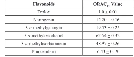
lowest antioxidant capacity. We proceeded to perform this test in heterogeneous media. The values obtained in this environment are summarized in Table 4.
Table 4. Summarizes the obtained ORACFL value in heterogeneous media
for the antioxidant studied.
Flavonoids ORACFL Value
Trolox 1.0 + 0.01
Naringenin 12.20 + 0.16
3-o-methylgalangin 19.53 + 0.25
7-o-methyleriodictiol 62.54 + 0.32 3-o-methylisorhamnetin 48.97 + 0.26
Micellar ORACFL values have the following high to low antioxidant capacity trend: 7-o-methyleriodictiol > 3-o-methylisorhamnetin > 3-o-methylgalangin > naringenin > pinocembrin. In order to explain these
results in heterogeneous media (specifically we used triton X-100 micelle) we calculated free fluorescein concentration [FL].15
We obtained a value of 74% of free fluorescein. This indicates that the fluorescein is not a good alternative as a probe to evaluate the antioxidant
capacity in heterogeneous media using the Triton X-100 micelle, and only
26% of fluorescein would be entering to the micelle. Probably there is not a
complete interaction of the probe molecule with the antioxidant, which may
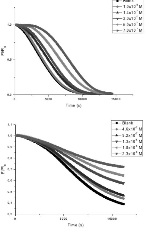
explain the extensive delays that exists in the decrease in fluorescence intensity over time. Figure 2 shows the behavior previously described for naringenin and
pinocembrin compounds in different medium.
Table 5. Summarizes the values obtained for the selected antioxidants
studied in ESR in homogeneous media.
Flavonoids k2 (M-1s-1)
Naringenin 0.11 + 0.01
7-o-methyleriodictiol 1.75 + 0.14
Table 6 shows the decay values constants for the above mentioned compounds using heterogeneous medium. It is possible to realize that the trend
obtained in a homogeneous medium remains when using the heterogeneous medium.
Table 6. Summarizes the values obtained for the selected antioxidants
studied in ESR in heterogeneous media.
Flavonoids k2 (M-1s-1)
Naringenin 0.17 + 0.02
7-o-methyleriodictiol 10.54 + 0.26
Figure 3 shows the kinetic profile in both medium for 7-o-methyleriodictiol.
These profiles were used to obtain the values of the constants summarized in Table 5 and 6 respectively.
Figure 2. Left side shows the kinetic profile for naringenin in homogeneous media. Right side show the kinetic profile with extensive delays
for pinocembrin in micellar media.
Kinetic ESR Assays for DPPH
Decay kinetics assays were performed for compounds: Naringenin and 7-o-methyleriodictiol in the presence of the DPPH radical. The following experimental conditions were used: DPPH (0.052 mM) and compound
concentrations of 5.5 mM and 10.50 mM in the case of naringenin.
Table 5 shows the the decay values constants obtained. It is clearly, that the decay constant is much greater for 7-o-methyleriodictiol compared to
naringenin. Indicating that 7-o-methyleriodictiol has a higher reactivity with
DPPH radical. corresponds to a homogeneous media and right side shows the profile using Figure 3. Kinetic profile for 7-o-methyleriodictiol using ESR. Left side
Hydroxyl radical scavenging assay using electron spin resonance (ESR) The capacity of the studied compounds to quench the hydroxyl radical reactivity using a non-catalytic method was also determined. Table 7 summarizes the values expressed as percentage of decrease with respect to a
blank solution.
Table 7. Summarizes the signal decrease expressed as percentage.
Flavonoids % Decrease OH
Naringenin 47%
3-o-methylgalangin 59%
7-o-methyleriodictiol 70%
3-o-methylisorhamnetin 68%
Pinocembrin 24%
It is observed that the compound 7-o-methyleriodictiol quenched in a 70% the hydroxyl radical signal, proving to be the one with a higher reactivity
towards this radical, being followed by 3-o-methylisorhamnetin with a very close value to 68%. Is possible to establish a trend in decreasing order of reactivity of quenching: 7-o-methyleriodictiol > 3-o-methylisorhamnetin > 3-o-methylgalangin > naringenin > pinocembrin. Figure 4 shows the spectra obtained when naringenin was used as antioxidant.
Figure 4. Upper shows the spectra for the blank solution. Lower shows the
decrease of the signal when naringenin was added as antioxidant.
Based on the obtained results in homogeneous media, we propose the following decreasing order of antioxidant capacity for the compounds under
study: 7-o-methyleriodictiol > 3-o-methylisorhamnetin > 3-o-methylgalangin
> naringenin > pinocembrin. We explain these results mainly based on the
number of phenolic hydroxyl groups which help to increase the antioxidant capacity. It is then noted that the studied compounds possess three hydroxyl
groups (naringenin, 7-o-methyleriodictiol and 3-o-methylisorhamnetin)
and two hydroxyl groups (3-o-methylgalangin and pinocembrin). However, the number of hydroxyl groups is not sufficient to explain the improved
antioxidant capacity, thus the presence of an o-dihydroxy structure in the B
ring, confers more stability to radical form enhancing the delocalization of
electrons. A double bond in conjunction with the 4-oxo function of the C ring.
Hydroxyl substituents at position 3 and 5 with 4-oxo function on the rings
A and C necessary to increase the antioxidant capacity. The presence of the double bond between carbons 2 and 3 of the C ring, because it enhances the
resonance stabilization of the radical formed.
Thus, it is observed that pinocembrin is one of those that has less hydroxyl groups, these being located on C5 and C7 position of ring A. It has
no double bond between C2-C3 carbons of the ring C, which would explain a lower capacity as an antioxidant. Naringenin bears a greater number of
hydroxyl groups located at C5 and C7 of ring A and in position 4’ of the ring B. This lower attributed to the radical resonance stabilization formed.
Instead, 3-o-methylgalangin has less hydroxyl groups but has a double bond between positions C2 and C3 of the C ring, in addition hydroxyl groups at
C7 and C3 position. The latter could be stabilized with the methoxy group at
the C3 position enhancing antioxidant capacity, which would explain a better antioxidant capacity compared to naringenin.
Compound 3-o-methylisorhamnetin exhibits improved antioxidant
capacity with respect to the aforementioned compounds because it has a double bond between carbons 2 and 3, it has a methoxy group at position C3 and
finally a hydroxyl group at the C5 position. This latter group presents a lower influence with respect to the catechol group located in the B ring of compound 7-o-methyleriodictiol. This substituent improves antioxidant properties due to a higher stabilization of the radical caused by the abstraction of H atom at position C4’, forming a hydrogen bond with the hydroxyl group at C3’ position.
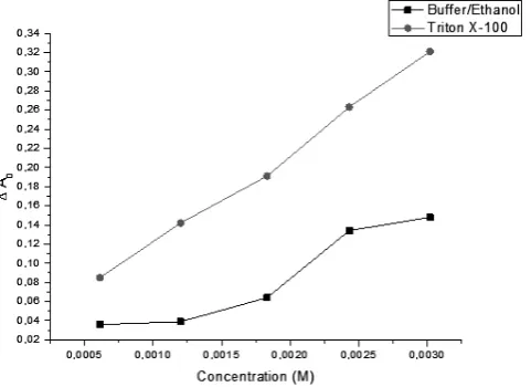
In order to rationalize the results between the different media used, we analyzed the effects of the medium used in the different measurements. It is possible to observe that the IC50 values obtained for the flavonoid do not allow for the differentiation of the antioxidant activity between the media used. Therefore, to compare the activity in the different media we used the variation of the initial absorbance (defined as ΔA0 = A0-A10)
This parameter allows for the estimation of the initial velocity of DPPH radical consumption. The analysis of this parameter gives some idea of the efficiency of the compounds as antioxidants in different media. We determined the initial absorbance variation (during the first 10 seconds) depending on the concentration of the compounds. Thus, it is possible to observe a difference of ΔA0 parameter throughout the concentration range studied in the different media used, demonstrating the influence of environment on the process speed. Figure 5 shows the initial absorbance variation for naringenin in both medium.
Figure 5. Initial absorbance variation (ΔA0) for naringenin in both medium.
Then, these compounds are favored by the micelle, improving the process speed of reaction between these compounds and the DPPH radical. Most favored compounds using heterogeneous medium are: Naringenin,
3-o-methylgalangin and pinocembrin. For compounds 7-o-methyleriodictiol and 3-o-methylisorhamnetin it was not possible to determine their IC50 values.
From the results obtained through the ORACFL method, we can argue that the
tendency of antioxidant capacity found is maintained independently of the medium used. Finally, ESR measurements show an increase in speed when
switching from homogeneous to heterogeneous medium environment.
CONCLUSIONS
Studied flavonoids from H. sinuatum showed an antioxidant activity which can be measured with different methods such as: DPPH, ORACFL and ESR.
DPPH assay in terms of IC50 indicated that the flavonoid 7-o-methyleriodictiol has the highest antioxidant capacity against this radical.
In contrast, using the DPPH assay in heterogeneous medium demonstrated that
the IC50 method is not an accurate parameter to evaluate the antioxidant capacity of these flavonoids. In the ORACFL assay, it is important that the fluorescent probe does not have a high affinity for the micelle, which can affect the results.
ESR assay demonstrates that there is an increase in the decay rate and therefore
in the reactivity of the flavonoids in a micellar medium. Reactivity towards the hydroxyl radical of the flavonoids studied presents the same trend obtained
with the different assays used to determine the antioxidant capacity.
In addition, it is observed that these compounds conserve their antioxidant
properties independent of the method and medium used.
ACKNOWLEDGEMENTS
Germán Barriga González thanks financial support by FONDECYT grant
N° 3120241. Eduardo Chamorro thanks financial support by FONDECYT N° 1140343, Nucleo UNAB DI-219-12N and The Millenium Science Initiative Nucleus No. 120082.
REFERENCES
1. K. B. Pandey; S. I. Rizvi, Oxid. Med. Cell. Longev.2, 270, (2009) 2. R. Torres; B. Modak; L. Villaroel; A. Urzúa; F. Delle-Monache; F.
Sánchez-Ferrando, Bol. Soc. Chil. Quím.41, 195, (1996)
3. M.-L. Tomassoni; D. Amori; M. V. Magni, Biochem. Biophys. Res. Commun.258, 476, (1999)
4. T. G. Niranjan; T. P. Krishnakantha, Mol. Cell. Biochem.204, 57, (2000) 5. P. I. Oteiza; A. G. Erlejman; S. V. Verstraeten; C. L. Keen; C. G. Fraga,
Clin. Dev. Immunol.12, 19, (2005)
6. A. M. Pisoschi; G. P. Negulescu, Biochem. Anal. Biochem.1, 1, (2011) 7. L. Yu; S. Haley; J. Perret; M. Harris; J. Wilson; M. Qian, J. Agric. Food
Chem.50, 1619, (2002)
8. J. Rodríguez; C. Olea-Azar; C. Cavieres; E. Norambuena; T. Delgado-Castro; J. Soto-Delgado; R. Araya-Maturana, Bioorg. Med. Chem.15,
7058, (2007)
9. R. Torres; F. Faini; B. Modak; F. Urbina; C. Labbé; J. Guerrero,
Phytochemistry67, 984, (2006)
10. B. Ou; M. Hampsch-Woodill; R. L. Prior, J. Agric. Food Chem.49, 4619,
(2001)
11. M. J. Matos; F. Pérez-Cruz; S. Vazquez-Rodriguez; E. Uriarte; L. Santana; F. Borges; C. Olea-Azar, Bioorg. Med. Chem.21, 3900, (2013)
12. M. Espinoza; C. Olea-Azar; H. Speisky; J. Rodríguez, Spectrochim. Acta Mol. Biomol. Spectros.71, 1638, (2009)
13. Y. Yoshimura; T. Inomata; H. Nakazawa; H. Kubo; F. Yamaguchi; T. Ariga,
J. Agric. Food Chem.47, 4653, (1999)
14. M. Polovka; V. Brezová; A. Staško, Biophys. Chem.106, 39, (2003) 15. C. Lucas-Abellán; I. Fortea; J. A. Gabaldón; E. Núñez-Delicado, J. Agric.