Recent Advances in the Synthesis and Applications of Collagen Based
Hydrogels: A Review
Jesús A. Claudio-Rizoa*, Laura Espíndola-Sernab, Juan J. Becerra-Rodriguezb, Lucia F. Cano-Salazara and Tirso E. Flores Guíaa.
b Ingeniería en Biotecnología, Universidad Politécnica de Pénjamo, Carretera Irapuato-La Piedad Km 44, C.P. 36921, Pénjamo, GTO, México.
Article Received: 19 February 2018 Article Accepted: 07 March 2019 Article Published: 06 April 2019
1. INTRODUCTION
Collagen hydrogels have demonstrated to have properties that make them candidates for application in
BTERM; however, their rapid degradation and weak mechanical properties limit their use. An accurate
control of the structural characteristics and properties of the collagen based hydrogels could provide the key
to regulate their behavior for in BTERM improved applications. Collagen has been widely used to prepare
biomaterials, since it represents the majority of the composition of the animal connective tissues [1]. From a
biochemical point, the main constituent of the extracellular matrix (EC M) is the collagen; this molecule is
formed in vivo by polymerization reactions in stages with enzymatic regulation among the conformer amino
acids [2]. To date, more than 20 types of collagen have been identified, varying in their amino acid sequence,
mainly [3]. The collagen polypeptide chains have a primary structure consisting of the sequence
Gly-X-Y-Gly-Pro-Hyp-Gly-X-Y where every third amino acid is glycine (Gly). Glycine comprises about
33% of the chain of amino acids while proline (Pro) and hydroxyproline (Hyp) comprise about 20%. The rest
of the percentage is composed of the polar amino acids, both acidic and basic, represented as X and Y [4].
The polypeptide chain of the collagen has amino functional groups ( -NH2), carboxylic acids (-COOH) and
hydroxyls (-OH) as substituents, which together with the amide bonds of the polymeric backbone represent
reactive centers. The secondary structure is defined by the local configuration of the chains resulting from
the satisfaction of stereochemical angles and the capacity of interaction via hydrogen bonding. A B S T R A C T
Collagen is a triple helical protein present in the connective tissue and bones of mammals. The collagen has the ability to polymerize by adjusting the pH and temperature, generating fibrillar matrices in the hydrogel state. Collagen hydrogels have shown great potential for applications related to biomedicine, tissue engineering and regenerative medicine (BTERM), mainly due to their high biocompatibility and ease of processing. However, collagen hydrogels have poor mechanical properties and high rate of proteolytic degradation, limiting their applicability for in vivo tests that demand control in said properties. In order to combat the aforementioned disadvantages, systems of collagen-based hydrogels have been designed in combination either with natural polymers, synthetic polymers, inorganic particles and/or complex biological molecules, demonstrating to regulate the self-assembly processes of the collagen molecules, forming crosslinked and/or interpenetrated polymeric networks based hydrogels. In addition, the physicochemical interactions among the collagen molecules and the functionalization agents regulate the structure and properties of the 3D hybrid networks generated, enhancing their application in specific fields of BTERM. This work summarizes the most recent strategies for the synthesis and applications of collagen-based hydrogels with diverse chemical components that have been designed for BTERM. Finally, this work also illustrates the importance of the properties of collagen-based hydrogel systems and their potential use in other future biotechnological applications.
Keywords: Collagen hydrogels, chemical functionalization, biomedicine, Tissue engineering, Biotechnology
The tertiary structure gives rise to the collagen molecule consisting of three polypeptide chains arranged in
a triple helix configuration. The collagen molecules form a supramolecular structure comprising five
molecules packed in a specific arrangement, generating a collagen microfibril. The microfibrils are coupled
in specific series forming a collagen fiber; these fibers generate bundles of fibers to give rise to much larger
collagen fibers [5].
The collagen molecule, also called tropocollagen, has approximately a total of 240 ε -amino groups as well as
230 carboxylic groups and 300 nm in length [5]. Under physiological conditions these groups are charged. In
its natural state, collagen configurations are maintained by covalent chemical bonds, hydrogen bridge-type
interactions, ionic and hydrophobic interactions. The functional groups in the collagen interact with each
other to form intra or intermolecular bonds providing stability to the collagen fibers. Under these co nditions,
only a small number of charged groups are free but the electrostatic state of the collagen can be altered by the
change in pH of the medium, resulting in the weakening of intra - or intermolecular electrostatic interactions
[6]. The ends of the collagen triple helices are terminals with –NH2 and –COOH functional groups without
helical configuration called N- and C-terminal telopeptides, respectively, which produce crosslinking for the
stabilization of collagen and the formation of fibers. These non -helical ends are also related with the
formation of immune response reactions [4].
The versatility of collagen as a building biomaterial is mainly due to its complex hierarchical structure
originated from its molecular sequence. The tropocollagen molecu les are assembled into complex
supramolecular aggregates by a polymerization process called fibrilogenesis; this is a process driven by
entropy, this process has also been presented for other protein self -assembly systems [7]. It is driven by the
loss of solvent molecules from the surface of the protein molecules and results in assemblies of longer
fibrillar structures with conical ends of circular cross section, which minimize the surface area to volume
ratio of the final assembly. The interactions that direct the association between tropocollagens are mostly
electrostatic and hydrophobic [8]. The stabilization of such interactions is ensured by the existence of
molecular crosslinking, which it occurs among the telopeptides of the collagen molecules with th e main
chain of the helix [9]. The process of fibrilogenesis can be carried out in situ by extracting collagen of
diverse ECMs to generate collagen-based hydrogels. The hydrogels are polymeric materials swollen in water
that maintain a specific three-dimensional structure. They are very suitable materials for BTERM
applications due to their good interaction with living tissues, since on the one hand they show good
biocompatibility properties, mainly due to their soft, elastic consistency and water content. Besides, their
characteristic of swelling brings them the property of absorbing, retaining and releasing under controlled
conditions quantities of water that regulates their structural conformation, desirable properties for BTERM
Specifically, the hydrogels formed from the partial digestion, solubilization and in situ polymerization of
decellularized tissues can retain the biological activity of the native ECM. Therefore, these hydrogels have
complex biochemical compositions, fibrous protein content (e.g., collagen, fibrin, elastin), proteoglycans
(PGs), and glycosaminoglycans (GAGs) that closely mimic the native 3D tissue environment, compared to
simple composition hydrogels [12]. The remaining ECM molecules after extracting the coll agen have a
direct relationship with the in situ polymerization process, structure and properties of the generated
hydrogels. A higher content of matrix proteins improves the mechanical properties and proteolytic
degradation rate, exhibiting enhanced biocompatibility properties [13]. The main ECMs used to extract
collagen and to generate hydrogels are: rat tail tendon [14], bovine Achilles tendon [15], bovine pericardium
[13], porcine intestinal submucosa [16], porcine bladder [17], fish skin [18], and porc ine skin [19].
These collagen based hydrogels have shown bioactivity and biocompatibility associated with their residual
composition. Recently, a review has been reported about diverse ECMs employed in the design of
biomedical hydrogels, describing the main role of collagen based hydrogel with characteristic composition
on the improved applicability [20]. Generally, the collagen hydrogels have been studied as substrates for
ophthalmology, sponges for burns/wounds, systems for controlled delivery of functio nal molecules or
nanoparticles, and matrices for 3D cell culture. They are also investigated for tissue engineering including
skin replacement, bone substitutes, and artificial blood vessels and heart valves [21].
Although collagen hydrogels show excellent biocompatibility and biological performance, there is the
challenge to tailor their physicochemical properties for an enhanced application in BTERM, as they ha ve
poor mechanical properties and fast degradation, limiting their use as cargo delivery systems or long -term
dressings, among others [22].
Therefore, it is still necessary to investigate methods to regulate the characteristics and to expand the use of
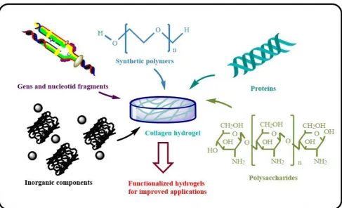
collagen based hydrogels in the medical biotechnology field. Recent approaches to combat this need include
functionalization with organic phases, such as biopolymers or biological molecules with specific activity,
synthetic polymers of defined chemical architecture and inorganic phases, such as nanoparticles, nanotubes,
bioglasses (Fig. 1).
The presence of these coupling phases on the collagen polymerization process has been shown to control the
kinetics and microstructure of the hybrid fibrillated matrices i n the hydrogel state, where the concentration
of the coupling agent is determinant in the tailoring of the physicochemical and biological properties [23]. In
this sense, it is important to indicate that the alteration of the properties of the collagen base d hydrogels is
associated with the generation of interpenetrating network systems (IPNs), covalent crosslinking bonds and
inter- and/or intra-molecular interactions of the collagen fibers with the coupling molecules [24]. Few
reviews have been reported indicating strategies to generate functionalized collagen hydrogels with
diversity of chemical phases; therefore, the objective of this work is to show the relationship of the
composition of these hybrid matrices, detailing the strategies of the process of sy nthesis, crosslinking
methods, highlighting the improved properties of the systems and their application in some BTERM field.
Finally, we also mention the challenges, possibilities and perspectives of application of these hydrogel
systems in different areas of the biotechnological field.
2. COLLAGEN CROSSLINKING
The generation of collagen hydrogels with a variety of coupling agents requires strategies to reinforce their
basic properties for specific applications in the biotechnological field. Structurally, the fibrous collagen
network must be reinforced mechanically to support the load of the coupling materials. Generally,
crosslinking strategies have been developed for this purpose. Crosslinking, in a specific way, consists in the
generation of points of union among the triple helical chains of collagen. These int ra- or inter-molecular
interactions can be established physically; by means of programmed freezing -drying cycles to achieve a high
entanglement of the collagen fibers, considerably improving their mechanical properties and altering the
swelling capacity of the generated hydrogel [24]. In this strategy, the control of the degradation rate of the
hydrogel is not affected, so that the time of use of the materials under proteolytic conditions is limited [25].
The physical crosslinking can also carry out by the variation of the pH, temperature, electrical fields or other
physical stimuli [26]. This type of crosslinking offers advantages such as, relatively easy manufacture and
the rate of collagen degradation and improvement in its mechanical properties are obtained by using
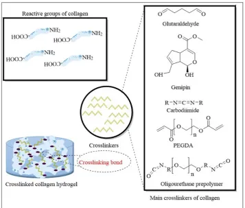
chemical crosslinking approaches (Fig. 2).
The chemical crosslinking approaches are based on the generation of covalent bonds amon g the functional
groups amine or carboxylic acid of the collagen with exogenous molecules. These methods increase the
resistance of the hydrogel toward both chemical and enzymatic degradation, reduce its immunogenicity,
sterilize and improve its mechanical properties. However, a direct evidence between the concentrations of
chemical crosslinkers with the biocompatibility has been determined in several works. In this sense, it is
advisable to use low concentrations of crosslinker in order not to reduce the c haracteristic biocompatibility
of collagen. The collagen crosslinkers most studied are the glutaraldehyde (GLU), carbodiimides
(specifically, 1-Ethyl-3-(3 -dimethylaminopropyl) carbodiimide, a water-soluble carbodiimide) (EDC),
genipin (GEN), 1,3-phenylenediacetic acid (APH), polyethylene glycol diacrylate (PEGDA) and aqueous
polyurethane prepolymers (PPU). In the collagen hydrogels crosslinked with GLU, the ε -amine groups of
collagen yield an imine bond (so-called as Schiff base); the crosslinking reaction is relatively fast; reacting
with most ε-amine groups, improving both mechanics and degradation resistance, but it has been observed a
drastic reduction of the biocompatibility [28].
Figure 2. Approaches to chemically crosslink to the collagen to gener ate hydrogels with improved
properties.
When EDC is used as crosslinker, results as effective catalyst in the condensation of collagen carboxylic
crosslinking bonds, thus the degradation products of these hydrogels do not show cytotoxic character;
however, the crosslinking reaction is not taken out at physiological conditions (pH 7.4, 37 °C) [29].
Diverse strategies for the preparation of collagen hydrogels are based on the use of GEN as chemical
crosslinker, as a spontaneous crosslinking by formation of Schiff base is produced by a Michael reaction
involved in this process. The structure and properties of hydrogels show a direct relationship with the GEN
concentration; a notable disadvantage to consider is the generation of blue residues during the preparation of
hydrogels, limiting their transparence and use as 3D culture systems [30].
Hydrogels were successfully formed via functionalization of collagen with A PH as novel and bifunctional
segment crosslinker; the functionalization with the aromatic segment provided the formation of a triple
helical covalent network with tunable crosslinking density and swelling ratio, whereby enhanced mechanical
properties. Particularly based on their bioactivity and full biocompatibility at low APH concentrations, these
hydrogels have significant appeal for applications in mineralized tissue regeneration [31].
The use of synthetic polymers with reactive ends to generate covalent crosslinking bonds is an interesting
approach to tailor the properties of the collagen hydrogels and to functionalize with other components.
Strategies using PEGDA to induce photo crosslinking based on the formation of covalent linkages among the
functional groups acrylamide with the collagen-amines. Under this approach, the collagen hydrogels show
enhanced hydrolytic stability and susceptibility to collagen enzymatic degradation; showing also direct
dependency of the mechanical properties on time of UV irradiation. Limitations related with to use of UV
irradiation for applications to formation of hydrogel in situ or cell encapsulation must be considered [32].
Crosslinking agents with high solubility in water are required to potentiate the chemical crossli nking
reactions with hydrolyzed collagen precursors, thus avoiding the use of solvents or phases that can decrease
the biocompatibility. In this sense, recently the use of PPU based on poly(ethylene glycol) (PEG) and
aliphatic diisocyanates as crosslinker of the collagen chains has been reported. The process involves the
formation of urea linkages between end-blocked isocyanate of PPU and collagen amines. The crosslinking
process is taken out at physiological conditions. The structure and properties of coll agen hydrogels show a
direct relationship with the chemical structure of PPU. The use of soluble PPU around 15 % weight
accelerates the collagen polymerization forming consistent hydrogels in 20 minutes. Higher concentrations
of PPU inhibit the collagen polymerization, and decrease its biocompatibility [33].
The main component of the synthetic collagen crosslinkers discussed is PEG. Recently, a review of the state
of the art of physicochemical modifications made by PEG in collagen -based hydrogels has been established
[20]. This work highlights the use of PEG derivatives, widely used in formulations of pharmaceutical
products or in synthesis of biomedical polyurethanes, as a strategy to modulate both physical and biological
properties of natural ECM hydrogels, providing a comparison of the characteristics of hybrid hydrogels in
3. TYPES OF COLLAGEN-BASED HYDROGELS
The crosslinking process of the collagen chains is important because it allows to generate functionalized
hydrogels with a variety of coupling agents, such as: natural polymers, synthetic polymers, inorganic
particles and complex biological molecules. This section covers the state of the art in the dev elopment and
study of the properties of these hybrid hydrogels in the field of BTERM, highlighting the innovation in the
formulation, the crosslinker used, the characterization techniques, and the main advantages offered for their
applications in some area of BTERM.
3.1 Natural polymers
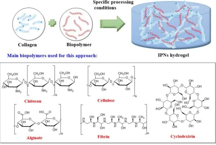
The high both biocompatibility and bioavailability represented by natural polymers, such as chitosan, fibrin,
elastin, alginate, cellulose, cyclodextrin, poly(lactic acid) PLA, among others, represents significant
importance to develop systems of collagen-coupled hydrogels (Fig. 3).
Figure 3. Production of collagen hydrogels functionalized with natural polymers.
Natural polymers can be extracted from biomass, generating systems of materials with natural sustainability,
novel properties that can be exploited in the biotechnological field, ensuring low economic cost in their
production. The physicochemical combination of the hydrolysed collagen chains with biopolymer molecules
can generate hydrogels of IPNs. In this type of system, collagen chains are physically crosslinked, producing
entanglements that modify the properties of collagen and that of the coupling agent . If the coupling
the properties of the hybrid hydrogel. In table 1, the main systems of collagen hydrogels coupled with natural
polymers are summarized. Degradation rate control represents a major challenge in this hydrogels system,
since the components are naturally degraded. If the hydrogel is used for tissue repair or regeneration, it is
important that the hydrogel is degraded as the new tissue is formed. The by-products of degradation do not
show a cytotoxic character related to the biocompatibility of biopolymers. For long -term performance
applications, it is important to control their rate of degradation; which is achieved using exogenous
crosslinking molecules that ensure the formation of covalent bonds, inhibiting the proteolytic degradation of
the hydrogel. The reinforcement of the mechanical properties of these hydrogels should also be considered
when it is required to incorporate molecules with biological interest, such as drugs or cytokines that benefit
metabolic pathways of interest in the BTERM area; again the crosslinking process is important to ensure
Collagen source
Coupling agent Collagen crosslinker
Processing conditions
Novel features Characterization methods
In vitro / In vivo
results Ref. Bovine Achilles tendon Thiolated hyaluronic acid
--- pH 7.4 at 37 °C, 5% CO2 for ∼1 h
The physical characteristics of hydrogel can be
modulated through the variation of Hyaluronic Acid (HA) concentration, which, directly influences the glioblastoma multiform cells (GBM) behavior.
The collagen-gel offer a 3D microenvironment to tumor cell studies. Mechanical analysis, confocal reflectance microscopy, SEM, cell spreading assay and migrations experiments.
The HA concentration of the hydrogel induces cellular morphological
changes and variation in spreading and migration of GBM cells.
[34]
Soft coral
Sarcophyto n
Alginate EDC-NHS 48 h, RT The stiffness, strength of alginate-collagen hydrogel was more elevated than alginate hydrogel.
Hyperelastic behavior similar to human tissues.
SEM, DIC and mechanical analysis
Hydrogel mechanical properties was dependent on the collagen concentration and orientation and arrangement of collagen fibers.
[35]
Rat tail tendon
Alginate, CaSO4 and Na2HPO4
--- 4°C for 20 min, pH 7, 37°C for 1.5 h.
Increased mechanical properties and osteoconductivity. The hydrogel contains collagen and apatite phosphate, components of natural bone.
SEM, DMA, TGA, XDR and FTIR
Injectable biocompatible hydrogel system with gelation time variable, from 5 to 10 minutes for bone tissue regeneration. [36] Bovine Achilles tendon Dialdehyde carboxymethyl cellulose (DCMC)
--- Centrifugate d at 2500 rpm, 4°C, 20
Spongy collagen cryogels of fast swelling rate.
Improved thermostability.
LLS spectroscopy, FTIR, SEM, DSC and swelling
The swelling is dependent of DCMC content and pH.
min, frozen at -20°C for 5 days and thawed at RT.
Good blood compatibility analysis. Increased DCMC in cryogel decreases hemolysis ratio and increases antithrombotic activity. Porcine dermis Microcrystalline cellulose and 1-butyl, 3-methylimidazolium chloride ([C4mim]Cl)
--- 100°C under N2 for 2 h
Structure macroporous 3D with actives amino groups to increased binding Cu(II). Reusable until 4
absorption/desorption cycles before their broken. Useful for wastewater bioremediation SEM, FTIR, mechanical analysis and absorption/desor ption assays for Cu(II)
quantification.
The absorption capacity is related to collagen/cellulose mass ratio. The pH optimum to
absorption is 6 in all study cases. The hydrogels were desorbed at 95% efficiency. [38] Rat tail tendon Bacterial cellulose Gluconoacetobacter xylinus
EDC-NHS RT for 24 h, dried to 37°C
The hydrogel induced cell growth for bone
regeneration purposes. Its uses do not show cytotoxic, genotoxic and mutagenic effect.
SEM, FTIR, 31P NMR, TGA, mechanical analysis and cytotoxicity, genotoxicity and mutagenic assay.
The hybrid hydrogel containing osteogenic growth peptide stimulated the early development of osteoblastic phenotype and enhanced cell growth.
[39]
Collagen (BD Biosciences )
Chitosan --- 37°C for 30 min in a humidified 5% CO2 incubator
Mechanical properties suitable for survival,
maturation and performance of cardiomyocytes.
Stable for 7 days in vivo
SEM, rheological analysis, live/dead, enzymatic degradation and LDH cytotoxicity assay, and histological and immunofluoresce Immobilizing of Angiopoietin-1 peptide, allowing to the cardiomyocytes can survive within the hydrogel both in vitro
and in vivo conditions. The hydrogel injected in Lewis rat model was
nt staining. localized in the injection site one week after. Fish skin Chitosan and gelatin
GLU
-20°C for 24hand lyophilize until dried.
The component influence to gel properties, so the largest swelling is for gel with higher content of chitosan, collagen confer less compressed and more flexibility while gelatin more compressible and less flexibility. SEM, Oscillatory rheology, mechanical analysis and biodegradation assay.
The gels with salmon components have the higher solubility and lower absorbency while African catfish shows more hardness, less flexibility and better action antioxidant. [41] Collagen (Mian Scientific suppliers Lahore)
Chitosan and 1 sugar (ribose, fucose or rhamnose)
Triethyl orthoformat e
pH 10, -20°C for 24 h, then washed in absolute ethanol and dried RT
2-deoxy-D-ribose sustainable and prolonged delivery of chitosan/collagen hydrogel promotes angiogenesis to wound healing. FTIR, SEM, microbiological and biochemistry analysis, and chick chorionic allantoic membrane (CAM) assay
Superficial wounds of rats was completely closed and the skin looked healthy after 17 days of treatment with 2-deoxy-D-ribose.
[42]
Rat tail tendon
Chitosan and silica of 210 nm and 438 nm
GEN pH 7.4, mix all
components by vortexed, and incubate at 37°C for 24 h
Hybrid hydrogels have pro-osteogenic properties without additional osteogenic inductor.
SEM, cell viability assay,
biochemical and molecular biology analysis
Collagen-chitosan-silic a gels promotes osteogenic differentiation of human bone marrow-derived mesenchymal stromal cells (hBMSCs) in vitro
cultures. The differentiation of hBMSCs is influenced for particle silica size.
[43]
Achilles tendon
(β-CD) multialdehydes
polyrotaxane stirred at 37 °C for 15 min, frozen at -20 °C for 48 h and thawed at 25°C
properties. The biogels exhibit good swelling, suitable for cell adhesion and proliferation. The biodegradation rate has been improved, and show lower cytotoxicity similar to gels with carbodiimide
FTIR, SEM, mechanical and biochemical analysis, enzymatic degradation and cytotoxicity assay effective biocrosslinker than conserve the
properties of collagen. L929 fibroblasts cell culturing in hydrogel had good adhesion, growing and proliferation after 5 days. Porcine dermis Chitosan Alginate Curcumin
EDC-NHS pH 7, 4°C for 2 h
The hydrogel permits sustained released of curcumin for diabetic wound healing.
Improved properties of hydrogel.
SEM, XRD, TGA mechanical analysis and cell viability
/morphology assays.
Wounds of treated in vivo with curcumin loaded hydrogel healed and closed faster than control and placebo scaffold groups. Also the hydrogel aids to reducing the persistent inflammation in wounds. [45] Rat tail tendon
Fibrin PEG ether tetrasuccini midyl glutarate (4S-StarPEG)
pH 7, 37°C and 5% CO2
Fibrin-collagen hydrogel maintains good cell viability and permits the astrocytes migration.
FTIR, SEM, cell viability/motility assays and molecular biology.
Astrocytes had good viability in hydrogels. The height and weight of gels did not change significantly after 9 days of cell culture. Fibrin regulates the astrocyte migration.
[46]
Porcine articular joint
Fibrin --- pH 7, 37°C for 30 min
Injectable fibrin hydrogel functionalized with ECM microparticles and
SEM,
biochemical, histological and
In vitro and in vivo
ECM-hydrogel was able to supported
transforming growth factor (TGF)-β3, promote
chondrogenesis and is chondroinductive, for articular cartilage regeneration immunohistoche mical analysis. chondrogenesis by controlated release of (TGF)-β3, generating more cartilage-like tissue.
Bovine skin Bovine fibrinogen and hidroxiapatite
(HAP)
--- Mix at 4°C, add HAP, 37 °C for 45 min and add cell
Hydrogels with low HAP concentration promote perfusion, growth cellular and vasculogenesis. The HAP improves the mechanical properties of hydrogels but its increased inhibits the endothelial network formation.
Mechanical, ultrasound imaging, 3-D LDPI, histologycal and immunohistoche mistry analysis.
In vitro, the
vasculogenesis was increased at low concentration of HAP of 5 mg ml-1, showing perfusion in the implant site, cellular growth, capillary formation and inflammatory reaction. [48] Porcine dermis
Silk fibroin and HAP
GLU
Gently stirred to gel formation, RT for 24 h, pH 7.0The hydroxyapatite particles with size ranging from 30 to 100 nm were uniformly distributed along the polymer matrix without regular crystallographic orientation.
Elastic modulus was improved with silk fibroin adding.
SEM, TEM, XRD, FTIR, mechanical analysis, cell viability assays.
Assays in vitro shows good biocompatibility between MG63 osteoblast-like cells and hydrogel. [49] Recombina t human collagen type III (RHCIII) produced 2-methacryloyloxyet hyl phosphorylcholine
EDC-NHS Mixed to 0°C, curated 100% humidity under nitrogen at
Soft hydrogel that contains 85% water showing robust mechanical properties, suitable to cutting with femtosecond laser and posterior microcontact SEM, fluorescence microscopy, optical and mechanical analysis,
The gels were cut with laser, fitting precisely and welding together with excised porcine corneas.
In vitro, fibronectin
in yeast (Pishia pastoris)
RT. printing. refractive indices
and cytotoxicity assays.
promoted cell adhesion and highest mitosis rate of GFP-HCEC cell line. Fish skin Pullulan Sodium
trimetaphos phate
pH 9.0, RT for 90 min and 50°C for 30 min, pH 7.0
Translucent, clear and soft super absorbent hydrogel. Improved mechanical properties and
biocompatible for wound healing
SEM, FTIR, TGA, DSC,
compatibility assay, swelling and biochemical analysis
The swelling ratio reached was 320%. The gels show good biocompatibility, enhanced adhesion and proliferation of fibroblast cell and promote angiogenesis in the chick
choriallantoic membrane. Rats wound was closed to 96% after 11±2 days.
[51]
Bovine Achilles tendon
Sodium hyaluronate Oxidized gellan (GellOx) or oxidized pullulan (PullOx)
Mix to pH 7, change to pH 8 to
pregelifield, 37°C for 30 min.
The hydrogels allowed encapsulation, maintained viability and proliferation of eukaryotic cell during ex vivo
temporarily storage. SEM, NMR, fluorescence microscopy, crosslinking analysis for photocolorimetry , rheological analysis, stability and cytotoxicity assay Fibroblasts and adipose-derived stem cells were encapsulated in oxidized polysaccharides-hydro gels. The best cell viability was for PullOx-hydrogel. The hydrogel with GellOx limit the cell metabolic activity. [52] Genetically engineered silk–collage Genetically engineered silk–collagen-like
--- Mixed for 4h, gelation for overnight at
Stability for long time, self-healing behavior and tunable mechanical
FTIR, rheological analysis, oxidized degree, cell
Rat bone MSC cultured in direct contact with the
n-like copolymer produced by Pichia pastoris
copolymer produced by Pichia pastoris
RT, pH 7.4 properties. viability/proliferat ion assay and molecular biology
hydrogels are viable for 21 days of assay, however proliferation and mineralization were lower than controls. Bovine Achilles tendon Aldehyde-functionali zed dextran
--- 4°C, pH 7.0 and gelation to 37°C, 24h
Aldehyde-functionalized Dextran/Collagen hydrogel are stronger, more
thermostable and with a better regular structure than pristine collagen hydrogels. Biocompatible with
fibroblast and Hela cell.
NMR, SEM, rheological and mechanical analysis oxidized degree and cytotoxicity assay Fibroblast culture shows cytoplasmic extensions. Cell cultures are alive on the surface of hydrogels, without differences between ones. [54] Grass carp (Ctenophar yngodon idella) skin
Polydopamine Dopamine hydrochlorid e
pH 7.4, 4°C and
centrifuged for10 min. 37°C for 4 h to initiate gelation
Improved thermal stability and swelling, improved resistant to enzymatic degradation For use in biomedical applications
FTIR, XDR, AFM, physicochemical and biocompatibility analysis and enzymatic stability L929 fibroblasts cultures were able to adhered and
proliferate for 5 days. [55] Collagen I (Biolad) Poly(γ-glutamic acid) (γ-PGA) Fibronectin-li ke engineered protein
4°C to mix and storage.
Injectable scaffold, whose gelation occurs at body temperature in
approximately 2 min. Suitable for regenerative medicine, by delivery of MSCs and anti-oxidant drugs into injured sites.
CD and UV-VIS spectroscopy, optical and fluorescence microscopy, SEM and rheological analysis
The composition does notaffect the growth of mMSCs in the hydrogel at 4 days of culture.
In vivo mMSCs were able to remain in the injection site and have protective effects against renal dysfunction.
[56]
Table 1. Collagen hydrogels coupled with biopolymers collagen
(HLC) expressed in
Escherichia coli
butanediol diglycidyl ether
for 30 min, sol-gel transition at 50°C for 3h, washed in hot bath containing pyrogen-free water and stirred for 72 h, refreshing water every 2 h.
properties, high swelling capacity, increased cell adherence and cell viability with anti-inflammatory effects. This soft hydrogel is biocompatible,
cytocompatible and
non-biodegradable, suitable to tissue engineering.
mechanical analysis and swelling behavior
were attachment and transfected, the cells viability increased, without cytotoxic effect. In vivo were proved
anti-inflammatory effects,
3.2 Synthetic polymers
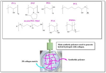
Diverse formulations of collagen-based hydrogels functionalized with synthetic polymers have been
developed (Fig. 4). Synthetic polymers, such as PEG, polycaprolactone (PCL), PPU, polyacrylamide
(PAM), polymethylmethacrylate (PMMA), polyvinyl alcohol (PVA) , polyvinylpyrrolidone (PVP), among
others, are characterized by giving control in the degradation, mechanics and swelling of hybrid hydrogels;
however, these polymers significantly decrease the biocompatibility associated with collagen.
Figure 4.
Synthesis of collagen based hydrogels coupled with synthetic polymers.
Chains of synthetic polymers can alter the entanglement of collagen chains, producing IPNs hydrogels, and
chemical crosslinking systems if they have reactive groups. The mass ratio of the synthetic polymer to the collagen
is an important variable for generating hybrid hydrogels with improved properties for applications in BTERM. In
concentrations of around 10 to 15% of weight of synthetic polymer, the biocompatibility of collagen is not
significantly affected, ensuring control in the mechanics and degradation of the hybrid material. Table 2 shows the
collagen hydrogels formulations using synthetic polymers as coupling agents. The systems of hydrogels with
properties of charge-controlled release of molecules with biological interest are based on collagen matrices
reinforced with synthetic polymers; this mainly because the mechanical properties and swelling can be adapted,
promoting a controlled release, thus improving their applicability in BTERM. Within this system, PEG derivatives
have been shown to be excellent regulators of the properties of collagen hydrogels, indicating a direct relationship
strategies for the tailoring of signaling and cellular fates required in BTERM strategies, as reported in a recent
review [20]. Various synthetic chemical routes can be designed to polymerize and / or copolymerize the monomers
of synthetic polymers with hydrolyzed collagen molecules, generating complex architectures of hydrogels with
adjusted properties; however, in these approaches it is important to control that the polymerization processes are
Collagen source Coupling agent Collagen crosslinker Processing conditions
Novel features Characterization methods
In vitro / In vivo results Ref.
Rat tail tendon Hydroxylam ine hydrochlori de –methacryla ted Methacrylic anhydride (MA), hydroxylamine hydrochloride and EHC-NHS Mix vigorously stirred for 10 min and 24 h to reaction, precipitation with ethanol for 8 h.
The hydroxylamine
hydrochloride -methacrylated collagen conjugate with integrated matrix metalloproteinase
(MMP)-modulating capability hydrogel are suitable for regulation of
inflammation-response, stable in structure and chemical composition, without cytotoxic effect
CD and UV-vis spectroscopy, mechanical and biochemical analysis
Hydrogels induced the reduction of MMP-9 and MMP-3 up to ~13 and ~32 RFU% respectively for 4 days of incubation
in vitro.
G292 osteosarcoma cell culture on hydrogel did not show toxic effects during 4 days of incubation. [58] Porcine knees Methacrylat ed solubilized decellulariz ed cartilage (MeSDCC) Glycidyl methacrylate
pH 7.0 mixed centrifuged at 3000 rpm, and stored at 4 °C overnight
The material entirely derived from native cartilage ECM with similar properties to native cartilage. Suitable to related issues of cartilage tissue.
1 H NMR, biomechanics, bioactivity, biochemistry, histological and molecular biology analysis.
These materials were able to induced chondrogenesis and matrix synthesis. The MeSDCC concentration affects the cellular behavior, promoting the ECM synthesis.
[59]
Bovine skin Gelatin-met hacrylate (GelMA)
MA 50°C to
completely mix until 24 h, pH 7.0, at 37°C for 30 min to polymerize, UV exposure at 365 nm, 37°C for 5 min
The GelMA-hydrogel contains a IPN of gelatin-methacrylate GelMA that permits wide variation of shear modulus while retaining the fibrillary structure of collagen.
Appropriate for studies of cell behavior. Oscillatory rheology, confocal microscopy, mechanical, biochemical and image analysis and cell viability/morphol ogy assay,
BAEC cells and
MDA-MB-231 cells were adhered to collagen fibers of GelMA hydrogel.
BAEC cell exhibited a spread and fibroblastic morphology in both the absence and presence of collagen fibers, although increased of stiffness
decreased sprouting Equine knee GelMA,
cartilage derived matrix (CDM)
MA 50°C for 1 h, stirring and add MA, dialyzed for 7 days at 40°C, pH 7.4, freeze-dried.
The hydrogel stimulates the formation of cartilage template via endochondral, in vivo is biodegradable and reusable into mineralized bone new-tissues.
Confocal-vis-micr oscopy, XRD, µCT, mechanical and image analysis, and cell viability assay
Chondrocytes and MSCs cells showed high viability and chondrogenesis in GelMA-MSC-hydrogel regardless of the present or absence of CDM, after 6 weeks of culture. [61] Porcine articular cartilage Type II collagen methacryla mide (Col-II-MA)
MA Dissolution of Col-II-Ma for 12h, add photoinitiator, pH 7.0, 4°C, and UV light (8W )
Photo-crosslinkable type II collagen methacrylamide (Col-II-MA) hydrogel presented gelatinous form with improved mechanical strength.
Gel-II-MA hydrogel promote the growth, proliferation and chondrogenic differentiation of BMSCs cells.
1
H NMR, FTIR, CD, DMA, CLSM and cell viability/ morphology/proli feration assay.
After 2 weeks of subcutaneous
implantation of hydrogel in athymic mice,
histological samples showed more formation of cartilage lacuna and cartilaginous matrix. [62] Collagen from Mingrang Biological Science-Tec hnology Company Nano-hydro xyapatite (n-HAP) and PEG-PCL-PE G (PECE)
PEG and poly ε-caprolactone (PLC)
PECE solution at 60°C for 2 min, ice-water bath for 5 min, add collagen, and HAP, mix and
ultrasonicated at least for 30min
Injectable thermo-sensible, biocompatible and
biodegradable for bone regeneration. SEM, rheological analysis, optical microscopy, XRD, µ-CT, and histological assay. Histological observation of composite implanting into dorsal muscle pouches of rats showed degradation of implant and complete
disappearance of inflammation after 14 days of treatment. New bone tissue were formed at 20 weeks of the surgery in rat skulls.
[63]
Achilles tendon 2-Pyridineca rboxaldehyd e and fibronectin
for 12-16 h anchor full-length proteins of ECM High-resolution electrospray ionization mass spectrometry (HR-ESI-MS), fluorescence microscopy, SEM, biochemical and rheological analysis.
osteosarcoma cells were able to assembled and remodeled large collagen fiber. Collagen type I (without specified origin)
MA PEG-thiol pH 8.0 - 8.2, the time with gelation time variable.
Versatile hydrogel for uses in tissue engineering either as implantable scaffolds or injectable hydrogel. NMR, UV-Vis/CD spectroscopy, SEM, structural and mechanical analysis.
The cell HCECs were able to attachment and proliferate for 5 days of culture.
After 3 days of culture the cells CPCs are viable and spreading, showing elongated morphology.
[65]
Porcine intestinal submucosa
PPU PPU subunits pH 7.4, 37 ºC for 30 min
The polymerization rate and the gel network parameters such as fiber diameter, porosity, crosslinking degree, mechanics, swelling, in vitro degradation and cell proliferation, keep a direct relationship with the oligourethane concentration. FTIR, SDS-PAGE, SEM, UV-Vis turbidimetry, crosslinking assay and oscillatory rheology.
The hybrid hydrogels formulated with 15 wt% of oligourethane exhibit enhanced storage modulus and
degradation resistance, while maintaining the cell viability and impeding the fibroblast-induced contraction. [22] Type I collagen solution PVA and HAP
--- RT for 1h. Increased mechanical
properties. The composites are micro-porous sponge like structure, suitable to corneal
XRD, SEM, mechanical analysis and cell attachment-
Embryonic
keratocyto-like cells were able to attached, spread and proliferated
Table 2. Use of synthetic polymers to design hybrid hydrogels with collagen.
cell adhesion and proliferation. proliferation assays.
on PVA-COL-HAP composites after 7 day culture.
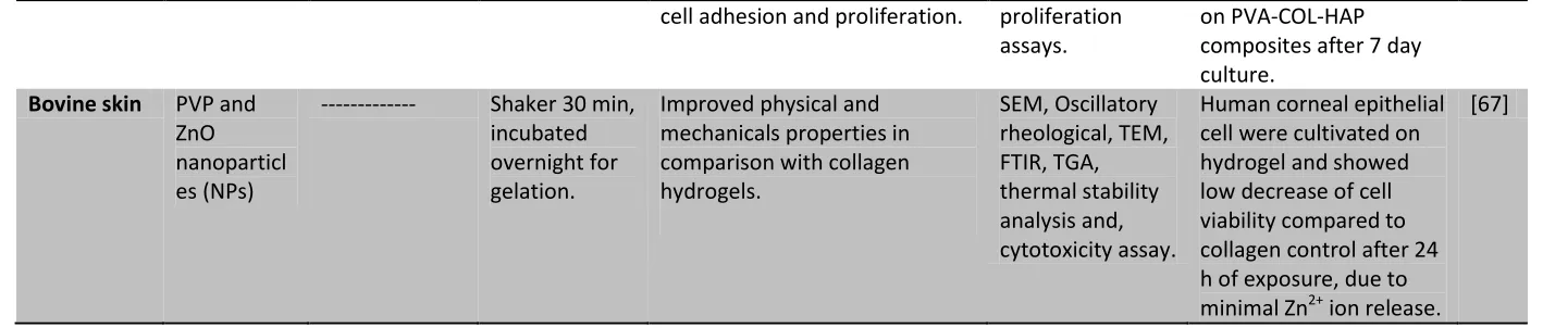
Bovine skin PVP and ZnO nanoparticl es (NPs)
--- Shaker 30 min, incubated overnight for gelation.
Improved physical and mechanicals properties in comparison with collagen hydrogels.
SEM, Oscillatory rheological, TEM, FTIR, TGA, thermal stability analysis and, cytotoxicity assay.
Human corneal epithelial cell were cultivated on hydrogel and showed low decrease of cell viability compared to collagen control after 24 h of exposure, due to minimal Zn2+ ion release.
76 | Page
3.3 Inorganic phases
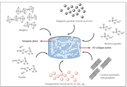
Composite hydrogels have been generated by coupling inorganic phases to the 3D collagen matrix (Fig. 5). The
mechanical properties of the collagen matrix must be optimized using crosslinking strategies, in order that the
inorganic phase be adequately supported, avoiding deformation of the network in the hydrogel state. The main
inorganic phases that have been coupled in collagen-based hydrogels are: hydroxyapatite, Zn, Ag, Si, Fe, Ti
nanoparticles, bioglasses, carbon nanotubes, graphene and clays such as kaolinite.
Figure 5. Design of collagen composite hydrogels using different inorganic phases.
The table 3 lists the collagen-based hydrogel systems coupled with inorganic phases. The presence of these inorganic
phases in the 3D fibrous matrix has generated biomaterials with high osteoinductive capacity, systems with potential
to be disease detection devices, biosensors and materials with antimicrobial capacity. The design of these
biocomposites in the hydrogel state requires that the charge of the inorganic phase inside the collagen matrix be well
dispersed, without significant modification of the typical fibrous structure. High concentrations of inorganic phase
generate brittle granular matrices, decreasing the biocompatibility of collagen. The magnetic properties of Fe
77 | Page
tissue generation. The different crystalline phases that the inorganic particles present also show a relationship in the
regulation of cell fate and cytotoxicity.
Specifically, Ti nanoparticles, in anatase phase, have shown excellent bone biocompatibility, while rutile phase
increased cytotoxicity has been observed. Si particles with mesoporous structure have shown excellent
biocompatibility for applications in tissue repair, modulating cell fate and phenotype. In addition, the intracellular
adsorption of this type of particles is necessary to direct specific metabolic pathways. It has been reported that the
heater the size of the inorganic Si particle, the intracellular degradation products show increment cytotoxicity,
limiting their use in BTERM applications. The introduction of carbon nanotubes in collagen hydrogels allows the
generation of materials with controlled electrical potential that can be used as detection systems for cellular
biochemical processes related to diseases. The antimicrobial capacity of the Ag nanoparticles has been exploited to
generate collagen hydrogels with bacterial infection control, generating biomaterials for applications related with the
reparation of dermal tissue. The encapsulation of molecules with biological interest within inorganic particles that are
coupled to collagen hydrogels represents another research approach. The swelling of the hydrogel would lead to a
controlled release of the loaded particles, and the subsequent hydrolysis would release the encapsulated molecules to
78 | Page Collagen
source
Coupling agent Collagen crosslinker
Processing conditions
Novel features Characterization methods
In vitro / In vivo
results Ref. Bovine Achilles tendon Magnetic nanoparticles based on ferrite
--- pH 7.0, RT, under 162G to 2110G magnetic fields.
Control gel orientation dynamically and remotely in situ.
Oscillatory rheology, TEM SEM,
Neurons within the 3D magnetically induced gels exhibited normal electrical activity and viability.
[68]
Rat tail tendon
Silica nanoparticles and sodium silicate
--- pH 7.0, RT Significantly improved the mechanical and thermal stability.
Oscillatory rheology, ICP-AES, SEM, DSC.
Favor the metabolic activity of immobilized human dermal fibroblasts while decreasing the hydrogel contraction. Colonizing and vascularizing without inducing strong inflammatory response in murine model. [69] Bovine dermis Bioglass® (45)SiO2-(24.5)Na2O-(2 4.5)CaO-(6)P2O5
--- pH 7.0, 37 °C, 30 min.
Bioactive and osteoinductive properties, as indicated by the formation of CHA in SBF and bone-like material in an ectopic environment, in vivo.
FTIR, XRD, SHG, SEM, µ-CT
Collagen remodeling associated with mineralization in subcutaneously injected scaffolds into rats.
[70]
Rat tail tendon
HAP --- pH 7.0, 25 °C for 1 h and 40 °C for 22.5 h.
Preserves the structural integrity and great tensile strength of collagen. Unidirectional aligned
79 | Page macro-pores with improved mechanical and thermal stability.
Porcine dermis
Bioglass
45% SiO2, 24.5% CaO, 24.5%
Na2O and 6% P2O5 mol%
GEN pH 3.0, RT 24 h, -20 °C for 16 h and -80 °C for 5 h.
Improve angiogenesis through differentiating endometrial stem cells (EnSCs) toward endothelial lineage and increasing level of vascular endothelial growth factor secretion.
FTIR, DSC-TGA, XRD and SEM
EnSCs could be programmed into cardiomyocyte linage and considered a suitable cell source for myocardial
regeneration
[72]
Rat tail tendon
Superparamagnetic iron oxide particles
--- pH 7.0, 37 °C, 20 min.
Great potential to visualize hMSCs and track their migration after
transplantation for articular cartilage repair.
MR imaging, histological analysis and molecular biology.
Did neither influence the viability nor the proliferation potential of hMSCs.
Furthermore, iron incorporation did not affect hMSCs in undergoing adipogenic, osteogenic or chondrogenic differentiation.
[73]
Chicken sternum cartilage
Iron oxide nanoparticles
--- pH 5.0, 37 °C using
electrodepos ition process.
Tailored swelling behavior and flexibility.
Osteogenic differentiation activity being promoted with alteration on mechanical properties.
SEM, VSM, ICP-MS, CLSM and mechanical analysis.
Enhanced osteogenic differentiation of the stimulated MC3T3-E1 cells originated from magnetically actuated mechanotransduction signaling pathway, embodying the upregulated
80 | Page
expression of osteogenic-related and
mechanotransduction-related genes
Rat tail tendon
Fullerol derivatives nanoparticles Gd@C82(OH)22
--- pH 7.4, RT for 1 h.
Enhanced bioactivity of is highly related with its surface-structure relation.
SEM, AFM, TR-PCR, oscillatory rheology and turbidity assays.
Greatly suppress the malignant progression of cancer cells in vitro, and the
metallofullerol can efficiently reduce the mechanical property of collagen matrix.
[75]
Rat tail tendon
TiO2 nanoparticles GEN pH 7.4, 37 °C for 1 h.
Significant influence on the swelling properties and their impact is strongly dependent on the concentration of nanoparticles.
Successfully induced formation of apatite-like structures.
Oscillatory rheology and SEM.
The materials are biocompatible as they can support
mitochondrial activity of MEFs as well as MG-63 cells.
[76]
Fish dermis
HAP / alendronate GEN pH 7.4, 37 °C for 2 h.
The gelation time of hydrogels ranged from 5 to 37 min. Notably, exhibited markedly improved mechanical property. Tunable degradation behaviors against collagenase
1
H NMR, FTIR, DRX, TGA.
Supported the adhesion and growth of murine MC3T3-E1 osteoblastic cells.
[77]
81 | Page tendon mesoporous
nanoactive glass
until gelation.
properties and mechanical stability
ATR-FTIR, TGA, N2 adsorption–desor ption, ICP-AES, XPS, Laser Doppler electrophoresis and DMA stabilization. Hydrolytic and enzymatic degradation reducing. Mesenchymal stem cell highly viable with unclear cell differentiation. Hydrolyz ed collagen (from Parvar Novin-E Tehran Co.)
Kaolin Acrylic acid (AA), ammonium persulfate (APS) N,N´-methyl ene bisacrylamid e (MBA), Mechanical stirred 200 rpm, 80°C, for 20 min, at 55°C.
The maximal swelling capacity of superabsorbent polymer (SAP) hydrogel is 674 g/g (g water/ g dried gel).
FTIR, SEM, swelling capacity and absorbency under load (AUL).
APS concentration is the factor that most effect over swelling capacity.
The time for the highest AUL is reached a 150 min.
[79]
Rat tail tendon
Chitosan, Iron (II) sulfate heptahydrate, iron (III) chloride hexahydrate
Dialdehyde starch (DAS)
RT for 1h, adding magnetic particles and stirred for 20 min and frozen for 2 days.
3D hydrogels have highly swelling capacity and are hydrophilic with magnetic properties. Its rigidity increases according to an increase in the concentration of magnetic particles.
SEM, TEM, ATR-FIR mechanical, physical, swelling and moisture content analysis. The composite hydrogels in PBS decreased the swelling capacity, Young’s Modulus and moisture content decrease with increasing of magnetic particles. [80] Porcine dermis ZnO, 2-Acrylamido-2-methy l-1-propanesulfonic acid sodium salt (PNaAMPS), PEGDA and Irgacure (2959).
EDC-NHS TR for 16 h, then 37°C for 5 h.
Improved mechanical strength and stability. Reduced cytotoxicity of ZnO due to add of PNaAMPs, useful in ophthalmic research as a possible novel
FTIR, fluorescence spectroscopy, enzymatic degradation and molecular
The expression of reporter genes in cells of rabbit corneal anterior stromal fibroblasts (RCFBF) contained in
82 | Page
corneal substitute. biology. composite hydrogels is efficient after 5 days of culture, offering resistance to degradation of hydrogel by inhibition of collagenase.
Rat tail tendon
Mesoporous silica nanoparticles (MSNs)
--- 37°C for 30 min and CO2 incubated.
The system is useful to load and deliver large protein molecules such as a nerve growth factor (NGF).
TEM, N2
adsorption–desor ption, FTIR, LDE, Brunauer–Emmet t–Teller (BET) method, confocal microscopy, cell viability assay and molecular
biology.
A NGF was linked to MSNs and embedded in collagen hydrogel. The system
NGF/MSN/Collagen was able to release sustainably the NGF for over a week following a similar pattern of NGF-MSN, improving the
outgrowth of neurites and stimulating the neuritogenesis.
[82]
Bovine Achilles tendon
Silica with either mineral phaseHAP, calcium carbonate (CaCO3), strontium phosphate (Sr3(PO4)2 or strontium
carbonate (SrCO3)
--- Orbital stirring at 37°C, 95% relative humidity for 3 days
Morphology rougher. The osteoblast resorption was carried out in
Silica/Collagen and
Ca+2/Silica/Collagen xerogels.
SEM, confocal microscopy, fluorescence spectroscopy, complexometry assays,
biochemical and histological analysis.
Monocytes was differentiated to osteoclasts in the surface of all xerogels and in the presence of osteoclast
differentiation factors (M-CSF and RANKI). The xerogel with
83 | Page
strontium components stopped the activity of osteoclast, while calcium favored. Rat tail
tendon
12 nm Silica nanoparticles or 80 nm Silica
nanoparticles
--- pH 7.0, RT, adding cell suspension and medium with PBS.
The hydrogels are suitable for fibroblast growth and cell viability.
SEM, TEM, DSC, Oscillatory rheology,
inmunodetection techniques, biochemical and histological analysis
Fibroblasts entrapped in gel were able to perform vital functions such as metabolism and cell division.
[84]
Rat tail tendon
300 nm bare silica nanoparticles
--- Acid pH, RT exposed to ammonium vapors overnight then pH 7.4, RT
The drug-loaded hydrogel has antimicrobial effect due to the prolonged release of antibiotics. Did not provoke irritation or inflammation in test realized, for uses in development of cutaneous wound healing systems.
SEM, FTIR and microbiological analysis
Streptomycin and rifampicin was homogeneously loaded in
silica-collagen gel, allowing sustained antibacterial effect against Staphylococcus aureus for 10 days and
in vivo decreasing the bacterial population in a model of infected wound.
[85]
Rat tail tendon
Silica nanoparticles of several sizes
--- pH 7, 37°C Increased mechanical properties, major proteolytic stability in fibroblast for silica of 500 nm.
SEM, TEM, Oscillatory rheology, enzymatic degradation, histological
The increase in silica size, decreased the cytotoxicity of fibroblast.
Gentamycin-loaded in the gel was
84 | Page assays, microbiological, biochemical and cytotoxicity analysis. sustainable releases for 7 days showed efficiently
antibacterial activity to 500 nm silica particles. Rat tail
tendon
Carboxyl-functionalize d multiwalled carbon nanotubes
--- pH 7.0, and mix, then 37°C for 15 min for gelation
Improved mechanical strength electrical conductivity and native submicron fibrous structure of hydrogels.
TEM, SEM, AFM, electrical properties analysis and cell viability assay
Improved mechanical and electrical
properties of
hydrogel. Cultures of rat cardiomyocytes showed increased rhythmic contraction after 5 days of culture.
[87] Type I collagen (Sigma Aldrich, CAS: 9007-34-5) Multiwalled carbon nanotube, chitosan and HAP.
--- Frozen by cooling to -40 °C at 0.9 °C/min, lyophilization at 0 °C, 200 mtorr for 48h
3D matrix highly porous (> 95%), improvement in its mechanical properties and biomineralization. High bioactivity in vitro,
appropriate for use in bone tissue.
XRD, SEM, EDS, FTIR, TGA, BET, optical
microscopy, mechanical analysis, and cell viability assay.
MG-63 cell culture into hydrogel did not show a significant difference of cell viability compared with control group.
[88]
Bovine Achilles tendon
Silver doped bioactive glass Bovine fibrinogen Mix all solution components and centrifuge 4°C, 600 rpm for 5min and gelation to 37°C.
Induction of proliferation and differentiation of dental pulp stem cells (DPSC) encapsulated. It showed strong bacteria inhibition. For applications in tissue regeneration. Reflectance confocal microscope, and antimicrobial analysis
DPSCs encapsulated in microbeads were viable, spread, proliferated and changed to fibroblastic morphology both in vitro as in vivo assays. Microbeads showed antibacterial activity,
85 | Page Table 3. Inorganic phases coupled inside collagen based hydrogels.
but the presence of Ag increase the microbial inhibition
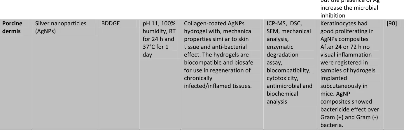
Porcine dermis
Silver nanoparticles (AgNPs)
BDDGE pH 11, 100% humidity, RT for 24 h and 37°C for 1 day
Collagen-coated AgNPs hydrogel with, mechanical properties similar to skin tissue and anti-bacterial effect. The hydrogels are biocompatible and biosafe for use in regeneration of chronically
infected/inflamed tissues.
ICP-MS, DSC, SEM, mechanical analysis,
enzymatic degradation assay,
biocompatibility, cytotoxicity, antimicrobial and biochemical analysis
Keratinocytes had good proliferating in AgNPs composites After 24 or 72 h no visual inflammation were registered in samples of hydrogels implanted
subcutaneously in mice. AgNP
composites showed bactericide effect over Gram (+) and Gram (-) bacteria.
3.4 Biological molecules with complex functionality
The modulation of the biological response required for improved applications in BTERM using collagen hydrogels
can be achieved when a biomolecule with specific biochemical function is coupled in the 3D matrix. In these
approaches, complex and selective hydrogels have been designed by coupling peptides of specific functionality,
growth factors and another proteins. Table 4 exemplifies these strategies. In general, each complex biological
molecule can control cell signaling processes by inhibiting or segregating of important cytokines from biochemical
pathways involved in BTERM strategies. The modulation of cell signaling processes represents an effective key for
successful application in the biotechnology area. However, the extraction and purification of the biological
molecules with functionality of interest represents a determining challenge for the reproducibility of these
strategies. The cell-hydrogel interaction is also benefited if there are biochemical components that allow to the cells
to regulate their metabolism within the matrix allowing them to grow, reproduce and proliferate. Peptides of
different proteins of interest can be coupled in collagen-based hydrogels to mimic cell- extracellular matrix
interactions, favoring the cell growth and proliferation. To guarantee the desired performance in these systems with
controlled biological functionality, the hydrogel should be stable at periods of cellular recognition, should have
antimicrobial capacity and tailored degradation and swelling. The strategies reviewed in the previous sections
Collagen source
Coupling agent Collage n crosslin ker
Processing conditions
Novel features Characterization methods
In vitro / In vivo results Ref.
Streptococc al
collagen-lik e 2 (Scl2) protein, expressed in Escherichia coli BL21-DE3 (HIHA-Scl2) Matrix metalloproteinase 7 (MMP7) and aggrecanase (ADAMTS4) cleavable peptides
Acrylate Mix all component at RT until gelling.
The HIHA-Scl2 hydrogel, is biodegradable, contains binding sites to HA, heparin (H) and integrase (I) that serves as attractors to carry out chondrogenesis from embedded hMSCs and cleavable peptides that act for degradation and remodeling of collagen.
FTIR, SEM, fluorescent spectroscopy, mechanical, swelling, biochemical, histology and immunohistochemi stry analysis, and molecular biology.
hMSCs embedded in HIHA-Scl2 hydrogel showed high metabolic activity and higher gene expression of chondrogenetic markers from 2 to 6 weeks Accumulation of GAGs were reached and more elevated synthesis of collagen type II than collagen type I or type X, while mechanical integrity were maintained. [91] Recombina nt Scl2 protein expressed in Escherichia coli BL21-DE3
HA or chondroitin sulfate or RGDS peptides PEG acrylate -NHS and MMP7-cleavag e peptide
RT, pH 8.5 and mix in several points.
Biocompatible, bioactive and biodegradable hydrogels with possible application for articular cartilage regeneration.
FTIR, CD, (CP-MAS) solid-state 13C NMR, fluorescence confocal microscopy, mechanical, biochemical, histology and immunohistochemi stry and gene expression analysis.
Hydrogels contained MMP7-cleavage peptide were degraded for recombinant human MMP7, the weight lost were ~75% at 7 days. hMSCs embedded into hydrogel had good viability and underwent enhanced chondrogenic
differentiation. The hydrogel promoted the endogenous MMP7 activity.
[92]
Types I and III
Globular domains of C-terminal
--- 37°C for 2h, and 2h for binding
The CLG3/CLP hydrogel promote la survival of
CD, phase-contrast optical microscopy,
A laminin-derived cell adhesive heterodimer
Table 5. Biological molecules of specific functionality coupled in collagen hydrogels atelocollage
n (Nippon Meat Packers, Inc., Osaka, Japan)
region of laminin alfa1 chain (LG3)-9-redisue peptide of the C-terminal region of the laminin γ1 chain (LP). with collagen binding peptide
(CLG3/CLP)
CLG3/CLP NSCs SDS-PAGE and cell
viability/adhesion/ proliferation assay.
(CLG3/CLP) was binding to collagen hydrogel and promote adhesion, survival and proliferation of Neural stem cells (NSCs) for 7 days of assay
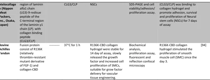
Bovine Achilles Tendon
Fusion protein consist of R136K (relatively
thrombin-resistant mutant derivative of FGF-1) and collagen-CBD
--- 37°C for 1 h R136K-CBD collagen hydrogel were stable for 14 day of assay, slowly released the growth factor and increased cell proliferation of SMCs, suitable for grow factor delivery for vascular tissue engineering.
Biochemical analysis,
proliferation assay, fluorescent and reflection confocal microscopy
R136K-CBD collagen hydrogel stimulated the proliferation of smooth muscle cell (SMC) since the day 3.
It is worth mentioning that the cell-hydrogel interaction is a determining factor in the modulation of the biological
response, favoring the applicability in BTERM areas. The chemical composition of the hydrogel can also direct cell
signaling processes, thus it is important to select the ideal components that will form the 3D matrix, in order to
potentiate the effects on the modulation of the cellular response required for an efficient application in BTERM.
4. PERSPECTIVES AND CONCLUSIONS
The diversity of collagen-based hydrogel systems varying the coupling agent has been widely exploited in BTERM
strategies. The biocompatibility of collagen, inherent to its properties and functions of structural protein and
signaling of the extracellular matrix, is the main reason to include it in these hydrogels systems. The coupling with
natural polymers, synthetic polymers, inorganic phases and / or complex biological molecules has allowed adapting
the properties of these hydrogels to improve their performance in BTERM applications. Mainly, the regularization
of the degradation rate and the control of the mechanical properties of the 3D fibrillar network of collagen are
achieved producing hybrid systems. These systems can be structurally as interpenetrated networks, presenting
physical and / or chemical crosslinking that modify the entanglement of collagen fibers and their physicochemical
properties.
These novel systems of hybrid hydrogels could be applied in diverse biotechnological strategies, not only in
BTERM, due to the adaptation of their properties, which it has been studied in detail using diverse physicochemical
techniques. This work visualizes successful performance of collagen-based hydrogels in the biotechnological field,
being applied as: i) substrates for vegetable tissue, ii) hydrogels for encapsulation of microorganisms for
environmental bioremediation, iii) matrices for heavy metal ion adsorption for remediation of water, iv) 3D
matrices to regulate and study the growth of microorganisms with biotechnological interest, v) base substrates for
the formulation of products in the food and/or cosmetological areas and vi) matrices for solid state and semisolid
fermentation.
Collagen-based hydrogels for application as substrates for vegetal tissues could be developed using cellulose,
alginate and / or dextran derivatives as coupling polymers. The degradation and swelling of these materials can be
regulated controlling the concentration of the natural polymer. The diffusion of nutrients and moisture would be
regulated modifying the composition of the hybrid hydrogel. The growth of vegetal tissue could be studied varying
the structure of the hydrogels, and new substrates could be generated with potential application in the agroindustrial
sector. Hydrogels with genes or encapsulated plasmids mimic the behavior of a virus, controlling the cellular
metabolism.
Collagen as a protein, represents a source of C and N, for microorganisms that have potential application in the area
of bioremediation of soils and waste water contaminated by heavy metals and/or environmentally dangerous
chemical substances. These microorganisms could be encapsulated in hybrid matrices with controlled properties