R E S E A R C H
Open Access
Macrophage colony-stimulating factor
(M-CSF) is an intermediate in the process
of luteinizing hormone-induced decrease
in natriuretic peptide receptor 2 (NPR2)
and resumption of oocyte meiosis
Wenchao Sun
1, Chang Liu
2, Ying Feng
3, Guangchao Zhuo
4, Wenjing Zhou
3, Xiaoyang Fei
1and Zhifen Zhang
5*Abstract
Background:Luteinizing hormone (LH) regulation of the ligand, natriuretic peptide precursor type C, and its receptor, natriuretic peptide receptor 2 (NPR2), is critical for oocyte maturation; however, the mechanism is not fully understood. Macrophage colony-stimulating factor (M-CSF) has recently been shown to be involved in oocyte maturation and ovulation. In the present study we determined whether or not M-CSF plays a role in the intermediate signal that mediates LH regulation of NPR2 in resumption of oocyte meiosis.
Methods: Immature female C57BL/6 mice were injected i.p. with 5 IU of equine chorionic gonadotropin (eCG) to stimulate follicle development. After 44–48 h, the eCG-stimulated mice were injected i.p. with an ovulatory dose of 5 IU of human chorionic gonadotropin (hCG). The ovaries were excised at selected times. Pre-ovulatory follicles (POFs) and cumulus-oocyte complexes were cultured in different media. Immunohistochemical and quantitative real-time PCR analyses were used to assess the expression of M-CSF, M-CSF receptor (M-CSF-R), and NPR2. The presence of germinal vesicle breakdown (GVBD) was examined under a stereomicroscope to morphologically evaluate resumption of oocyte meiosis.
Results: NPR2 was mainly expressed in cumulus cells of pre-ovulatory follicles, while M-CSF and M-CSF-R were expressed in both mural granulosa and cumulus cells. The levels of M-CSF/M-CSF-R and NPR2 decreased within 4 h after treatment of hCG. M-CSF not only reduced the expression of NPR2 mRNA via its receptor (M-CSF-R), but also increased the proportion of GVBD in oocytes.
Conclusion: M-CSF serves as an intermediate signal, thus inducing a vital decrease in the NPR2 levels in cumulus cells, and regulates the process of LH-induced resumption of meiosis.
Keywords:Luteinizing hormone, Macrophage colony-stimulating factor, Meiosis resumption, Natriuretic peptide receptor 2, Oocyte
* Correspondence:[email protected]
5Department of Gynecological Endocrinology, Hangzhou Obstetrics and
Gynecology Hospital, Nanjing Medical University, 369 Kunpeng Road, Hangzhou 310008, China
Full list of author information is available at the end of the article
Background
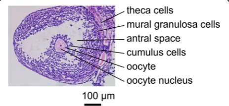
In female mammals, oocytes grow and undergo meiosis over a prolonged period of time [1, 2]. Once the growing follicles reach the early antral stage, oocytes acquire mei-otic competence [3]; however, oocytes are arrested at the diplotene stage of the first meiotic prophase because sig-nals from the surrounding granulosa cells (GCs) prevent the machinery required for resumption of meiosis [3, 4]. Throughout prophase arrest, the oocyte is situated in a follicle where the oocyte is encircled by GCs (Fig. 1). The essence of signals maintaining meiotic arrest has been demonstrated as a complicated interaction between cyclic adenosine 3′,5′-monophosphate (cAMP) and cyc-lic guanosine 3′,5′-monophosphate (cGMP) signaling [5–10]. Cyclic AMP is generated by the oocyte via the activation of Gs G-protein by the G-protein-coupled re-ceptor and adenylyl cyclases. Cyclic GMP, synthesized in surrounding GCs, diffuses into the oocyte through the network of gap junction communications, and inhibits oocyte cAMP-phosphodiesterase (PDE) 3A activity and hydrolysis of cAMP to maintain meiotic arrest [2–7, 9, 11, 12]. Subsequent studies have indicated that gener-ation of cGMP is stimulated by a paracrine loop, which includes natriuretic peptide receptor 2 (NPR2) and the ligand, natriuretic peptide precursor type C (NPPC) [13]. NPPC, produced by mural GCs, activates NPR2, which is produced mainly by cumulus cells surrounding the oocyte, increases cAMP and cGMP levels in the oocyte, and prevents spontaneous (gonadotropin-independent) resumption of oocyte meiosis [13, 14]. In mice defi-cient in the ligand, NPPC, or its cognate receptor, NPR2, oocytes precociously re-enter the meiotic cell cycle as soon as the oocytes reach the early antral follicle stage [13, 15].
Oocytes resume meiosis when the luteinizing hormone (LH) surge causes dramatic changes in pre-ovulatory fol-licles (POFs) [16, 17]. It has been shown that LH causes a spectacular decrease in NPPC in the follicles of a wide variety of mammalian species, including mice, rats, pigs, and humans [4, 5, 13, 18, 19]. Decreased NPPC in turn reduces the amount of NPR2 and cGMP, and meiosis
resumes in oocytes [4, 5]. Recently, when the kinetic curve of cytokines was further studied, NPPC was not shown to be decreased until 2 h after the LH surge, whereas the decrease in cGMP was first detected at 15– 20 min [14]. NPR2 also undergoes a rapid decrease in activity within 10 min after LH exposure, when the NPPC concentration is constant [20, 21]. This phenomenon (that the receptor is motivated before ligand activation) gave rise to the hypothesis that multiple pathways mediate LH regulation of NPR2 and downstream cGMP signaling in the ovarian follicle [21]. These multiple pathways include the phosphoprotein phosphatase signaling pathway [20], which has recently been associated with ovulation process. Multiple pathways are known to include the epidermal growth factor receptor (EGFR) signaling pathway [21–26], but recent findings have shown new relationships. The exact number of these multiple pathways is unknown.
Macrophage colony-stimulating factor (M-CSF), a hemopoietic growth factor with a classic function of controlling the proliferation and differentiation of mac-rophages, has recently been shown to be involved in oocyte maturation and ovulation [27–30]. We have pre-viously reported that M-CSF is implicated in follicular GC function [27], and M-CSF can modulate the gener-ation of NPPC, which may regulate ovulgener-ation triggered by LH [31]. In the present study we determined whether or not M-CSF is included in the aforementioned “mul-tiple pathways” that mediate LH regulation of NPR2 in ovarian follicles.
Methods
Animals and hormone treatments
Immature (22–25 days old) female C57BL/6 mice (Zhejiang Academy of Medical Sciences, Hangzhou, China) were injected i.p. with 5 IU of equine chorionic gonadotropin (eCG) to stimulate follicle development. After 44–48 h, the eCG-stimulated mice were injected i.p. with an ovulatory dose of human chorionic gonado-tropin (hCG; 5 IU). The ovaries were excised at selected times after injection and processed for immunohisto-chemical analysis and quantitative real-time PCR. For cell culture of POFs and cumulus-oocyte complexes (COCs), the eCG-stimulated mice were euthanized and the ovaries were excised without hCG injection. All che-micals were purchased from Sigma-Aldrich (St. Louis, MO, USA) unless otherwise stated. All animal proce-dures were approved by the guidelines of the Nanjing Medical University Administrative Panel on Laboratory Animal Care.
Culture of POFs
The POFs were dissected stereomicroscopically from the ovaries of eCG-stimulated mice, as previously de-scribed [32, 33]. The POFs were placed in minimum
essential media (MEM)-α supplemented with 100 mg/ ml of fetal bovine serum (FBS), 100 U/ml of penicillin G, and 100 μg/ml of streptomycin sulfate. After equilibration, follicles (10–15 per group) were cul-tured at 37 °C in an atmosphere of 5% O2, 5% CO2,
and 90% N2 for the indicated time in the presence or absence of hCG (5 IU/ml). At the end of the culture period, follicles were collected for quantitative RT-PCR analysis to measure hCG regulation of NPR2 transcript levels.
Culture of COCs
COCs were obtained by puncturing the POFs in the ovaries from eCG-stimulated mice. After isolation, COCs were washed in the medium and cultured for 2 h. The culture medium was MEM-α supplemented with 100 mg/ml of FBS, 100 U/ml of penicillin G, and 100 μg/ml of streptomycin sulfate with or with-out 30 nM NPPC. At least 10 COCs per treatment group were cultured. Cultures were maintained under a controlled atmosphere of 5% O2, 5% CO2, and 90% N2 at 37 °C. After culture, COCs were collected for quantitative RT-PCR to calculate the NPR2 transcript levels. The presence of germinal vesicle breakdown (GVBD) was examined under a stereomicroscope.
Table 1Primer sequences, forward (F) or reverse (R), used for quantitative RT-PCR
Gene F or R Primer sequence
NPPC F GGGAGCCAATCTCAAGGGAG
R GTTGCCGCCTTTGTATTTGC
NPR2 F GCATTGTCACCGAGTATTGTCC
R CAGACCGTAATCTGTTATTTTGAGC
M-CSF F TGATTGGGAATGGACACCTG
R AAAGGCAATCTGGCATGAAGT
M-CSF-R F GGTGGCTGTGAAGATGCTAAAG
R AGGCTCCCAAGAGGTTGACTAT
Rpl19 F CCGCTGCGGGAAAAAGAAG
R CAGCCCATCCTTGATCAGCTT
RNA isolation, reverse transcription, and quantitative real-time PCR
Mouse ovaries, cultured follicles, and COCs were collected in 350μl of RNeasy lysis buffer. The tissues and cells were stored at−80 °C until analysis for mRNA expression. Total RNA was isolated from frozen samples using the RNeasy micro-RNA isolation kit (Qiagen, Valencia, CA, USA), as recommended by the manufacturer’s instructions. Reverse transcription and real-time PCR was then carried out to quantify the steady-state mRNA levels of NPPC, NPR2, M-CSF, and M-CSF-R using an ABI 7500 real-time PCR in-strument (Applied Biosystems, Foster City, CA, USA). The housekeeping gene, Rpl19, was considered the internal con-trol. The primers for real-time PCR of NPPC, NPR2, M-CSF, M-CSF-R, and Rpl19 are listed in Table 1. The levels of NPPC, NPR2, M-CSF, and M-CSF-R mRNA were first normalized to the level of Rpl19 expression, then demon-strated relative to a control group in which the level of ex-pression was set at 1. Each experiment was repeated independently at least three times.
Immunohistochemistry
The excised ovaries were fixed in 10% formalin. After dehydration, the fixed ovaries were embedded in paraplast, then sectioned at 5-μm intervals onto Superfrost Plus microscope slides. For immunohisto-chemical staining, sections of ovaries were deparaffi-nized and rehydrated, treated with 3% H2O2 for 20 min to inactivate intrinsic peroxidase activity, and incubated with ethylene dinitrilo tetraacetic acid buf-fer for 10 min for antigen retrieval. Washes were carried out with automation phosphate buffer. Sec-tions were incubated for 2 h at 4 °C with rabbit anti-mouse antibody diluted 1:200 in buffer contain-ing 5% bovine serum albumin. Sections were next incubated with horseradish peroxidase-labeled goat anti-rabbit antibody for 50 min at room temperature. Staining was achieved using diaminobenzidine chromo-gen. The staining reactions were stopped with distilled water, and sections were dehydrated and mounted with neutral balsam.
Image analysis of densitometry
Slides were examined under a microscope with a 200 × ob-jective. The obtained images were captured and examined by Image Pro Plus 6.0 software (Media Cybernetics, Inc., Washington, USA). The integrated optical density (IOD) was calibrated, and the area of interest was set. The mean optical density was defined as the IOD divided by the total area examined.
Statistical analysis
Statistical analyses were carried out using SPSS software (ver-sion 16.0: SPSS, Inc., Chicago, IL, USA). Data are presented as the mean ± SEM. Differences between experimental and control groups were analyzed by ANOVA test. Statistical significance was set atPvalue of less than 0.05.
Results
Localization of M-CSF, M-CSF-R, and NPR2 in POFs
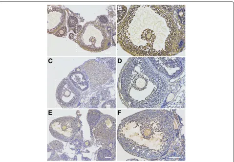
To investigate if M-CSF, M-CSF-R, and NPR2 signaling is involved in regulation of oocyte meiosis in POFs. We analyzed M-CSF, M-CSF-R, and NPR2 localization in ovarian sections from eCG-stimulated mice. Immuno-histochemistry analysis revealed that CSF and M-CSF-R are expressed in both mural GCs and cumulus cells (Fig. 2a-d), while the expression of NPR2 in POFs was mainly detected in cumulus cells and was also observed in peri-antral mural GCs (GCs located on the lining of antral spaces, [Fig. 2e and f]).
Changes in expression of M-CSF, M-CSF-R, and NPR2 in the ovaries of mice injected with gonadotropin
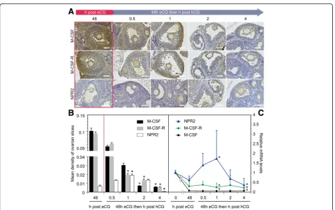
We further elucidated the importance of M-CSF, M-CSF-R, and NPR2 signaling in LH-induced resumption of oo-cyte meiosis. Using immunohistochemical techniques, we determined the changes in expression of M-CSF, M-CSF-R, and NPR2 in the ovaries of eCG-stimulated mice at 0 (48 h after eCG treatment), 0.5, 1, 2, and 4 h after injec-tion with hCG (Fig. 3a). The mean optical density was cal-culated and showed that M-CSF and M-CSF-R expression was gradually decreased within 4 h after hCG treatment, which was administered 48 h after eCG injection (Fig. 3b). NPR2 expression peaked at 1 h, but an obvious reduction in expression was detected at 4 h (Fig. 3b). Expression of M-CSF, M-CSF-R, and NPR2 mRNA was also detected (Fig. 3c) and was consistent with the results of immuno-histochemistry and densitometry analysis.
Kinetic curve of NPR2 mRNA levels controlled by hCG in vitro
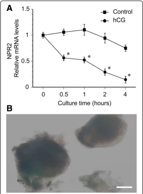
To study the effect of hCG on the kinetic curve of NPR2 mRNA levels in POFs, we examined the expression of NPR2 mRNA after 0, 0.5, 1, 2, and 4 h of culture after hCG treatment. The hCG (5 IU/ml) significantly de-creased NPR2 mRNA levels by one-half at 0.5 h, and
nearly 90% at 4 h of culture. In the control group with-out hCG treatment, the levels of NPR2 mRNA in POFs were slightly increased after POFs were cultured for 1 h. Then, the NPR2 mRNA levels were only slightly de-creased after POFs were cultured for 4 h (Fig. 4a). The image of cultured POFs is presented in Fig. 4b. The re-sults are in agreement with the speculation that hCG regulates NPR2.
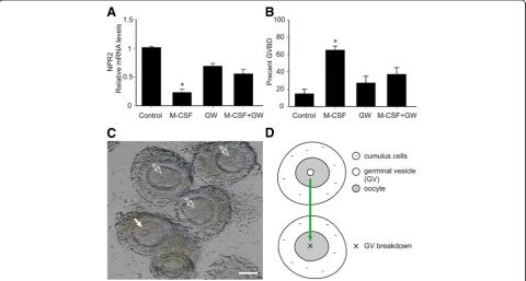
M-CSF/M-CSF-R signaling decreases the level of NPR2 mRNA in cumulus cells and contributes to resumption of meiosis in oocytes
The effect of M-CSF/M-CSF-R signaling on NPR2 mRNA expression and oocyte maturation was determined using cultured COCs isolated from POFs of eCG-stimulated mice. Isolated COCs spontaneously resume meiosis be-cause mural GCs containing the inhibitory molecule, NPPC, is removed. Therefore, a culture system supple-mented with 30 nM NPPC was adopted to maintain meiotic arrest in oocytes [23]. M-CSF (200 ng/ml) signifi-cantly decreased the level of NPR2 mRNA when COCs
were cultured for 2 h (Fig. 5a). M-CSF stimulated GVBD in approximately 65% of oocytes at 2 h of culture (Fig. 5b and c). GVBD marks the onset of meiotic resumption and is the key event in oocyte maturation (Fig. 5d). Further-more, the M-CSF-induced decrease in NPR2 and resump-tion of oocyte meiosis was partially inhibited by GW2580 (a selective M-CSF-R inhibitor purchased from Selleck-chem, Houston, TX, USA; Fig. 5a and b), suggesting that M-CSF functioned via the activity of the M-CSF receptor.
Discussion
In the present study, we focused on the effect of M-CSF in the regulation of oocyte meiosis, and identified some crucial roles, including: (a) NPR2 was mainly expressed in cumulus cells of POFs, while M-CSF and M-CSF-R were expressed in both mural GCs and cumulus cells; (b) the levels of M-CSF/M-CSF-R and NPR2 decreased within 4 h after hCG treatment; and (c) M-CSF not only reduced the expression of NPR2 mRNA via its receptor (M-CSF-R), but also increased the proportion of GVBD of oocytes, which indicates that M-CSF is an intermedi-ate signal, inducing a vital decrease in NPR2 levels in cu-mulus cells, and regulates the process of LH-induced resumption of meiosis.
M-CSF extensively participates in the processes of ovulation [27, 29]. Female mice lacking the coding re-gion for the M-CSF gene (M-CSF deficient) have remarkably lower ovulation rates compared to the wild-type counterparts. Indeed, administration of M-CSF from birth to reinstate circulating M-CSF levels could reverse these defects [27]. In humans, high serum con-centrations of M-CSF were related to successful oocyte retrieval during in-vitro fertilization and embryo transfer cycles [28]. LH surge triggers dramatic changes in cyto-kines during ovulation [16]. Although the changes in M-CSF and M-M-CSF-R 24 h after a LH surge are clear [29], the alteration in M-CSF/M-CSF-R within a short period of time (4 h) following the LH surge is unknown. There-fore, we studied the changes in M-CSF/M-CSF-R expression in the ovaries of mice injected with gonado-tropin. The data from our study showed that M-CSF/M-CSF-R, expressed in both mural GCs and cumulus cells, was gradually decreased within 4 h after hCG treatment. We hypothesized that this change is related to estradiol (E2). Reportedly, the expression of M-CSF was enhanced by E2in luteinized GCs in a dose-dependent manner in vitro [27]. E2maintains cumulus cell expression of NPR2 and inhibits the resumption of meiosis in mouse oocytes in vitro [34], and the level decreased during ovulation.
Thus, decreased E2has a certain role to the resumption of oocyte meiosis and ovulation. Therefore, the de-creased level of M-CSF after hCG treatment may be due to the decreased levels of E2. Although the hypothesis is theoretically feasible, it is still necessary to further inves-tigate whether or not a decrease in M-CSF during ovula-tion is related to a decrease in the E2level.
Recent studies have revealed that cGMP stimulated by NPR2 from cumulus cells diffuses into oocytes via gap junctions and controls cAMP concentration through in-hibition of PDE3A activity, indicating that higher NPR2 levels in cumulus cells is responsible for oocyte mei-otic arrest by maintaining high cAMP levels in oo-cytes [1, 4–7]. Our results showed that NPR2 is primarily expressed in cumulus cells surrounding oocytes, which is consistent with the literature. The results pre-sented herein support the thought that control of NPR2, which is expressed in cumulus cells, is essential for main-taining meiotic arrest in pre-ovulatory oocytes [4, 13]. LH reduces the activity of NPR2 during ovulation, and pro-motes resumption of meiosis in oocytes [14]. In particular, our findings demonstrated that NPR2 is also expressed in peri-antral mural GCs (GCs situated on the antral side), which substantiates the viewpoint that some mGCs are activated by NPPC in an autocrine process to raise cGMP levels [24, 34]. NPR2 was down-regulated following a spe-cific time curve after hCG injection. The kinetic curve of NPR2 after hCG treatment in our study was similar to that reported in the literature [23], but there were some differences. In our research, NPR2 expression peaked 1 h after hCG treatment, and an obvious reduction of ap-proximately 85% in expression was detected at 4 h in vivo. The rise in NPR2 expression within 2 h after LH treat-ment was different from that reported in the literature, likely due to the long half-life (40 ~ 120 h) of eCG admin-istered 48 h before hCG injection to stimulate follicle de-velopment [22, 24]. The eCG promoted the up-regulation of NPR2 during follicle growth [33]. To clarify the NPR2 expression change within 2 h after LH treatment, cultured POFs were used to examine the kinetic curve of NPR2 mRNA levels controlled by hCG in vitro. The data from our study were consistent with the literature [23]. We did not observe a significant increase in expression of NPR2 in hCG-containing media in vitro, possibly because in vitro culture eliminated the residual effect of eCG. To fur-ther declare the effect of M-CSF/M-CSF-R signaling on NPR2 mRNA expression and oocyte maturation, COCs isolated from POFs were cultured. The results showed that the M-CSF/M-CSF-R signaling reduced levels of NPR2 mRNA in cumulus cells and promoted resumption of oocyte meiosis. Then, we added GW2580 to restrict the effect of M-CSF signaling in vitro. The results showed that the effect of M-CSF at 4 h was partially abolished.
Conclusion
We conclude that M-CSF is an intermediate signal, in-ducing a vital decrease in NPR2 levels in cumulus cells, and regulates the process of LH-induced resumption of meiosis. Although further research is needed, our find-ings bring forth powerful evidence to interpret“multiple pathways” that mediate LH regulation of NPR2 in the process of resuming oocyte meiosis.
Abbreviations
cAMP:cyclic adenosine 3′,5′-monophosphate; cGMP: cyclic guanosine 3′,5′ -monophosphate; COCs: Cumulus-oocyte complexes; E2: Estradiol;
eCG: Equine chorionic gonadotropin; EGFR: Epidermal growth factor receptor; FBS: Fetal bovine serum; GCs: Granulosa cells; GVBD: Germinal vesicle breakdown; hCG: Human chorionic gonadotropin; IOD: Integrated optical density; LH: Luteinizing hormone; M-CSF: Macrophage colony-stimulating factor; M-CSF-R: M-CSF receptor; MEM: Minimum essential media; NPPC: Natriuretic peptide precursor type C; NPR2: Natriuretic peptide receptor 2; PDE: Phosphodiesterase; POFs: Pre-ovulatory follicles
Acknowledgements
We acknowledge core support provided by the National Natural Science Foundation of China (grant no. 31470078).
Funding
The study was funded by core support provided by the National Natural Science Foundation of China (grant no. 31470078).
Availability of data and materials
All data generated or analyzed during this study are included in this published article. The datasets used during the current study are available from the corresponding author on reasonable request.
Authors’contributions
YF, GZ and WZ did experiments, performing data analysis and assisted with manuscript preparation. WS, CL and XF cooperated with sample collection and preparation. XF and ZZ edited the manuscript. ZZ designed study and prepared manuscript. All authors read and approved the final manuscript.
Ethics approval
All animal procedures were approved by the guidelines of the Nanjing Medical University Administrative Panel on Laboratory Animal Care. The study does not include any human samples.
Consent for publication
Not applicable.
Competing interests
The authors declare that they have no competing interests.
Publisher’s Note
Springer Nature remains neutral with regard to jurisdictional claims in published maps and institutional affiliations.
Author details
1Center of Reductive Medicine, Hangzhou Obstetrics and Gynecology
Received: 30 May 2017 Accepted: 26 September 2017
References
1. Mehlmann LM. Stops and starts in mammalian oocytes: recent advances in understanding the regulation of meiotic arrest and oocyte maturation. Reproduction. 2005;130:791–9.
2. Solc P, Schultz RM, Motlik J, Prophase I. Arrest and progression to metaphase I in mouse oocytes: comparison of resumption of meiosis and recovery from G2-arrest in somatic cells. Mol Hum Reprod. 2010;16:654–64.
3. Holt JE, Lane SI, Jones KT. The control of meiotic maturation in mammalian oocytes. Curr Top Dev Biol. 2013;102:207–26.
4. Zhang M, Xia G. Hormonal control of mammalian oocyte meiosis at diplotene stage. Cell Mol Life Sci. 2012;69:1279–88.
5. Zhang M, Ouyang H, Xia G. The signal pathway of gonadotrophins-induced mammalian oocyte meiotic resumption. Mol Hum Reprod. 2009;15:399–409. 6. Norris RP, Ratzan WJ, Freudzon M, Mehlmann LM, Krall J, Movsesian MA,
et al. Cyclic GMP from the surrounding somatic cells regulates cyclic AMP and meiosis in the mouse oocyte. Development. 2009;136:1869–78. 7. Vaccari S, Weeks JL 2nd, Hsieh M, Menniti FS, Conti M. Cyclic GMP Signaling
is involved in the luteinizing hormone-dependent meiotic maturation of mouse oocytes. Biol Reprod. 2009;81:595–604.
8. Tripathi A, Kumar KV, Chaube SK. Meiotic cell cycle arrest in mammalian oocytes. J Cell Physiol. 2010;223:592–600.
9. Vaccari S, Horner K, Mehlmann LM, Conti M. Generation of mouse oocytes defective in cAMP synthesis and degradation: endogenous cyclic AMP is essential for meiotic arrest. Dev Biol. 2008;316:124–34.
10. LaPolt PS, Leung K, Ishimaru R, Tafoya MA, You-hsin Chen J. Roles of cyclic GMP in modulating ovarian functions. Reprod BioMed Online. 2003;6:15–23. 11. Sasseville M, Cote N, Guillemette C, Richard FJ. New insight into the role of
phosphodiesterase 3A in porcine oocyte maturation. BMC Dev Biol. 2006;6:47. 12. Richard FJ, Tsafriri A, Conti M. Role of phosphodiesterase type 3A in rat
oocyte maturation. Biol Reprod. 2001;65:1444–51.
13. Zhang M, YQ S, Sugiura K, Xia G, Eppig JJ. Granulosa cell ligand NPPC and its receptor NPR2 maintain meiotic arrest in mouse oocytes. Science. 2010; 330:366–9.
14. Robinson JW, Zhang M, Shuhaibar LC, Norris RP, Geerts A, Wunder F, et al. Luteinizing hormone reduces the activity of the NPR2 guanylyl cyclase in mouse ovarian follicles, contributing to the cyclic GMP decrease that promotes resumption of meiosis in oocytes. Dev Biol. 2012;366:308–16.
15. Kiyosu C, Tsuji T, Yamada K, Kajita S, Kunieda T. NPPC/NPR2 signaling is essential for oocyte meiotic arrest and cumulus oophorus formation during follicular development in the mouse ovary. Reproduction. 2012;144:187–93. 16. Coticchio G, Dal Canto M, Mignini Renzini M, Guglielmo MC, Brambillasca F,
Turchi D, et al. Oocyte maturation: gamete-somatic cells interactions, meiotic resumption, cytoskeletal dynamics and cytoplasmic reorganization. Hum Reprod Update. 2015;21:427–54.
17. Conti M, Hsieh M, Zamah AM, Oh JS. Novel signaling mechanisms in the ovary during oocyte maturation and ovulation. Mol Cell Endocrinol. 2012; 356:65–73.
18. Kawamura K, Cheng Y, Kawamura N, Takae S, Okada A, Kawagoe Y, et al. Pre-ovulatory LH/hCG surge decreases C-type natriuretic peptide secretion by ovarian granulosa cells to promote meiotic resumption of pre-ovulatory oocytes. Hum Reprod. 2011;26:3094–101.
19. Zhang W, Chen Q, Yang Y, Liu W, Zhang M, Xia G, et al. Epidermal growth factor-network signaling mediates luteinizing hormone regulation of BNP and CNP and their receptor NPR2 during porcine oocyte meiotic resumption. Mol Reprod Dev. 2014;81:1030–41.
20. Egbert JR, Shuhaibar LC, Edmund AB, Van Helden DA, Robinson JW, Uliasz TF, et al. Dephosphorylation and inactivation of NPR2 guanylyl cyclase in granulosa cells contributes to the LH-induced decrease in cGMP that causes resumption of meiosis in rat oocytes. Development. 2014;141:3594–604. 21. Liu X, Xie F, Zamah AM, Cao B, Conti M. Multiple pathways mediate
luteinizing hormone regulation of cGMP signaling in the mouse ovarian follicle. Biol Reprod. 2014;91:9.
22. Panigone S, Hsieh M, Fu M, Persani L, Conti M. Luteinizing hormone signaling in preovulatory follicles involves early activation of the epidermal growth factor receptor pathway. Mol Endocrinol. 2008;22:924–36.
23. Wang Y, Kong N, Li N, Hao X, Wei K, Xiang X, et al. Epidermal growth factor receptor signaling-dependent calcium elevation in cumulus cells is required
for npr2 inhibition and meiotic resumption in mouse oocytes. Endocrinology. 2013;154:3401–9.
24. Tsuji T, Kiyosu C, Akiyama K, Kunieda T. CNP/NPR2 signaling maintains oocyte meiotic arrest in early antral follicles and is suppressed by EGFR-mediated signaling in preovulatory follicles. Mol Reprod Dev. 2012;79:795–802. 25. Park JY, YQ S, Ariga M, Law E, Jin SL, Conti M. EGF-like growth factors as
mediators of LH action in the ovulatory follicle. Science. 2004;303:682–4. 26. Norris RP, Freudzon M, Nikolaev VO, Jaffe LA. Epidermal growth factor receptor
kinase activity is required for gap junction closure and for part of the decrease in ovarian follicle cGMP in response to LH. Reproduction. 2010;140:655–62. 27. Zhang Z, Fang Q, Wang J. Involvement of macrophage colony-stimulating
factor (M-CSF) in the function of follicular granulosa cells. Fertil Steril. 2008; 90:749–54.
28. Takasaki A, Ohba T, Okamura Y, Honda R, Seki M, Tanaka N, et al. Clinical use of colony-stimulating factor-1 in ovulation induction for poor responders. Fertil Steril. 2008;90:2287–90.
29. Salmassi A, Zhang Z, Schmutzler AG, Koch K, Buck S, Jonat W, et al. Expression of mRNA and protein of macrophage colony-stimulating factor and its receptor in human follicular luteinized granulosa cells. Fertil Steril. 2005;83:419–25.
30. Salmassi A, Mettler L, Jonat W, Buck S, Koch K, Schmutzler AG. Circulating level of macrophage colony-stimulating factor can be predictive for human in vitro fertilization outcome. Fertil Steril. 2010;93:116–23.
31. Xu S, Zhang Z, Xia L-X, Huang J. Role of macrophage colony-stimulating factor (M-CSF) in human granulosa cells. Gynecol Endocrinol. 2016;32:1005–8. 32. Jaffe LA, Norris RP, Freudzon M, Ratzan WJ, Mehlmann LM. Microinjection of
follicle-enclosed mouse oocytes. Methods Mol Biol. 2009;518:157–73. 33. Sato Y, Cheng Y, Kawamura K, Takae S, Hsueh AJ. C-type natriuretic peptide
stimulates ovarian follicle development. Mol Endocrinol. 2012;26:1158–66. 34. Zhang M, Su YQ, Sugiura K, Wigglesworth K, Xia G, Eppig JJ. Estradiol promotes
and maintains cumulus cell expression of natriuretic peptide receptor 2 (NPR2) and meiotic arrest in mouse oocytes in vitro. Endocrinology. 2011;152:4377–85.
• We accept pre-submission inquiries
• Our selector tool helps you to find the most relevant journal
• We provide round the clock customer support
• Convenient online submission
• Thorough peer review
• Inclusion in PubMed and all major indexing services
• Maximum visibility for your research
Submit your manuscript at www.biomedcentral.com/submit