R E S E A R C H
Open Access
Wavefront-optimized surface retreatments
of refractive error following previous laser
refractive surgery: a retrospective study
Kevin M. Broderick
1*, Rose K. Sia
2, Denise S. Ryan
2, Richard D. Stutzman
1, Michael J. Mines
1, Travis C. Frazier
3,
Mark F. Torres
3and Kraig S. Bower
4Abstract
Background:Retreatments are sometimes necessary to correct residual or induced refractive errors following refractive surgery. Many different combinations of primary treatment methods and retreatment techniques have been studied, however, few studies have investigated wavefront-optimized (WFO) technology for retreatment following primary refractive surgery. This study aimed to report the outcomes of WFO photorefractive keratectomy (PRK) retreatments of refractive error following previous laser refractive surgery with PRK, laser in situ keratomileusis (LASIK), or laser-assisted subepithelial keratectomy (LASEK).
Methods:We reviewed records of patients who underwent WFO PRK retreatments using the Allegretto Wave Eye-Q 400 Hz Excimer Laser System (Alcon Surgical) between January 2008 and April 2011 at Walter Reed Army Medical Center and Madigan Army Medical Center. Outcomes were recorded in terms of uncorrected distance visual acuity (UDVA), manifest refraction spherical equivalent (MRSE), corrected distance visual acuity (CDVA), and complications at 1 month (M), 3 M, and 6 M post-op.
Results:Seventy-eight patients (120 eyes) underwent WFO PRK retreatment during the study period. Primary surgery was surface ablation in 87 eyes (78 PRK, 9 LASEK) and LASIK in 33 eyes. The mean spherical equivalent before retreatment was−0.79 ± 0.94 D (−3.00 to 1.88 D). UDVA was≥20/20 in 69 eyes (60.0 %) at 1 M, 54 eyes (71.1 %) at 3 M, and 27 eyes (73.0 %) at 6 M follow-up. MRSE was within ±0.50 D of emmetropia in 78 eyes (67.8 %) at 1 M, 59 eyes (77.6 %) at 3 M, and 25 eyes (67.6 %) at 6 M follow-up. CDVA was maintained within ±1 line of pre-op in 113 of 115 eyes (98.3 %) at 1 M, 74 of 76 eyes (97.4 %) at 3 M, and 37 eyes (100 %) at 6 M follow-up.
Conclusion:Although follow-up was limited beyond 3 M, WFO PRK retreatments in patients with residual refractive error may be a safe and effective procedure. Further studies are necessary to determine the long-term safety and stability of outcomes.
Keywords: Wavefront-optimized, PRK, LASIK, LASEK, Retreatment, Enhancement
* Correspondence:kevin.m.broderick2.mil@mail.mil
Meeting presentation: Portions of this material were presented at the 2011 Refractive Surgery Subspecialty Day Meeting of the International Society of Refractive Surgery (ISRS) October 21–22, 2011 in Orlando, FL, USA (Abstract #: RP101345). The material has not otherwise been presented or published. Kevin M. Broderick first author and Kraig S. Bower senior author.
1Ophthalmology Service, Walter Reed National Military Medical Center, 8901
Wisconsin Avenue, Bethesda, MD 20814, USA
Full list of author information is available at the end of the article
Background
Laser refractive surgery is one of the most commonly performed eye surgeries worldwide and has been estab-lished to be successful in correcting refractive errors. With conventional refractive surgery, a patient may not achieve his or her maximal visual quality potential post-operatively as a result of changes in corneal shape and subsequent induction of ocular aberrations [1]. Several studies have shown that conventional methods result in the induction of higher order aberrations, including spherical aberration and coma, which can lead to nega-tive visual outcomes [2, 3]. The advent of wavefront technology has greatly improved our understanding of ocular aberration, and with its incorporation into re-fractive surgery techniques, patients are experiencing better visual outcomes and greater satisfaction when compared with conventional methods [1, 4, 5]. Even with these improved outcomes, however, retreatments are sometimes necessary to correct residual or induced refractive error following refractive surgery.
Retreatment following refractive surgery is a subject that has been extensively studied and has been observed in many different combinations of primary treatment methods and retreatment methods. The overall rate for retreatment varies widely in the literature with the aver-age rate ranging from 5.5 % to 8.3 % for primary myopic laser in situ keratomileusis (LASIK) [5]. Prior to the advent of wavefront technology, flap-lift retreatments following conventional LASIK was shown to be safe and effective in multiple studies [6–9]. Jin and Merkley [10] illustrated that retreatment using conventional LASIK is safe, effective, and predictable following wavefront-guided (WFG) and standard myopic LASIK. They also compared conventional to WFG retreatments following primary conventional LASIK treatments in another study [11]. Kanellopoulous and Lawrence [12], along with several other groups, illustrated similar findings following WFG retreatments of primary conventional LASIK. Kashani et al. [5] observed that WFG retreat-ments following primary WFG surgeries in both myopes and hyperopes had favorable outcomes with respect to safety, predictability, and efficacy. Few studies, however, have reported outcomes specifically of wavefront-optimized (WFO) retreatments following refractive surgery.
To our knowledge, the only recent study addressing retreatment data using WFO technology was a study by Randleman et al. [13], which looked primarily at retreat-ment rates after WFO ablation and examined potential risk factors for retreatment, including age, sex, corneal characteristics, and environmental factors. All patients involved in that study underwent either WFO photore-fractive keratectomy (PRK) or LASIK for both their pri-mary and retreatment surgeries. In this study, we report
the visual outcomes of WFO PRK retreatment following PRK, LASIK, and laser-assisted subepithelial keratec-tomy (LASEK).
Methods
We performed a retrospective database review of patients who underwent WFO PRK retreatments for re-sidual refractive error following previous laser refractive surgery at either the Walter Reed Army Medical Center (WRAMC) Center for Refractive Surgery (CRS) or the Madigan Army Medical Center (MAMC) Refractive Surgery Center (RSC) between January 2008 and April 2011. As part of the U.S. Army Warfighter Refractive Eye Surgery Program (WRESP), each refractive center maintains separate outcomes databases as computerized spreadsheets (Microsoft Excel, Redmond, Washington) to track patient outcomes and follow-up. In addition to pa-tient demographic information, these databases list pre-operative refractive error, primary treatment performed, type of retreatment performed, if any, outcomes, and any complications. Data from patients identified as having undergone WFO PRK retreatments were extracted from these databases and compiled in a new computerized spreadsheet for analysis. Prior to review of these data-bases, approval for the study was obtained from the institutional review board (Department of Clinical In-vestigation) at both WRAMC and MAMC.
All patients were active duty personnel participating in the WRESP who gave informed consent to undergo the retreatment procedure for residual refractive error. WRESP patients are typically 20 to 50 years old and predominantly (74.4 %) male. All active duty soldiers with at least 18 months of service obligation remaining at the time of surgery are potentially eligible for partici-pation in the program, with preference given to combat arms soldiers [14]. Pre-operative evaluation of refractive candidates included uncorrected distance visual acuity (UDVA), corrected distance visual acuity (CDVA), manifest and cycloplegic refractions, keratometry and ultrasound corneal pachymetry, contact tonometry, and comprehensive ophthalmic exam. Patients with active ophthalmic disease, keratoconus, glaucoma, ocular herpes simplex or herpes zoster, significant corneal neo-vascularization, clinically significant lens opacity, medical conditions that may impair healing (e.g. collagen vascular disease, autoimmune disease, immunodeficiency disease), lack of refractive stability within 6 months prior to sur-gery, suspicious corneal topography, and pregnancy were excluded from treatment. There were no set criteria in de-termining the need for a retreatment procedure; this was generally decided by the patient and physician.
retreatment, each patient obtained corneal tomography to assess for and rule out evidence of ectasia or forme-fruste keratoconus. If concerning corneal tomography was present, the patient was no longer considered a candidate for refractive surgery. No patients included in the study showed evidence (either pre or post-operatively) of corneal ectasia during the study period.
All retreatments were WFO ablations with Allegretto Wave Eye-Q 400 Hz Excimer Laser System (Alcon Surgical, Fort Worth, Texas). The epithelium was deb-rided mechanically using either a 9.0 mm Amoils rotating scrubber brush (Innovative Excimer Solutions, Toronto, Canada) or with a dilute solution of 20 % alcohol for approximately 30 s, depending on surgeon preference. Prophylactic mitomycin-C (MMC) was used in 69 (57.5 %) retreatment cases, at either 0.01 % or 0.02 % con-centration, and was applied to the stromal bed immedi-ately following ablation for times ranging between 15 and 60 s, depending on surgeon preference.
The post-operative management of patients varied little between surgeons and generally consisted of the following treatment plan: topical moxifloxacin hydro-chloride 0.5 % ophthalmic solution (Vigamox, Alcon Laboratories, Fort Worth, Texas) four times daily for one week; topical prednisolone acetate 1 % ophthalmic so-lution (Pred Forte, Allergan Inc., Irvine, California) every two hours for the first three days followed by four times daily for the remainder of the first week followed by a six-week taper; topical ketorolac tromethamine 0.4 % ophthal-mic solution (Acular-LS, Allergan Inc., Irvine, California) up to four times daily during the first 48 h as needed for pain; and frequent lubrication with preservative-free artifi-cial tears. A high oxygen transmissible soft contact lens was placed on all eyes at the time of surgery and removed after complete re-epithelialization, typically between four and seven days. Postoperative data from regularly sched-uled follow-up visits at one, three, and six months in-cluded UDVA, manifest refraction, manifest refraction spherical equivalent (MRSE), CDVA and complications, including but not limited to corneal haze, dry eyes, and steroid response ocular hypertension or glaucoma.
Visual outcomes were documented in terms of stand-ard measures of efficacy, safety predictability and stabil-ity. Efficacy was measured as the number (%) of eyes achieving UDVA 20/20. Safety was measured in terms of maintenance of CDVA within one line of preopera-tive. Loss of more than one line CDVA from any cause was considered a complication. Predictability was mea-sured as the number (%) of eyes with MRSE within ±0.50 diopter (D) of emmetropia. Refractive stability was measured as the number (%) of eyes with < 0.50 D change in MRSE over a minimum six month follow-up period. Safety index was calculated as the ratio of mean postoperative CDVA over mean preoperative CDVA.
Efficacy index was measured by ratio of mean postoper-ative UDVA over mean preoperpostoper-ative CDVA. Visual out-comes were also graphically presented using a standard format [15].
Results
Between January 2008 and April 2011, 78 patients (120 eyes) underwent WFO PRK retreatment for residual re-fractive error at either WRAMC or MAMC. Table 1 summarizes the preoperative clinical and demographic characteristics of these patients. The primary surgery was surface ablation in 87 eyes (78 PRK, 9 LASEK) and LASIK in 33 eyes. MMC was used in 69 (57.5 %) of the retreatment cases at either 0.01 % or 0.02 % concentra-tion for time periods ranging between 15 and 60 s. Sur-face epithelium was debrided by the Amoils rotating scrubber brush in 22 eyes and 20 % alcohol in 98 eyes. There were no intraoperative complications. Follow-up rate was 115 out of 120 eyes (95.8 %) at one month, 76 eyes (63.3 %) at three months, and 37 eyes (30.8 %) at six months.
UDVA was≥20/20 in 69 eyes (60.0 %) at one month, 54 eyes (71.1 %) at three months, and 27 eyes (73.0 %) at six months follow-up. CDVA was maintained within ±1 line of pre-op in 113 of 115 eyes (98.3 %) at one month, 74 of 76 eyes (97.4 %) at three months, and 37 eyes (100 %) at six months follow-up. MRSE was within ±0.50 D of emmetropia in 78 of 115 eyes (67.8 %) at one month, 59 of 76 eyes (77.6 %) at three months, and 25 of 37 eyes (67.6 %) at six months follow-up. Based on available data at one and six months postoperatively,
Table 1Preoperative characteristics of patients who underwent WFO PRK retreatments following previous laser refractive surgery with PRK, LASEK, or LASIK between January 2008 and April 2011 at Walter Reed Army Medical Center or Madigan Army Medical Center
78 patients (120 eyes)
Age ± SD (years) 40.3 ± 8.3 (26–58)
Gender (% Female) 20 (25.6 %)
Sphere ± SD (D) −0.46 ± 1.08 (−3.00 to 2.00)
Cylinder ± SD (D) −0.67 ± 0.64 (0 to−2.50)
MRSE ± SD (D) −0.79 ± 0.94 (−3.00 to 1.88)
UDVA ± SD (logMAR) 0.31 ± 0.17 (0 to 1.00)
Snellen equivalent (20/x) 40 (20 to 200)
CDVA >20/20 (% of eyes) 100 %
Average Keratometry ± SD (D) 41.33 ± 1.91 (36.85 to 46.05)
Central Corneal Thickness ± SD (μm) 493 ± 46 (392 to 623)
Primary surgery (% surface ablation) 57 (73.1 %)
Epithelial Removal Technique (20 % ethanol)
81.7 %
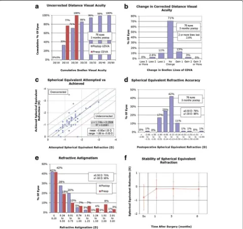
MRSE changed < 0.5 D between one and six month follow-up in 21 of 35 eyes (60.0 %). The three month postoperative visual outcomes are shown in Fig. 1. Of the 76 eyes followed at three months postoperatively, 59 eyes (77.6 %) had UDVA within one line of their pre-operative CDVA. Safety and efficacy indices progres-sively improved over six months postoperatively (Table 2). Dry eye was the most commonly reported post-operative complication throughout the follow-up period: 16 eyes (13.9 %) at one month, 16 eyes (21.1 %) at three months, and 2 eyes (5.4 %) at six months. No clinically significant corneal haze developed in any of the
Fig. 1Three-month outcomes of wavefront-optimized PRK retreatment.aUncorrected distance visual acuity.bChange in corrected distance
visual acuity.cSpherical equivalent attempted vs. achieved.dSpherical equivalent refractive accuracy.eRefractive astigmatismfStability of spherical equivalent refraction
Table 2Safety and efficacy indices of wavefront-optimized PRK retreatment
Follow up (month) Safety Index Efficacy Index
1 0.91 0.73
3 1.00 0.81
6 1.03 0.84
eyes post-operatively. Corneal haze of grade 1+ or less was noted in 13 eyes (11.3 %) at one month and 6 eyes (7.9 %) at three months. All cases of corneal haze re-solved without surgical intervention by six months. One patient (2 eyes) had loss of two lines of CDVA at one month follow-up, but subsequently regained them at three month follow-up without intervention. A separate patient (2 eyes) had no complications at one month follow-up but had loss of two lines of CDVA at three months, and subsequently was lost to further follow-up. Table 3 summaries all complications over the six month follow-up period.
Primary LASIK
Thirty-three eyes had LASIK as primary treatment. The mean spherical equivalent before retreatment was −0.70 ± 0.86 D (−1.75 to 1.25 D). UDVA was≥ 20/20 in 12 of 31 eyes (38.7 %) at one month, 18 of 24 eyes (75.0 %) at three months, and 5 of 6 eyes (83.3 %) at six months follow-up. CDVA was main-tained within ±1 line of pre-op in all eyes at one month (31 eyes), three months (24 eyes), and six months follow-up (6 eyes). MRSE was within ±0.50 D of emmetropia in 17 of 31 eyes (54.8 %) at one month, 19 of 24 eyes (79.2 %) at three months, and 4 of 6 eyes (66.7 %) at six months follow-up. Based on available data at one and six months postopera-tively, MRSE changed < 0.5 D between one and six month follow-up in 2 of 6 eyes (33.3 %).
Primary surface ablation
Eighty-seven eyes had surface ablation (78 PRK, 9 LASEK) as primary treatment. The mean spherical equivalent before retreatment was−0.83 ± 0.97 D (−3.00 to 1.88 D). UDVA was≥20/20 in 57 of 84 eyes (67.9 %) at one month, 36 of 52 eyes (69.2 %) at three months, and 22 of 31 eyes (71.0 %) at six months follow-up. CDVA was maintained within ±1 line of pre-op in 82 of 84 eyes (97.6 %) at one month, 50 of 52 eyes (96.2 %) at three months, and 31 eyes (100 %) at six months follow-up. MRSE was within ±0.50 D of emmetropia in 61 of 84 eyes (72.6 %) at one month, 40 of 52 eyes (76.9 %) at three months, and 21 of 31 eyes (67.7 %) at six months follow-up. Based on available data at one and six months postoperatively, MRSE changed < 0.5 D between one and six month follow-up in 19 of 29 eyes (65.5 %).
Discussion
Wavefront technology has greatly improved our under-standing of ocular aberration, and with its incorporation into refractive surgery techniques, patients are experien-cing better visual outcomes and greater satisfaction when compared to conventional methods [2, 5]. In mul-tiple studies, both WFG and WFO refractive surgeries have been found to achieve essentially similar visual out-comes [2, 4, 16]. The use of WFG technology for refract-ive enhancement has also been shown to be safe and effective [5]. However, the visual outcomes following WFO retreatment have not yet been fully established.
From our review, retreatment using WFO PRK retreat-ment following primary refractive surgery with PRK, LASEK, or LASIK appears to be a safe, effective, and predictable treatment method for correcting residual or induced refractive error. Though our reduced long-term follow-up rate limits our ability to comment on the long-term stability of retreatment cases, our results indi-cate that retreatments appear stable through to the six month post-operative period. The results from our study are consistent with results from the WFO retreatment study by Randleman et al., although their study focused primarily on the rate of retreatment and the factors in-fluencing that rate, as opposed to visual outcomes from the retreatment procedure [13]. Their study also briefly noted WFO retreatment outcomes (UDVA), but did not comment on the safety of this platform as a retreatment method or on any post-retreatment complications [13]. In our study, there were no major adverse events follow-ing WFO PRK retreatments. Dry eye was the most com-mon complication seen in this study, but we were unable to determine if the dry eye symptoms were worse prior to retreatment or if these symptoms became chronic given the limited follow-up duration. Our study was limited primarily by low patient follow-up after three months. This was most likely due to patients’
Table 3Post-operative complications
Post-op complications 1 M (n= 115) 3 M (n= 76) 6 M (n= 37)
No. % No. % No. %
None 79 68.7 53 69.7 35 94.6
Dry eye 16 13.9 16 21.1 2 5.4
Corneal haze (trace) 10 8.7 4 5.3 0 0.0
Corneal haze (1+) 3 2.6 2 2.6 0 0.0
Corneal haze (2+ or >) 0 0.0 0 0.0 0 0.0
Corneal scar 0 0.0 0 0.0 0 0.0
Residual astigmatism 6 5.2 2 2.6 0 0.0
Induced astigmatism 6 5.2 1 1.3 0 0.0
Undercorrected 3 2.6 3 3.9 2 5.4
Overcorrected 11 9.6 1 1.3 1 2.7
Regression 0 0.0 0 0.0 0 0.0
Corneal abrasion 0 0.0 0 0.0 0 0.0
Delayed epithelial healing 0 0.0 0 0.0 0 0.0
Recurrent corneal erosion 0 0.0 0 0.0 0 0.0
Steroid responder 0 0.0 0 0.0 0 0.0
Corneal infiltrate 0 0.0 0 0.0 0 0.0
various active duty requirements, such as training exer-cises and deployments, which precluded patients from attending follow-up appointments. Ideally, more of our patient population would return for regular re-visits and for a longer follow-up duration to better assess refractive outcomes; however, with all of our subjects being active duty military personnel, a large portion of our patients were moved to other military facilities, sent for pro-longed training assignments, or deployed to Afghanistan or Iraq for 12–15 month-long tours, during the follow-up period. As a result, our ability to comment on the re-fractive stability beyond the 6 month period is limited. Further limitations to our study include surgical variabil-ity between individual retreatment cases. Surgical vari-ables that varied largely based on surgeon preference included the method of epithelial debridement (brush or alcohol), the decision to use MMC, and if used, the con-centration and exposure time of MMC. The actual effect of these surgical variables on visual outcomes has widely been debated. The concentration of MMC used, the stromal exposure time to MMC, and the correct situ-ation to use MMC varies considerably between practices and surgeons. A true consensus on the optimal use of MMC had not yet been established in either primary or retreatment cases.
The visual and refractive outcomes from this study are comparable to the outcomes from other retreatment studies. Jin and Merkley [10] compared visual outcomes of conventional LASIK retreatments following either primary WFG LASIK or primary conventional LASIK: 75.0 % of eyes in both of their study groups had UDVA > 20/20 following retreatment. In their WFG group, 91.0 % of eyes were within ±0.50 D of emmetropia, and of their standard primary group, 87.0 % of eyes were within ±0.50 D of emmetropia. In addition, they re-ported that no eyes lost≥2 lines of CDVA, which is also true in our LASIK population subset. Within our study, 3.3 % of eyes (4 eyes of two patients) lost 2 lines of CDVA; both patients were from our PRK subset group. One of these patients (two eyes) improved and regained CDVA at the next month of follow-up, and the second patient maintained ±1 line CDVA until the last month of follow-up. Our rate of 2.6 % is actually lower than that reported in another Jin and Merkley [11] study where outcomes of conventional and WFG myopic LASIK retreatment were assessed. In their study, they reported that 17 % of eyes in their WFG retreatment group lost 2 lines of CDVA.
Many studies to date have shown very similar and often excellent refractive outcomes following both primary and retreatment cases utilizing either WFG or WFO plat-forms. [1, 3, 5, 10, 16] Additionally, many studies have shown that following WFG or WFO primary treatments, there is a similar increase in the amount of induced higher
order aberrations [1, 17]. Given similar refractive out-comes and induction of higher order aberrations between the two platforms, albeit following primary refractive pro-cedures, our experience was that the WFO platform was best utilized for patients with higher refractive errors or with a significant amount of astigmatism given the faster laser ablation and more peripheral treatment profile. Most retreatment cases, however, will not have markedly high amounts of residual refractive error or astigmatism, so either platform could be utilized by the surgeon.
During the study period, we performed 110 enhance-ment procedures at WRAMC, which comprised 1.8 % of the total number of refractive surgeries performed at this center during the study period. Though our study did not primarily focus on establishing a retreatment rate during the study period, our experience with retreatment cases appeared to be similar to others cited in the litera-ture. Randleman et al. reported an overall retreatment rate of 6.3 % following WFO PRK and LASIK. Kashani et al. reported a retreatment rate of 3.1 % in their WFG LASIK retreatment study; Netto and Wilson reported a retreatment rate of 14 % in their standard LASIK retreatment study.
Conclusion
While our study only provides a descriptive narrative of WFO PRK retreatment outcomes, it demonstrates potential outcomes and variables to be investigated in future prospective studies. Our study also tentatively establishes the safety of WFO PRK retreatments fol-lowing any primary refractive surgery. Since the safety of WFG retreatments has been established in several studies, and WFO retreatment outcomes appear to match those of WFG retreatments, our study allows clinicians to have treatment options when faced with patients needing retreatment. Our study also lays the groundwork for a randomized controlled trial com-paring the two platforms in retreatment cases.
Competing interests
The authors have no competing interests.
Authors’contributions
KMB analyzed and interpreted the data and drafted the manuscript. RKS, DSR, RDS, MJM, TCF, MFT and KSB have made substantial contributions in the conception and design of the study, acquisition of the data and critically revised the manuscript for intellectual content. All authors read and approved the final manuscript.
Financial support
There was no financial support, public or private, used to fund this study.
Disclaimer
Author details
1Ophthalmology Service, Walter Reed National Military Medical Center, 8901
Wisconsin Avenue, Bethesda, MD 20814, USA.2Warfighter Refractive Eye
Surgery Program and Research Center, Ft. Belvoir, VA, USA.3Ophthalmology Service, Madigan Army Medical Center, Tacoma, WA, USA.4The Wilmer Eye
Institute, Johns Hopkins University, Baltimore, MD, USA.
Received: 4 September 2015 Accepted: 22 January 2016
References
1. Miraftab M, Seyedian MA, Hashemi H. Wavefront-guided vs wavefront-optimized LASIK: a randomized clinical trial comparing contralateral eyes. J Refract Surg. 2011;27(4):245–50.
2. Durrie DS, Smith RT, Waring 4th GO, Stahl JE, Schwendeman FJ. Comparing conventional and wavefront-optimized LASIK for the treatment of hyperopia. J Refract Surg. 2010;26(5):356–63.
3. Padmanabhan P, Mrochen M, Basuthkar S, Viswanathan D, Joseph R. Wavefront-guided versus wavefront-optimized laser in situ keratomileusis: contralateral comparative study. J Cataract Refract Surg. 2008;34(3):389–97. 4. Kashani S, Rajan M, Gartry D. Wavefront-guided retreatment after primary
wavefront-guided laser in situ keratomileusis in myopes and hyperopes: long-term follow-up. Am J Ophthalmol. 2009;147(3):417–23.e2.
5. Smadja D, Reggiani-Mello G, Santhiago MR, Krueger RR. Wavefront ablation profiles in refractive surgery: description, results, and limitations. J Refract Surg. 2012;28(3):224–32.
6. Netto MV, Wilson SE. Flap lift LASIK retreatment in eyes with myopia. Ophthalmology. 2004;111:1362–7.
7. Hersh PS, Fry KL, Bishop DS. Incidence and associations of retreatment after LASIK. Ophthalmology. 2003;110:748–54.
8. Mulhern MG, Condon PI, O’Keefe M. Myopic and hyperopic laser in situ keratomileusis retreatment: indications, techniques, limitations, and results. J Cataract Refract Surg. 2001;27:1278–87.
9. Brahma A, McGhee CN, Craig JP, Brown AD, Weed KH, McGhee J, et al. Safety and predictability of laser in situ keratomileusis enhancement by flap re-elevation in high myopia. J Cataract Refract Surg. 2001;27:593–603. 10. Jin GJ, Merkley KH. Retreatment after wavefront-guided and standard
myopic LASIK. Ophthalmology. 2006;113:1623–8.
11. Jin GJ, Merkley KH. Conventional and wavefront-guided myopic LASIK retreatment. Am J Ophthalmol. 2006;141:660–8.
12. Kanellopoulos AJ, Pe LH. Wavefront-guided enhancements using the wavelight excimer laser in symptomatic eyes previously treated with LASIK. J Refract Surg. 2006;22:345–9.
13. Randleman JB, White AJ, Lynn MJ, Hu MH, Stulting RD. Incidence, outcomes, and risk factors for retreatment after wavefront-optimized ablations with PRK and LASIK. J Refract Surg. 2009;25(3):273–6.
14. Hammond MD, Madigan Jr WP, Bower KS. Refractive surgery in the United States Army, 2000–2003. Ophthalmology. 2005;112(2):184–90.
15. Reinstein DZ, Waring 3rd GO. Graphic reporting of outcomes of refractive surgery. J Refract Surg. 2009;25(11):975–8.
16. Stonecipher KG, Kezirian GM. Wavefront-optimized versus wavefront-guided LASIK for myopic astigmatism with the ALLEGRETTO WAVE: three-month results of a prospective FDA trial. J Refract Surg. 2008;24:S424–30. 17. Perez-Straziota CE, Randleman JB, Stulting RD. Visual acuity and
higher-order aberrations with wavefront-guided and wavefront-optimized laser in situ keratomileusis. J Cataract Refract Surg. 2010;36(3):437–41.
• We accept pre-submission inquiries
• Our selector tool helps you to find the most relevant journal • We provide round the clock customer support
• Convenient online submission • Thorough peer review
• Inclusion in PubMed and all major indexing services • Maximum visibility for your research
Submit your manuscript at www.biomedcentral.com/submit