R E S E A R C H
Open Access
In vitro
expression of
Streptococcus pneumoniae
ply
gene in human monocytes and pneumocytes
Da-Kang Hu
1, Yang Liu
2, Xiang-Yang Li
3*and Ying Qu
1*Abstract
Background:Streptococcus pneumoniaeis one major cause of pneumonia in human and contains various virulence factors that contribute to pathogenesis of pneumococcal disease. This study investigated the role of pneumolysin, Ply, in facilitatingS. pneumoniaeinvasion into the host blood stream.
Methods:S. pneumoniaestrains were isolated from clinical blood and sputum samples and confirmed by PCR. Expression ofplygene was assessed by infecting human monocytes and pneumocytes.
Results:A total of 23 strains of S. pneumoniae isolated from blood (n = 11) and sputum (n = 12) along with S. pneumoniaeATCC49619 were used to infect human monocyte (THP-1) and Type II pneumocyte (A549) cell lines. All clinical strains ofS. pneumoniaeshowed higher expression ofplymRNA than the American Type Culture Collection (ATCC) strain. Among the clinical strains, blood isolates showed higher expression ofplygenes than sputum isolates, i.e., 21.5- to 21.6-folds in THP-1 and 20.4- to 24.9-folds in A549 cell lines.
Conclusions:The data from the current study demonstrated thatplygene of blood- and sputum-derivedS. pneumoniae is differentially expressed in two different cell lines. Under survival pressure,plyis highly expressed in these two cell lines for blood-derivedS. pneumoniae, indicating that ply gene may facilitateS. pneumoniae invasion into the host blood system.
Keywords:Streptococcus pneumoniae, Infection, Virulence gene, Pneumolysin (ply)
Background
Streptococcus pneumoniae (S. pneumoniae) is a gram-positive diplococcus that is the main pathogenic bacter-ium of community-acquired pneumonia, otitis media, meningitis, abscesses, and other infections, particularly in infants and the elderly [1]. In developing countries, up to 1 million deaths annually are caused by pneumo-nia in children less than 5 years of age, in whichS. pneu-moniaeis the major pathogenic bacterium [2]. Normally, S. pneumoniae is colonized in the respiratory tract of asymptomatic carriers as an opportunistic bacterium [3]. And it is difficult to predict when it converts to patho-genic bacterium although Manso et al.[4] reported that such a phase variation consists of genetic rearrangements.
However, when the host immunosystem becomes weak, S. pneumoniae will be infectious. During S. pneumoniae conversion from an opportunistic bacterium to a par-ticularly pathogenic bacterium that causes severe inva-sive infections such as blood and cerebrospinal fluid (CSF) infections, the complex interaction will occur betweenS. pneumoniaeand the body’s immune system. In addition, infection could also be due to acquisition of virulent serotypes not previously part of the colonizing serotypes, which is named as transformation. Multiple components of S. pneumoniae, such as capsule and other virulence factors are involved in triggering the host immune responses [5-7], while a variety of cells in the host immune system (such as neutrophils, mono-cytes/macrophages, and dendritic cells) will be activated to defend against the infection by either killing the bacteria or releasing a variety of factors (such as IL-1α, IL-1β, IL-6, IFN-α, IL-8, and ICAM-1).
The septicemia and meningitis caused by S. pneumo-niae are much more fatal than that of other types of * Correspondence:[email protected];[email protected]
3Department of Laboratory Medicine, The Second Affiliated Hospital of Wenzhou Medical University, 109# College West Road, Wenzhou 325027, China
1
Department of Laboratory Medicine, Taizhou Municipal Hospital, 381# Zhongshan East Road, Taizhou 318000, China
Full list of author information is available at the end of the article
© 2015 Hu et al.; licensee BioMed Central. This is an Open Access article distributed under the terms of the Creative Commons Attribution License (http://creativecommons.org/licenses/by/4.0), which permits unrestricted use, distribution, and
infections [8]. However, few studies have been reported that focus on the mechanism responsible forS. pneumo-niae invasion into the host blood system and nervous system [7]. Mahdiet al. [9] confirmed the role of tran-scription factor SP_0927 in pathogenesis and viru-lence. Uchiyamaet al. [7] reported that neuraminidase A (NanA) is a virulence factor that facilitates S. pneu-moniae invasion into the CSF. Ricci et al. [10] re-ported that pneumococcal surface protein A (PspA) contributes to pneumococcal meningitis rather than pneumococcal surface protein C (PspC). Based on pre-vious studies on S. pneumoniae infection, different virulence factors in S. pneumoniae are able to induce multiple reactions from the host immune system. To date, there are more than 10 virulence factors discov-ered inS. pneumoniae, including NanA, capsular poly-saccharide synthesis A (CpsA), choline-binding protein A (CbpA), pneumococcal surface adhesion A (PsaA), PspA, PspC, pneumolysin (Ply), and so on [2]. Different virulence factors may have different functions in contribut-ing to the different types ofS. pneumoniaeinfections.
Furthermore, study onS. pneumoniaevirulence factors uses human cell lines, such as monocytes or lung epithe-lial cells. These types of cells are important in our body to prevent or defend against S. pneumoniae infection. The major role of monocytes is to kill bacteria and to trigger leukocyte chemotaxis for immunoreactions, while lung epithelial cells may function as an antigen presenter. In addition, type II pneumocytes have long been known to synthesize and secrete complement com-ponent C3, providing a target for S. pneumoniae adher-ence to those cells via CbpA. Thus, both types of cells are the most commonly used for study of the pathogenic mechanism of S. pneumoniae in vitro. To date, there is no report showing which specific virulence factor facili-tatesS. pneumoniaeinvasion into the host blood system. Possibly, different virulence factors play different roles in disease progression. In this paper, only plyexpression is shown. We first isolatedS. pneumoniae(23 strains) from clinical blood and sputum samples. They then infected monocytes or lung epithelial cell lines and the levels of plymRNA expression were analyzed.
Methods
The experimental protocol was established according to the ethical guidelines of the Helsinki Declaration and was approved by the Human Ethics Committee of Taizhou Municipal Hospital, China. Written informed consent was obtained from individual participants.
Isolation ofS. pneumoniaestrains
The standard S. pneumoniae strain ATCC49619 was provided by the Chinese National Center for Medical Culture Collections (Beijing, China). Twenty-three clinical
S. pneumoniae strains were isolated from different in-patients at the Second Affiliated Hospital of Wenzhou Medical University (Wenzhou, Zhejiang, China) in 2009, which were preserved in 20% glycerin Luria-Bertani Medium and stored at −80°C; among them, 11 strains were isolated from blood samples, called blood-derived S. pneumoniae, and the other 12 strains were isolated from sputum samples, called sputum-derived S. pneumoniae. All 23 strains had no duplication and only one strain was isolated from each selected patient. All S. pneumoniae strains were identified by the Gram-Positive Identification Card (GPI) of VITEK-32 automatic microorganism analyzer (bioMérieux Co., Marcy-Etoile, France) and confirmed by PCR according to du Plessis [11].
S. pneumoniaeculture
A total of 24 S. pneumoniae strains were cultured on blood agar plates at 37°C in a 5% CO2 incubator
(Thermo Electron Co., Waltham, MA, USA) and ad-justed to 1.0 McFarland in normal saline, which is used for infection experiments and RNA extraction.
Cell line and culture
Human monocytes THP-1 and type II pneumocytes A549 cell lines were obtained from the Cell Bank of Chinese Academy of Sciences (Shanghai, China) and cul-tured with RPMI 1640 and F-12 K medium (GIBCO, CA, USA), respectively, and 10% fetal bovine serum (Zhejiang Tianhang Biological Technology Co., Zhejiang, China) plus 1% strep-penicillin (Beijing Solarbio Science & Technology Co., Beijing, China) in a 37°C humidified incu-bator (Thermo Electron Co., Waltham, MA, USA) with 95% air and 5% CO2. THP-1 and A549 cells were prepared
at concentrations of 4.0 and 3.0 × 10⁸/L respectively for the infection experiments and such preparation was con-ducted between May 2011 and July 2011 at the Center Laboratory of Experiments, Wenzhou Medical University.
Infection of THP-1 and A549 cell lines withS. pneumoniae cultures
One milliliter of THP-1 cells was grown in 24-well cul-ture plates, which were 4.0 × 10⁸/L. One milliliter of S. pneumoniae cultures from clinical samples and American Type Culture Collection (ATCC) strains was then seeded into the 24-well culture plates, which was 1.0 McFarland. The blank control used normal saline instead of S. pneumoniae. The cells were maintained at 37°C in 5% CO2 for 4 h (25 wells) and 8 h (other
cells after the suspensions were harvested, whereas there was no trypsin used for THP-1 cell suspension culture. The experiments were repeated at least once and all the 24 S. pneumoniae strains were studied in these two cell lines.
Semi-quantitative RT-PCR
Total RNA from S. pneumoniae culture was isolated using a RNA extraction kit from Takara (Dalian, China) according to the manufacturer’s protocols. The purity of RNA was determined by the Nanodrop 2000 spectro-photometer (Thermo Electron Co., Waltham, MA, USA) and the optical density ratios of OD260/OD280 were all between 1.8 and 2.2. After that, these RNA samples were converted to cDNA using 2.0 μl of 5x PrimeScript Buffer, 0.5 μl PrimeScript Buffer RT Enzyme Mix I, 0.5 μl Oligo dT Primer (50 μM), 0.5 μl Random 6 mers (100 μM), 5.0 μl total RNA, and 1.5 μl RNase free ddH2O for 15 min at 37°C and then stopped at
85°C for 5 s. After that, real-time PCR analysis was performed by using 10.0 μl SYBR Premix Ex Taq II (Takara), 0.8μl PCR primer each (10 μM), 0.4μl ROX Reference Dye II (50x), 2.0 μl RT mixture, and 6.0 μl ddH2O for pre-degeneration at 95°C for 30 s and then
40 cycles of 95°C for 5 s and 60°C for 34 s in a 7,500 fluorescence quantitative PCR instrument (ABI, San Ramon, CA, USA). The ply primers were synthesized according to a previous study [12] (5′-GATGGCAAA TAAAGCAGTAAATGACT-3′ and 5′-TGATGCCAC TTAGCCAACAAATCG-3′). The 16S rRNA primers were synthesized according to reference [13]: (5′ -GGTGAGTAACGCGTAGGTAA-3′ and 5′-ACGATC CGAAAACCTTCTTC-3′). All the primers were syn-thesized at Shinegene Co. (Shanghai Shanjing, China). Levels of 16S rRNA were used as an internal refer-ence to quantify the level of ply expression in this study. The blank control was from non-infected cell lines to replace S. pneumoniae with normal saline. PCR amplification data were quantified by using ABI 7500 Software v2.0.1. while ‘Threshold’ and ‘Baseline’ were selected as ‘auto’.
Statistical analysis
SPSS17.0 statistical software (SPSS, Chicago, IL, USA) was used for all statistical analyses. The normality ana-lysis was performed by using the Kolmogorov-Smirnov test, and the homogeneity of variance analysis between two groups using Levene’s test. The mean difference be-tween clinical S. pneumoniae (including blood and spu-tumS. pneumoniaesamples) and control or ATCC49619 was analyzed using the single-sample t test, and the comparison of blood- and sputum-derived S. pneumo-niae samples using analysis of variance for factorial designs in infection of A549 cells. In addition, the
two-sample t test was used to determine whether there is a difference in virulence factor expressions between blood group and sputum group of S. pneu-moniae before the infection. The Cochran & Cox separate variance estimation t test was used among blood or sputum groups of S. pneumoniae after the infection of THP-1 cells. P > 0.10 in the normality test, P > 0.10 in the variance homogeneity test, and P < 0.05 in mean comparison were considered statis-tically significant.
Results
Expression of virulence geneplyinS. pneumoniaeafter infecting THP-1 and A549 cells
To detect the changes in expression of virulence geneply in theseS. pneumoniaesamples, we used theS. pneumo-niaesamples to infect THP-1 and A549 cells for 4 h and 8 h, respectively.
As shown in Table 1,ΔCt values of the virulence gene ply were significantly different after blood-derived S. pneumoniae infected THP-1 cells for 4 h and 8 h compared to pre-infection, but there was no significant difference between 4 h and 8 h infection. However, there was no statistically significant difference in expression of ply after sputum-derivedS. pneumoniaeinfected THP-1 cells for 4 h and 8 h compared to pre-infection, but the expression was significantly higher after 8 h infection. However, compared to the ATCC49619 control, before and after blood- or sputum-derived S. pneumoniae infected THP-1 cells,ΔCt values of virulence geneplyhad a significant difference, indicating that the expression of plywas weaker than ATCC49619 before infection, but ex-pression of ply was higher than ATCC49619 after infec-tion. Between blood- and sputum-derivedS. pneumoniae, ΔCt ofplyexpression were significantly different after they infected THP-1 cells for 8 h: after infection in THP-1 cells for 8 h, ply expression of blood-derived S. pneumoniae was stronger than that of sputum-derived ones. Similar re-sults ofplyexpression were obtained from blood-derived or sputum-derivedS. pneumoniaeinfecting A549 cells for 4 h and 8 h (Table 1).
show a significant difference (F= 0.025, P = 0.874), ana-lyzed by a variance of factorial designs. However, inter-action between samples (including blood and sputum samples) and infection time points had statistical signifi-cance (F= 5.061,P= 0.030).
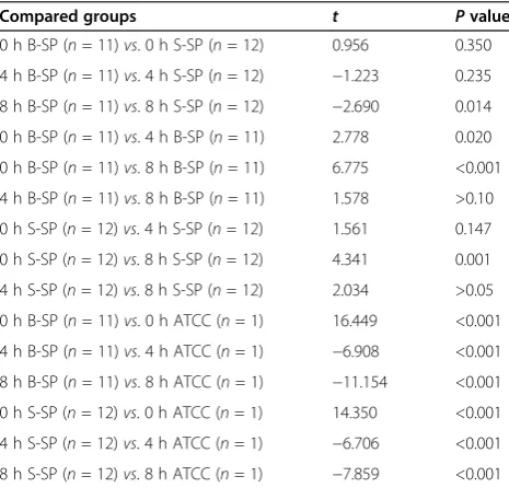
Table 2 showed P values between the following com-pared groups after S. pneumoniae infected THP-1 cells due to non-homogeneity of variance. Cochran & Cox separate variance estimationttest was used to compare blood and sputum groups of S. pneumoniae after the infection.
Changes in THP-1 and A549 cell morphology after
S. pneumoniaeinfection
As shown in Figure 1, the longer time of THP-1 cells culture with S. pneumoniae, the more deaths THP-1 cells suffered, levels of which were associated with differ-ent sources of S. pneumoniae isolations and amounts. Similar data were observed in A549 cell cultures of these S. pneumoniaesamples.
Discussion
In the current study, we first isolated and confirmed 23 S. pneumoniae strains from clinical blood and sputum samples. We then used these 23 S. pneumoniae strains and ATCC49619 to infect human monocytes and pneu-mocytes and then analyzed expression of theS. pneumo-niae virulence gene ply in these 23 strains and ATCC49619. We found that levels ofplymRNA expres-sion were much higher in clinical S. pneumoniae samples than in the ATCC49619 standard and in blood-derived S. pneumoniae samples than in sputum-derived ones. This finding demonstrated differential expression ofplymRNA levels in blood-derivedS. pneumoniaeafter infecting THP-1 and A549 cells compared to sputum-derived ones. This study provides indirect molecular evi-dence that the S. pneumoniae virulence gene ply may facilitate S. pneumoniae invasion into the host blood system.
Indeed, Ply is a pore-forming toxin with a molecular weight of 53 kD and is known as a cholesterol-binding cytolysin, a key virulence factor for clinical S. pneumo-niae strains. Ply protein can dissolve cell membranes in almost all eukaryotes. Specifically, Ply toxin will bind to a cholesterol-rich membrane using its pore-forming mechanism and then penetrate into the lipid bilayer of the cell membrane and oligomerize to form a perfor-ation on the cell membrane [14]. Ply can also activate macrophages to execute apoptosis without membrane pore formation [15]. Thus, Ply toxin has a wide range of biological activities, such as facilitation ofS. pneumoniae colonization and pathogenicity. Ply can also activate the classic complement pathway in the human body, inhibit cough and bactericidal activity and migration of white blood cells [16], stimulate IL-8 synthesis [17], and in-crease ICAM-1 and IL-1β secretions [18-20]. The Ply impact on the immune response to the pneumococcus is highly dependent on the strain background, thus, it is surely important of the interaction between specific virulence factors and other components of the genetic background of this organism [19]. A recent study also confirmed the role of plyin biofilm formation, which is separate from the hemolytic activity responsible for tissue damage during pneumococcal disease [21]. Our current data showed that after infecting THP-1 cells,
Table 1 Expression of virulence geneplyafterS. pneumoniaeinfected THP-1 or A549 cells
Groups n ply(△Ct,‾x ±s)
0 h 4 h (THP-1) 8 h (THP-1) 4 h (A549) 8 h (A549)
ATCC 1 16.2 31.5 27.9 21.8 19.1
Blood SP 11 29.2 ± 2.6 24.8 ± 3.2 23.0 ± 1.5 20.4 ± 5.1 18.2 ± 2.9
Sputum SP 12 28.1 ± 2.9 26.3 ± 2.7 24.6 ± 1.4 20.8 ± 3.0 23.3 ± 3.1
Note:△Ct = Mean Ct of tested gene−Mean Ct of control gene. Smaller△Ct means moreplyexpression. Level of 16S rRNA was used as a control, which expresses much higher than that ofply. SP,Streptococcus pneumoniae; ATCC, American Type Culture Collection.
Table 2Pvalues between the following compared groups afterS. pneumoniaeinfected THP-1 cells
Compared groups t Pvalue
0 h B-SP (n= 11)vs. 0 h S-SP (n= 12) 0.956 0.350
4 h B-SP (n= 11)vs. 4 h S-SP (n= 12) −1.223 0.235
8 h B-SP (n= 11)vs. 8 h S-SP (n= 12) −2.690 0.014
0 h B-SP (n= 11)vs. 4 h B-SP (n= 11) 2.778 0.020
0 h B-SP (n= 11)vs. 8 h B-SP (n= 11) 6.775 <0.001
4 h B-SP (n= 11)vs. 8 h B-SP (n= 11) 1.578 >0.10
0 h S-SP (n= 12)vs. 4 h S-SP (n= 12) 1.561 0.147
0 h S-SP (n= 12)vs. 8 h S-SP (n= 12) 4.341 0.001
4 h S-SP (n= 12)vs. 8 h S-SP (n= 12) 2.034 >0.05
0 h B-SP (n= 11)vs. 0 h ATCC (n= 1) 16.449 <0.001
4 h B-SP (n= 11)vs. 4 h ATCC (n= 1) −6.908 <0.001
8 h B-SP (n= 11)vs. 8 h ATCC (n= 1) −11.154 <0.001
0 h S-SP (n= 12)vs. 0 h ATCC (n= 1) 14.350 <0.001
4 h S-SP (n= 12)vs. 4 h ATCC (n= 1) −6.706 <0.001
8 h S-SP (n= 12)vs. 8 h ATCC (n= 1) −7.859 <0.001
expression of ply mRNA was significantly induced in blood-derived S. pneumoniae strains compared to sputum-derived ones, indicating that the increased ply expression may contribute to S. pneumoniae against defense of THP-1 cells, which may be one of the mecha-nisms for S. pneumoniae invasion into the host blood system. In the current study, we determinedply expres-sion after infecting two types of human cell lines and data are identical, which are consistent with a previous study [22]. Besides, as a whole, expression of psaA and cpsA was also studied in a previous study [23]. And different patterns of their expression clearly showed different roles of such three virulence genes. The ex-pression ofcpsA is the basis of the pathogenicity of SP and ply expression is more important than that of psaA in SP invasion into the blood system. Our current data demonstrated that inflammation induction of different SP sources tends to be consistent [20]. How-ever, the infection types are determined mainly by differ-ent sources of SP itself, especially its expression of virulence genes.
In the current study, we utilized a standard strain of S. pneumoniae ATCC49619 for comparison. We found that S. pneumoniae isolated from both blood and spu-tum samples were more virulent by expression of high levels of Ply toxin after infecting THP-1 and A549 cells. ATCC49619 is a standard S. pneumoniae strain and its physical and chemical properties are relatively stable. The S. pneumoniaeisolated from clinical samples were much more easily influenced by external environment than ATCC49619. Thus, compared to clinical S. pneu-moniae, the levels of virulence and viability of ATCC49619 are weaker, all of which may indicate that clinical S. pneumoniae strains are more infectious.
However, it remains to be determined whether S. pneu-moniae isolated from different individuals also have dif-ferent levels of infection potential.
Moreover, we also showed the interaction between S. pneumoniaeand THP-1 cells or A549 cells. Different sources ofS. pneumoniaeresulted in different outcomes, which could suggest that various strains of S. pneumo-niae have different abilities to infect the lung, blood, or even the CSF. However, this study is just a proof-of-principle and much more are needed to investigate other virulence factors ofS. pneumoniaeor their combinations in infecting the human body. Indeed, S. pneumoniae have many other virulence genes, such as nanA [7], which has been demonstrated to facilitateS. pneumoniae invasion into the CSF. Moreover, S. pneumoniae inva-sion into the host blood system is an important step and prerequisite for its further invasion into the CSF in most cases. The role of Ply toxin in S. pneumoniae invasion into the host blood system and its relationship with NanA are worthy of further investigation. In addition, the significance of thisin vitro study needs further con-firmation by anin vivostudy.
Conclusions
The current study demonstrated thatply gene of blood-and sputum-derived S. pneumoniae is differentially ex-pressed in two different cell lines. Under survival pressure, higher expression of ply gene is needed in these two cell lines for blood-derived S. pneumoniae, which indicates that ply gene may facilitate S. pneumo-niaeinvasion into the host blood system.
Competing interests
The authors declare that they have no competing interests.
Authors’contributions
DKH and XYL defined the research theme. YL designed methods and experiments and carried out the laboratory experiments. YQ analyzed the data and wrote the paper. All authors read and approved the final manuscript.
Acknowledgements
This work was supported in part by a grant from The Science and Technology Development Planning (#10275) funds of Jiaojiang District, Taizhou City, China.
Author details
1Department of Laboratory Medicine, Taizhou Municipal Hospital, 381# Zhongshan East Road, Taizhou 318000, China.2Department of Laboratory Medicine, The First Affiliated Hospital of Nanchang University, 17# Yong Wai Zheng Street, Nanchang 330006, China.3Department of Laboratory Medicine, The Second Affiliated Hospital of Wenzhou Medical University, 109# College West Road, Wenzhou 325027, China.
Received: 5 December 2014 Accepted: 27 April 2015
References
1. Jin P, Xiao M, Kong F, Oftadeh S, Zhou F, Liu C, et al. Simple, accurate, serotype-specific PCR assay to differentiate Streptococcus pneumoniae serotypes 6A, 6B, and 6C. J Clin Microbiol. 2009;47:2470–4.
2. Mitchell AM, Mitchell TJ. Streptococcus pneumoniae: virulence factors and variation. Clin Microbiol Infect. 2010;16:411–8.
3. Suzuki H, Ikeda K. Mode of action of long-term low-dose macrolide therapy for chronic sinusitis in the light of neutrophil recruitment. Curr Drug Targets Inflamm Allergy. 2002;1:117–26.
4. Manso AS, Chai MH, Atack JM, Furi L, De Ste Croix M, Haigh R, et al. A random six-phase switch regulates pneumococcal virulence via global epigenetic changes. Nat Commun. 2014;5:5055.
5. Cockeran R, Mitchell TJ, Feldman C, Anderson R. Pneumolysin induces release of matrix metalloproteinase-8 and−9 from human neutrophils. Eur Respir J. 2009;34:1167–70.
6. Morona JK, Morona R, Paton JC. Attachment of capsular polysaccharide to the cell wall of Streptococcus pneumoniae type 2 is required for invasive disease. Proc Natl Acad Sci U S A. 2006;103:8505–10.
7. Uchiyama S, Carlin AF, Khosravi A, Weiman S, Banerjee A, Quach D, et al. The surface-anchored NanA protein promotes pneumococcal brain endothelial cell invasion. J Exp Med. 2009;206:1845–52.
8. Weisfelt M, van de Beek D, Spanjaard L, Reitsma JB, de Gans J. Clinical features, complications, and outcome in adults with pneumococcal meningitis: a prospective case series. Lancet Neurol. 2006;5:123–9. 9. Mahdi LK, Ebrahimie E, Adelson DL, Paton JC, Ogunniyi AD. A transcription
factor contributes to pathogenesis and virulence in Streptococcus pneumoniae. PLoS One. 2013;8, e70862.
10. Ricci S, Gerlini A, Pammolli A, Chiavolini D, Braione V, Tripodi SA, et al. Contribution of different pneumococcal virulence factors to experimental meningitis in mice. BMC Infect Dis. 2013;13:444.
11. du Plessis M, Smith AM, Klugman KP. Rapid detection of penicillin-resistant Streptococcus pneumoniae in cerebrospinal fluid by a seminested-PCR strategy. J Clin Microbiol. 1998;36:453–7.
12. Walker JA, Allen RL, Falmagne P, Johnson MK, Boulnois GJ. Molecular cloning, characterization, and complete nucleotide sequence of the gene for pneumolysin, the sulfhydryl-activated toxin of Streptococcus pneumoniae. Infect Immun. 1987;55:1184–9.
13. Bentley RW, Leigh JA, Collins MD. Intrageneric structure of Streptococcus based on comparative analysis of small-subunit rRNA sequences. Int J Syst Bacteriol. 1991;41:487–94.
14. Palmer M. The family of thiol-activated, cholesterol-binding cytolysins. Toxicon. 2001;39:1681–9.
15. Bewley MA, Naughton M, Preston J, Mitchell A, Holmes A, Marriott HM, et al. Pneumolysin activates macrophage lysosomal membrane permeabilization and executes apoptosis by distinct mechanisms without membrane pore formation. mBio. 2014;5:e01710–4.
16. Paton JC, Ferrante A. Inhibition of human polymorphonuclear leukocyte respiratory burst, bactericidal activity, and migration by pneumolysin. Infect Immun. 1983;41:1212–6.
17. Rogers PD, Thornton J, Barker KS, McDaniel DO, Sacks GS, Swiatlo E, et al. Pneumolysin-dependent and -independent gene expression identified by cDNA microarray analysis of THP-1 human mononuclear cells stimulated by Streptococcus pneumoniae. Infect Immun. 2003;71:2087–94.
18. Thornton J, McDaniel LS. THP-1 monocytes up-regulate intercellular adhesion molecule 1 in response to pneumolysin from Streptococcus pneumoniae. Infect Immun. 2005;73:6493–8.
19. Harvey RM, Hughes CE, Paton AW, Trappetti C, Tweten RK, Paton JC. The impact of Pneumolysin on the macrophage response to Streptococcus pneumoniae is strain-dependent. PLoS One. 2014;9, e103625. 20. Zhang J, Hu DK, Wang DG, Liu Y, Liu CB, Yu LH, et al. Effects of clinical
isolates of Streptococcus pneumoniae on THP-1 human monocytic cells. Mol Med Rep. 2013;8:1570–4.
21. Shak JR, Ludewick HP, Howery KE, Sakai F, Yi H, Harvey RM. Novel role for the Streptococcus pneumoniae toxin pneumolysin in the assembly of biofilms. mBio. 2013;4:e00655–13.
22. Hirst RA, Yesilkaya H, Clitheroe E, Rutman A, Dufty N, Mitchell TJ, et al. Sensitivities of human monocytes and epithelial cells to pneumolysin are different. Infect Immun. 2002;70:1017–22.
23. Hu DK, Wang DG, Liu Y, Liu CB, Yu LH, Qu Y, et al. Roles of virulence genes (PsaA and CpsA) on the invasion of Streptococcus pneumoniae into blood system. Eur J Med Res. 2013;18:14.
Submit your next manuscript to BioMed Central and take full advantage of:
• Convenient online submission
• Thorough peer review
• No space constraints or color figure charges
• Immediate publication on acceptance
• Inclusion in PubMed, CAS, Scopus and Google Scholar
• Research which is freely available for redistribution