R E V I E W
Open Access
Current approaches to increase CAR T cell
potency in solid tumors: targeting the
tumor microenvironment
Irene Scarfò and Marcela V. Maus
*Abstract
Chimeric antigen receptor (CAR) T-cell therapy represents a revolutionary treatment for haematological
malignancies (i.e. B-ALL). However, the success of this type of treatment has not yet been achieved in solid tumors. One hypothesis is that the immunosuppressive nature of the tumor microenvironment (TME) influences and affects the efficacy of adoptive immunotherapy. Understanding the role of the TME and its interaction with CAR T-cells is crucial to improve the potency of adoptive immunotherapy. In this review, we discuss the strategies and potential combinatorial approaches recently developed in mouse models to enhance the efficacy of CAR T-cells, with particular emphasis on the translational potential of these approaches.
Keywords:Chimeric antigen receptor T-cells, Adoptive immunotherapy, Tumor microenvironment
Background
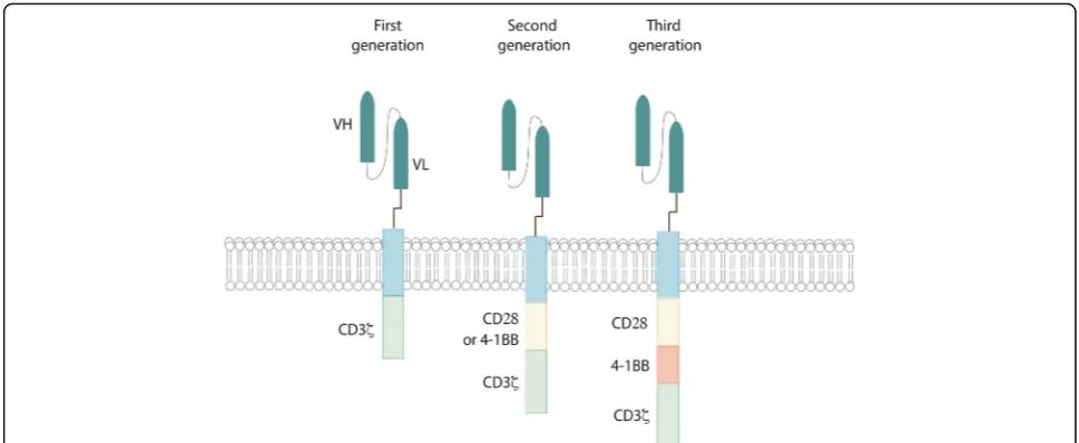
Adoptive cell therapy (ACT) is a novel tool in the fight against cancer. In particular T-cells engineered to express Chimeric Antigen Receptors (CARs) have demonstrated recent significant clinical efficacy with improvements in patient outcomes for a number of hematological malig-nancies [1–4]. CARs are synthetic molecules composed of an extracellular binding domain, a transmembrane do-main and an intracellular signaling/activation dodo-main. The extracellular component consists of the light and heavy chain regions derived from an antibody to form a single chain variable fragment (scFv), and serves to recognize and bind specific tumor-associated antigens (TAAs) in a MHC-independent manner. A hinge domain, typically derived from CD8 or IgG4 molecules, connects this module with the intracellular one. This last portion is formed by CD3ζ segment which is responsible to trigger T-cell activation. The first generation of CAR vectors was designed with CD3ζdomain alone. Second and third gen-erations added to CD3ζone or two costimulatory domains (CD28 and/or 4-1BB) respectively (Fig. 1). All these com-ponents are typically inserted using γ-retroviral or
lentiviral transduction systems. Although silencing of LTR-driven transgenes has been known to occur in other tissues, vector silencing was not observed in one study of human lymphocytes [5]. Interestingly, one study showed that efficacy of CAR T cells in vivo is a function of the density of CAR expression, and that this can have a sub-stantial impact on antitumor efficacy and persistence of CAR T cells both systemically and at the tumor site [6].
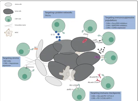
By combining the ability to avoid HLA restriction in anti-gen recognition with high specificity and potent activation, engineering these molecules to be expressed in T cells have emerged as one of the most promising approaches for can-cer treatment. However, attempts to recapitulate the suc-cess achieved with CAR T-cells in B-cells malignancies for solid tumors has been disappointing. The three main hur-dles encountered for the application of CAR T cell therapies to solid tumors are (1) the identification of proper tumor associated antigens, (2) the limited trafficking of adoptively transferred cells to tumor sites and (3) the immunosuppres-sive effect of tumor microenvironment. Here we will focus on approaches to address the third problem (Fig. 2); others have described approaches to the first two [7–13].
The complex and heterogeneous tumor microenviron-ment plays an essential role in tumor initiation progres-sion and therapeutic resistance. Recent studies have highlighted the importance of innate immune activation * Correspondence:[email protected]
Cellular Immunotherapy Program, Massachusetts General Hospital Cancer Center and Harvard Medical School, 149 13th Street, Room 7.219, Charlestown, Boston, MA 02129, USA
for the generation of spontaneous T cell responses to tumor-associated antigens and anti anti-tumor activity Woo and colleagues showed that type I IFN-mediated activation of the STING pathway of cytosolic DNA sens-ing is one of the major players in sustainsens-ing a T cell inflamed-tumor phenotype which represents a favorable marker of responsiveness to immunotherapies [14]. Acti-vation of the STING pathway contributes to actiActi-vation of Batf3 dendritic cells, which appear to be central to anti-tumor immunity. These orchestrating dendritic cells are recruited by chemokines to the tumor site, then mi-grate to the secondary lymphatic organs and interact with CD8+ T cells. After clonal expantion, the T cells can migrate back to the tumor microenvironment and carry out their effector functions against tumor cells [15]. Although a thorough evaluation of innate immune activators and an inflamed-tumor microenvironment in the setting of CAR T-cells has yet to be investigated, the role of the tumor microenvironment in modulating effi-cacy of CAR-T cells is expected to be significant at the level of effector T cell function if not antigen presenta-tion. Furthermore, the degree of intra-tumoral expansion and persistence that is required for clinical efficacy has not been determined for solid tumors. In this review, we discuss various aspects of the tumor microenvironment that could inhibit the efficacy of immune responses, and some of the approaches recently developed to reprogram the tumor microenvironment in order to enhance the ef-ficacy of CAR T-cell therapy; some of these may also serve to enhance tumor sensitivity to other forms of im-munotherapy. Indeed although the focus of this review is on CAR T cells, many of the principles herein
discussed apply to and have been elucidated from studies of adoptive cell therapy more broadly, including tumor infiltrating lymphocytes (TILs) and TCR-engineered T cells [16–18].
Targeting hypoxia, nutrient starvation, and metabolism
Hypoxia and nutrient starvation are major hallmarks of the tumor microenvironment [19]. The lack of nutrients, par-ticularly amino acids such as tryptophan, is able to activate the integrated stress response which regulates T-cell activity [20]. Indoleamine 2,3 dioxygenase (IDO) is an intracellular enzyme which catalyzes the degradation of tryptophan to kynurenine. Both tumor cells and myeloid cells within the tumor microenvironment can express IDO, leading to a block in the proliferation and survival of T-cells. Recently, Ninomiya and colleagues [21] demonstrated that IDO ex-pression can also inhibit CAR T cells. In particular, they showed that CD19 CAR T-cells fail to control progression of CD19+ IDO-expressing tumors. The accumulation of kynurenine inhibited expansion, cytotoxicity and cytokine secretion of CAR T-cells, suggesting a plausible mechanism underlying the resistance of IDO positive tumors to this type of treatment. Interestingly, they demonstrated that flu-darabine and cyclophosphamide administration improved the efficacy of CAR T-cells by decreasing the expression of IDO. The combination of CAR T-cells and IDO inhibitors may represent a valuable option for malignancies resistant to chemotherapy and immunotherapy.
Moreover it is possible that metabolic stress in the tumor environment is able to modulate T cell metabolism, differentiation and effector functions. Indeed, tumor
infiltrating lymphocytes modify their metabolism as a re-sponse to hypoxia, which is characteristic of solid tumors [22]. Interestingly, it has been demonstrated that CAR T-cells with different costimulatory domains utilize distinct metabolic pathways, which in turn reflect variable persist-ence within the tumor microenvironment [23]. Kawalekar and colleagues showed that 4-1BB CAR T-cells had in-creased mitochondrial biogenesis, which prompted a cen-tral memory phenotype and led to a survival advantage compared to CAR T cells with the CD28 signaling do-main. In contrast, CD28-CAR T-cells yielded effector memory phenotype and enhanced glycolysis. This study highlighted the significance of the design of engineered T-cells with regard to their survival and exhaustion in the immunosuppressive tumor microenvironment.
Altering metabolic components within the tumor microenvironment is only one possibility for maximiz-ing the potential of CAR T cells. In a more recent study Newick and colleagues [24] demonstrated simi-lar findings by inhibiting protein kinase A (PKA) acti-vation. PKA is the downstream effector of two other
immunosuppressive factors produced within the
tumor microenvironment: prostaglandin E2 (PGE2) and adenosine. Different studies have reported the role of these two molecules as potent inhibitors of T-cell proliferation and activity [25, 26]. The authors dis-rupted PKA anchorage to the lipid rafts by expressing RIAD (regulatory subunit I anchoring disruptor) pep-tide. This peptide is able to displace the association between PKA and ezrin, a protein necessary to tether-ing PKA close to adenylyl cyclase. Mesothelin-directed CAR-RIAD T-cells exhibited higher infiltration, per-sistence and antitumor activity in vivo compared to mesothelin-directed CAR T-cells. Moreover, the ex-pression of RIAD led to an increased chemotaxis, guided by an induction of CXCR3 expression, and to better adhesion. As this approach is translated to clin-ical trials, the immunogenicity of the peptide may be-come an issue.
Another way to reprogram the tumor microenviron-ment is to augmicroenviron-ment the antitumor response of CAR T-cells by facilitating the recruitment of an endogenous
immune response. This approach was validated by Curran and colleagues [27] through the constitutive expression of CD40L by CAR T-cells. They demon-strated that this strategy improves CAR T-cell cytotox-icity, decreases expression of PD-1, and improves DC antigen presentation function in a systemic model of CD40 positive follicular lymphoma. These mecha-nisms are likely to be synergistic with the STING pathways described by Gajewski and colleagues (as above).
Thus targeting tumor immunosuppressive populations such as PGE2 and/or T-cells response to the altered microenvironment represents an exciting opportunity to increase the efficacy of immunotherapy.
Targeting stroma
Enhancing the efficacy of anti-tumor immune therapies could also be accomplished by targeting the non-malignant cancer-associated stromal cells (CASCs). These cells secrete growth factors, cytokines, and chemokines that promote tumor growth, metastasis and angiogenesis. One attractive stromal candidate is the fibroblast activa-tion protein (FAP), a serine protease implicated in extra-cellular matrix remodeling and selectively expressed by CASCs in over 90% of epithelial cancers [28]. Three differ-ent groups have described the use of anti-FAP CAR T-cells with some contrasting results. Tran and colleagues [29] showed that despite anti-FAP CARs displaying spe-cific degranulation and production of effector cytokines in response to antigen stimulation in vitro, they didn’t medi-ate an efficient antitumor response in vivo, and unexpect-edly and alarmingly, anti-FAP CARs caused severe cachexia and lethal bone toxicities. The authors found that FAP protein is also expressed by multipotent bone mar-row stromal cells (BMSCs) and related the observed tox-icity to this expression. However, Kakarla and colleagues [30] using a FAP-CAR with a different scFv demonstrated antitumor efficacy without toxicities in a mouse model of human lung cancer. Even if T-cell persistence was limited, an improved response was achieved by co-injecting FAP-specific and tumor-FAP-specific T-cells. Schuberth and col-leagues [31] developed an intra-peritoneal model for the adoptive transfer of FAP-CARs in a mesothelioma xeno-graft. Their data showed an increased survival; however, their anti-human FAP scFv did not have cross-reactivity with mouse FAP, which limited their ability to evaluate the on-target/off-tumor toxicity. A paper by Wang and col-leagues [32] suggested that targeting FAP positive cells en-hanced antitumor immunity via epitope spreading. They showed activation of endogenous CD4+ T-cells after three days of anti-FAP CAR treatment in an immune-competent syngeneic mouse model of mesothelioma and lung cancer. CD4+ activation was followed by a later aug-mentation of endogenous CD69+, INFγ+ CD8+ T-cell
infiltration. This antitumor response did not occur in im-munodeficient mice, supporting the importance of the adaptive immune system. In another publication, the same group underlined the relevance of FAP inhibition by showing it also had an immune-independent antitumor effect. Using a weakly immunogenic and highly desmo-plastic tumor, pancreatic ductal adenocarcinoma, Lo and colleagues [33] demonstrated that FAP-CAR T-cells inhib-ited tumor stromagenesis, reduced tumor vascular density and disrupted spatial orientation of tumor cells.
The safety concerns generated by the work of Rosenberg et al. [29] may be related to the specificity and affinity of the scFv, given that the last two studies with CAR T cells with different scFvs that recognize highly positive FAP cells, have been reassuring. Given the potential for multi-modal anti-tumor effects of FAP targeting, rational and interesting combinations for future immunotherapeutic approaches in-clude anti-stroma CAR T-cells with either anti-tumor CAR T-cells or checkpoint blockade.
Beyond targeting FAP, another recent strategy for using CAR T cells in stroma-rich tumors is to target the enzyme heparanase (HPSE). This enzyme is responsible of the degradation of the heparan sulfate proteoglycans (HPGs), a fundamental process for the trafficking and accumulation of T-cells to the tumor site. Caruana and colleagues [34] demonstrated that CAR T-cells lose the expression of HSPE during their manufacturing process. This phenomenon leads to an impaired ability to de-grade the extracellular matrix, suggesting a compromise in migration capacity. The authors showed that the in-duction of HSPE expression enhanced tumor infiltration and improved survival in neuroblastoma xenograft models.
Targeting cytokine networks
IFNγlevels. In order to increase the safety and reduce un-desired toxicities that could be generated by constitutive, systemic high levels of IL-12, which have been toxic in clin-ical trials [41], they developed a tricistronic vector that en-codes the scFv specific for MUC-16ectoantigen, IL-12 and the truncated EGFR elimination gene (EGFRt) and admin-istered it locally. Peritoneal administration of CAR T-cells was significantly more effective compared with intra-venous administration. This paper paved the way for a phase I clinical trial in platinum-resistant ovarian cancer patients, which will determine the safety and feasibility of this approach [42]. The production and release of a trans-genic payload in a CAR should minimize toxicities in addition the “switch-off” system such as the EGFRt pro-vides an additional measure of control for safety.
Furthermore IL-12 expression has proved to be im-portant for the generation and efficacy of CAR T-cells from the umbilical cord blood (UCB). Pegram and col-leagues [43] described a novel technique to expand and genetically modify UCB T-cells. First, they demonstrated that adding IL-12 and IL-15 to the cultured activated UCB T-cells led to a 150-fold expansion of this popula-tion, which showed an ideal phenotype expressing both memory and effector markers. Second, including the ex-pression of IL-12 in the anti-CD19 CAR vector resulted in a significant increase in survival of mice bearing acute lymphoblastic leukemia (ALL), without the need for pre-treatment or Il-2 support. These data support the clin-ical translation of using ACT to further boost the graft-versus-leukemia effect in UCB-transplanted patients with high-risk, relapsed/refractory ALL.
Another feasibile strategy to modulate tumor microenvir-onment signalling is to directly inhibit TGFβ, IL-10, and/or IL-4 signaling. The secretion of the first molecule by cancer cells and cancer associated cells is a well know mechanism of tumor evasion, and IL-10 as well as IL-4 are potent im-munosuppressive cytokines. One approach is to force the overexpression of a dominant negative form of the receptor on T cells. Zhang and colleagues [44] demonstrated an in-creased antitumor efficacy in vitro and in vivo of T cells overexpressing TGFβDNRII in a melanoma tumor model. Despite the promising results, further studies have to be done in orther to validate this approach in the context of CAR T-cell therapy. An additional recent system developed by Mohammed and colleagues [45] for the treatment of prostate cancer, a tumor characterized by elevated IL-4 levels, consists in an inverted cytokine receptor (ICR). Spe-cifically, this 4/7 ICR is formed by the extracellular domain of the inhibitory IL-4 receptor linked to the intracellular immunostimulatory domain of IL-7. The co-expression of anti-PSCA first generation CAR vector and ICR resulted in an increased in vitro and in vivo antitumor activity. This approach transforms an inhibitory signal for T cells into a stimulatory one and at the same time deprives cancer cells
of an important growth factor. The combination of a sec-ond generation CAR with 4/7 ICR could be evaluated to improve on these results.
Targeting immune checkpoints
A major mechanism through which the
that combining CAR T-cells with the expression of a “dominant negative” form of PD-1 led to higher persist-ence, increased antitumor effects and prolonged survival in a mesothelioma xenograft model. The“dominant nega-tive”form of PD1 in this model was composed of the only the extracellular domain of PD1 (without a signaling do-main), which would presumably compete with endogen-ous PD1 for ligating PD-L1. One explanation for these divergent results could be the different type of tumor treated. Moreover, Cherkassky and colleagues showed that CAR T-cells with 4-IBB costimulatory domain were able to function at lower doses compared to CAR T cells that included CD28 signaling domains, and 4-1BB-signaling CARs were more resistant to PD-1 mediated exhaustion.
Additional tumor models are necessary to show that PD-1-mediated CAR T-cell suppression is a general in-hibitory mechanism, particularly immunocompetent mouse models. It will be also important to understand the role of the costimulatory domains embedded into CARs and their differential mechanisms in mediating re-sistance to inhibitory molecules and inducers of CAR T-cell exhaustion. Even if the safest and most efficacius checkpoint combinations have yet to be identified [52], altogether these pre-clinical data provide supportive evi-dence that combining immune checkpoint blockade with CAR T-cells is a logical therapeutic strategy to improve the clinical outcome of CAR T-cell therapy in patients.
Conclusions
Adoptive cell therapy using CAR T-cells has demon-strated impressive therapeutic potential for the treat-ment of certain B cell malignancies. Even though there have been recent exciting publications using TIL therapy targeting mutant KRAS metastatic colorectal cancer [53] and IL13Rα2-targeted CAR T cells in glioblastoma mul-tiforme [54], results in solid malignancies may be subject to various limitations, including the immunosuppressive tumor microenvironment. Indeed, the microenviron-ment non only creates a physical barrier decreasing the penetration of modified T-cells into the tumor mass, but also plays an active role in immune suppression through the up regulation of inhibitory signals. Innovative strat-egies have been developed to overcome these challenges (Fig. 2), including co-administration of CAR T cells with checkpoint blockade, and co-administration with other drugs, therapies, and CAR T cells that target the tumor stroma and immunosuppressive molecules. Many of these strategies have been tested in xenograft and syn-geneic mouse models, and clinical trials of these combi-nations are warranted and eagerly awaited. Pre-clinical experiments will define rational combinations of these approaches, based on a deeper understanding of the unique tumor characteristics and interplay among im-mune cells and the tumor environment. Translating the
optimal combinations is likely to require iterative clinical trials to determine the safest and most effective combi-nations for patients with solid tumors.
Abbreviations
ACT:Adoptive cell therapy; B-ALL: B-cells acute lymphoblastic leukemia; BMSCs: Bone marrow stromal cells; CAR: Chimeric antigen receptor; CASCs: Cancer-associated stromal cells; CD: Cluster of differentiation; CD40L: CD40 ligand; CTLA-4: Cytotoxic T-lymphocyte-associate protein 4; CXCR: C-X-C motif chemokine receptor; DC: Dendritic cell; EGFRt: Truncated epidermal growth factor receptor; FAP: Fibroblast activation protein; HLA: Human leukocyte antigen; HPGs: Heparan sulfate proteoglycans; HPSE: Heparanase; IDO: Indoleamine 2,3 dioxygenase; Ig: Immunoglobulin; IL: Interleukin; INFγ: Interferon gamma; MDSC: Myeloid-derived suppressor cells; MHC: Major histocompatibility complex; NK: Natural killer; PD-1: Programmed death-1; PD-LPD-1: PD-1 ligand; PGE2: Prostaglandin E2;
PKA: Protein kinase A; RIAD: Regulatory subunit I anchoring disruptor; scFv: Single chain variable fragment; TAAs: Tumor-associated antigens; TGFβ: Trasforming growth factor beta; TILs: Tumor-infiltrating lymphocytes; TME: Tumor microenvironment; TRUCK: T-cells redirected for universal cytokine-mediated killing; UCB: Umbilical cord blood
Acknowledgements
Irene Scarfò receives support from American-Italian Cancer Foundation Post-Doctoral Research Fellowship. Marcela Maus receives support from the NIH (NCI K08CA166039), the Gabrielle’s Angel Foundation, and the V Foundation.
Funding
Irene Scarfò receives support from American-Italian Cancer Foundation Post-Doctoral Research Fellowship. Marcela Maus receives support from the NIH (NCI K08CA166039), the Gabrielle’s Angel Foundation, and the V Foundation.
Availability of data and materials
Not applicable
Authors’contributions
IS and MVM wrote the manuscript. Both authors read and approved the final manuscript.
Competing interests
MVM is listed as an inventor on patents related to the use of T cell therapies; these patents are held by the University of Pennsylvania, and some of them are licensed to Novartis. None of these are relevant to this manuscript.
Consent for publication
Granted
Ethics approval and consent to participate
Not applicable
Received: 13 November 2016 Accepted: 24 February 2017
References
1. Maude SL, Frey N, Shaw PA, Aplenc R, Barrett DM, Bunin NJ, Chew A, Gonzalez VE, Zheng Z, Lacey SF, et al. Chimeric antigen receptor T cells for sustained remissions in leukemia. N Engl J Med. 2014;371:1507–17. 2. Porter DL, Hwang WT, Frey NV, Lacey SF, Shaw PA, Loren AW, Bagg A,
Marcucci KT, Shen A, Gonzalez V, et al. Chimeric antigen receptor T cells persist and induce sustained remissions in relapsed refractory chronic lymphocytic leukemia. Med Sci Transl. 2015;7:303ra139.
3. Garfall AL, Maus MV, Hwang WT, Lacey SF, Mahnke YD, Melenhorst JJ, Zheng Z, Vogl DT, Cohen AD, Weiss BM, et al. Chimeric antigen receptor T cells against CD19 for multiple myeloma. N Engl J Med. 2015;373:1040–7. 4. Lee DW, Kochenderfer JN, Stetler-Stevenson M, Cui YK, Delbrook C, Feldman
SA, Fry TJ, Orentas R, Sabatino M, Shah NN, et al. T cells expressing CD19 chimeric antigen receptors for acute lymphoblastic leukaemia in children and young adults: a phase 1 dose-escalation trial. Lancet. 2015;385:517–28. 5. Burns WR, Zheng Z, Rosenberg SA, Morgan RA. Lack of specific
receiving genetically engineered lymphocytes and activation upon lymphocyte restimulation. Blood. 2009;114:2888–99.
6. Frigault MJ, Lee J, Basil MC, Carpenito C, Motohashi S, Scholler J, Kawalekar OU, Guedan S, McGettigan SE, Posey Jr AD, et al. Identification of chimeric antigen receptors that mediate constitutive or inducible proliferation of T cells. Cancer Immunol Res. 2015;3:356–67.
7. Di Stasi A, De Angelis B, Rooney CM, Zhang L, Mahendravada A, Foster AE, Heslop HE, Brenner MK, Dotti G, Savoldo B. T lymphocytes coexpressing CCR4 and a chimeric antigen receptor targeting CD30 have improved homing and antitumor activity in a Hodgkin tumor model. Blood. 2009;113:6392–402. 8. Craddock JA, Lu A, Bear A, Pule M, Brenner MK, Rooney CM, Foster AE. Enhanced tumor trafficking of GD2 chimeric antigen receptor T cells by expression of the chemokine receptor CCR2b. J Immunother. 2010;33:780–8. 9. Peng W, Ye Y, Rabinovich BA, Liu C, Lou Y, Zhang M, Whittington M, Yang
Y, Overwijk WW, Lizee G, Hwu P. Transduction of tumor-specific T cells with CXCR2 chemokine receptor improves migration to tumor and antitumor immune responses. Clin Cancer Res. 2010;16:5458–68.
10. Moon EK, Carpenito C, Sun J, Wang LC, Kapoor V, Predina J, Powell Jr DJ, Riley JL, June CH, Albelda SM. Expression of a functional CCR2 receptor enhances tumor localization and tumor eradication by retargeted human T cells expressing a mesothelin-specific chimeric antibody receptor. Clin Cancer Res. 2011;17:4719–30.
11. Beatty GL, O’Hara M. Chimeric antigen receptor-modified T cells for the treatment of solid tumors: defining the challenges and next steps. Pharmacol Ther. 2016;166:30–9.
12. Zhukovsky EA, Morse RJ, Maus MV. Bispecific antibodies and CARs: generalized immunotherapeutics harnessing T cell redirection. Curr Opin Immunol. 2016;40:24–35.
13. Gill S, Maus MV, Porter DL. Chimeric antigen receptor T cell therapy: 25 years in the making. Blood Rev. 2016;30:157–67.
14. Woo SR, Fuertes MB, Corrales L, Spranger S, Furdyna MJ, Leung MY, Duggan R, Wang Y, Barber GN, Fitzgerald KA, et al. STING-dependent cytosolic DNA sensing mediates innate immune recognition of immunogenic tumors. Immunity. 2014;41:830–42.
15. Corrales L, Matson V, Flood B, Spranger S, Gajewski TF. Innate immune signaling and regulation in cancer immunotherapy. Cell Res. 2017;27:96–108. 16. Ahmadzadeh M, Johnson LA, Heemskerk B, Wunderlich JR, Dudley ME,
White DE, Rosenberg SA. Tumor antigen-specific CD8 T cells infiltrating the tumor express high levels of PD-1 and are functionally impaired. Blood. 2009;114:1537–44.
17. Kerkar SP, Muranski P, Kaiser A, Boni A, Sanchez-Perez L, Yu Z, Palmer DC, Reger RN, Borman ZA, Zhang L, et al. Tumor-specific CD8+ T cells expressing interleukin-12 eradicate established cancers in lymphodepleted hosts. Cancer Res. 2010;70:6725–34.
18. Chinnasamy C, St Clair Gibson A, Micklewright D. Effect of spatial and temporal cues on athletic pacing in schoolchildren. Med Sci Sports Exerc. 2013;45:395–402. 19. Pavlova NN, Thompson CB. The emerging hallmarks of cancer metabolism.
Cell Metab. 2016;23:27–47.
20. Howie D, Waldmann H, Cobbold S. Nutrient sensing via mTOR in T cells maintains a tolerogenic microenvironment. Front Immunol. 2014;5:409. 21. Ninomiya S, Narala N, Huye L, Yagyu S, Savoldo B, Dotti G, Heslop HE,
Brenner MK, Rooney CM, Ramos CA. Tumor indoleamine 2,3-dioxygenase (IDO) inhibits CD19-CAR T cells and is downregulated by lymphodepleting drugs. Blood. 2015;125:3905–16.
22. Zhang Y, Ertl HC. Starved and asphyxiated: how can CD8(+) T cells within a tumor microenvironment prevent tumor progression. Front Immunol. 2016;7:32. 23. Kawalekar OU, O’Connor RS, Fraietta JA, Guo L, McGettigan SE, Posey Jr AD,
Patel PR, Guedan S, Scholler J, Keith B, et al. Distinct signaling of coreceptors regulates specific metabolism pathways and impacts memory development in CAR T cells. Immunity. 2016;44:380–90.
24. Newick K, O’Brien S, Sun J, Kapoor V, Maceyko S, Lo A, Pure E, Moon E, Albelda SM. Augmentation of CAR T-cell trafficking and antitumor efficacy by blocking protein kinase a localization. Cancer Immunol Res. 2016;4:541–51.
25. Dannenberg AJ, Subbaramaiah K. Targeting cyclooxygenase-2 in human neoplasia: rationale and promise. Cancer Cell. 2003;4:431–6.
26. Sitkovsky MV, Lukashev D, Apasov S, Kojima H, Koshiba M, Caldwell C, Ohta A, Thiel M. Physiological control of immune response and inflammatory tissue damage by hypoxia-inducible factors and adenosine A2A receptors. Annu Rev Immunol. 2004;22:657–82.
27. Curran KJ, Seinstra BA, Nikhamin Y, Yeh R, Usachenko Y, van Leeuwen DG, Purdon T, Pegram HJ, Brentjens RJ. Enhancing antitumor efficacy of
chimeric antigen receptor T cells through constitutive CD40L expression. Mol Ther. 2015;23:769–78.
28. Garin-Chesa P, Old LJ, Rettig WJ. Cell surface glycoprotein of reactive stromal fibroblasts as a potential antibody target in human epithelial cancers. Proc Natl Acad Sci U S A. 1990;87:7235–9.
29. Tran E, Chinnasamy D, Yu Z, Morgan RA, Lee CC, Restifo NP, Rosenberg SA. Immune targeting of fibroblast activation protein triggers recognition of multipotent bone marrow stromal cells and cachexia. J Exp Med. 2013;210:1125–35.
30. Kakarla S, Chow KK, Mata M, Shaffer DR, Song XT, Wu MF, Liu H, Wang LL, Rowley DR, Pfizenmaier K, Gottschalk S. Antitumor effects of chimeric receptor engineered human T cells directed to tumor stroma. Mol Ther. 2013;21:1611–20.
31. Schuberth PC, Hagedorn C, Jensen SM, Gulati P, van den Broek M, Mischo A, Soltermann A, Jungel A, Marroquin Belaunzaran O, Stahel R, et al. Treatment of malignant pleural mesothelioma by fibroblast activation protein-specific re-directed T cells. J Transl Med. 2013;11:187.
32. Wang LC, Lo A, Scholler J, Sun J, Majumdar RS, Kapoor V, Antzis M, Cotner CE, Johnson LA, Durham AC, et al. Targeting fibroblast activation protein in tumor stroma with chimeric antigen receptor T cells can inhibit tumor growth and augment host immunity without severe toxicity. Cancer Immunol Res. 2014;2:154–66.
33. Lo A, Wang LC, Scholler J, Monslow J, Avery D, Newick K, O’Brien S, Evans RA, Bajor DJ, Clendenin C, et al. Tumor-promoting desmoplasia is disrupted by depleting FAP-expressing stromal cells. Cancer Res.
2015;75:2800–10.
34. Caruana I, Savoldo B, Hoyos V, Weber G, Liu H, Kim ES, Ittmann MM, Marchetti D, Dotti G. Heparanase promotes tumor infiltration and antitumor activity of CAR-redirected T lymphocytes. Nat Med. 2015;21:524–9. 35. King IL, Segal BM. Cutting edge: IL-12 induces CD4 + CD25- T cell activation
in the presence of T regulatory cells. J Immunol. 2005;175:641–5. 36. Steding CE, Wu ST, Zhang Y, Jeng MH, Elzey BD, Kao C. The role of
interleukin-12 on modulating myeloid-derived suppressor cells, increasing overall survival and reducing metastasis. Immunology. 2011;133:221–38. 37. Broderick L, Brooks SP, Takita H, Baer AN, Bernstein JM, Bankert RB. IL-12
reverses anergy to T cell receptor triggering in human lung tumor-associated memory T cells. Clin Immunol. 2006;118:159–69.
38. Kilinc MO, Aulakh KS, Nair RE, Jones SA, Alard P, Kosiewicz MM, Egilmez NK. Reversing tumor immune suppression with intratumoral IL-12: activation of tumor-associated T effector/memory cells, induction of T suppressor apoptosis, and infiltration of CD8+ T effectors. J Immunol. 2006;177:6962–73. 39. Watkins SK, Egilmez NK, Suttles J, Stout RD. IL-12 rapidly alters the
functional profile of tumor-associated and tumor-infiltrating macrophages in vitro and in vivo. J Immunol. 2007;178:1357–62.
40. Koneru M, Purdon TJ, Spriggs D, Koneru S, Brentjens RJ. IL-12 secreting tumor-targeted chimeric antigen receptor T cells eradicate ovarian tumors in vivo. Oncoimmunology. 2015;4:e994446.
41. Leonard JP, Sherman ML, Fisher GL, Buchanan LJ, Larsen G, Atkins MB, Sosman JA, Dutcher JP, Vogelzang NJ, Ryan JL. Effects of single-dose interleukin-12 exposure on interleukin-12-associated toxicity and interferon-gamma production. Blood. 1997;90:2541–8.
42. Koneru M, O’Cearbhaill R, Pendharkar S, Spriggs DR, Brentjens RJ. A phase I clinical trial of adoptive T cell therapy using IL-12 secreting MUC-16(ecto) directed chimeric antigen receptors for recurrent ovarian cancer. J Transl Med. 2015;13:102.
43. Pegram HJ, Purdon TJ, van Leeuwen DG, Curran KJ, Giralt SA, Barker JN, Brentjens RJ. IL-12-secreting CD19-targeted cord blood-derived T cells for the immunotherapy of B-cell acute lymphoblastic leukemia. Leukemia. 2015;29:415–22.
44. Zhang L, Yu Z, Muranski P, Palmer DC, Restifo NP, Rosenberg SA, Morgan RA. Inhibition of TGF-beta signaling in genetically engineered tumor antigen-reactive T cells significantly enhances tumor treatment efficacy. Gene Ther. 2013;20:575–80.
45. Mohammed S, Sukumaran S, Bajgain P, Watanabe N, Heslop HE, Rooney CM, Brenner MK, Fisher WE, Leen AM, Vera JF. Improving chimeric antigen receptor-modified T cell function by reversing the immunosuppressive tumor microenvironment of pancreatic cancer. Mol Ther. 2017;25:249–58. 46. Topalian SL, Drake CG, Pardoll DM. Immune checkpoint blockade: a common
denominator approach to cancer therapy. Cancer Cell. 2015;27:450–61. 47. John LB, Devaud C, Duong CP, Yong CS, Beavis PA, Haynes NM, Chow MT,
enhances the eradication of established tumors by gene-modified T cells. Clin Cancer Res. 2013;19:5636–46.
48. Moon EK, Wang LC, Dolfi DV, Wilson CB, Ranganathan R, Sun J, Kapoor V, Scholler J, Pure E, Milone MC, et al. Multifactorial T-cell hypofunction that is reversible can limit the efficacy of chimeric antigen receptor-transduced human T cells in solid tumors. Clin Cancer Res. 2014;20:4262–73. 49. Liu X, Ranganathan R, Jiang S, Fang C, Sun J, Kim S, Newick K, Lo A, June
CH, Zhao Y, Moon EK. A chimeric switch-receptor targeting PD1 augments the efficacy of second-generation CAR T cells in advanced solid tumors. Cancer Res. 2016;76:1578–90.
50. Prosser ME, Brown CE, Shami AF, Forman SJ, Jensen MC. Tumor PD-L1 co-stimulates primary human CD8(+) cytotoxic T cells modified to express a PD1:CD28 chimeric receptor. Mol Immunol. 2012;51:263–72.
51. Cherkassky L, Morello A, Villena-Vargas J, Feng Y, Dimitrov DS, Jones DR, Sadelain M, Adusumilli PS. Human CAR T cells with cell-intrinsic PD-1 checkpoint blockade resist tumor-mediated inhibition. J Clin Invest. 2016;126:3130–44.
52. Khalil DN, Smith EL, Brentjens RJ, Wolchok JD. The future of cancer treatment: immunomodulation, CARs and combination immunotherapy. Nat Rev Clin Oncol. 2016;13:394.
53. Tran E, Robbins PF, Lu YC, Prickett TD, Gartner JJ, Jia L, Pasetto A, Zheng Z, Ray S, Groh EM, et al. T-cell transfer therapy targeting mutant KRAS in cancer. N Engl J Med. 2016;375:2255–62.
54. Brown CE, Alizadeh D, Starr R, Weng L, Wagner JR, Naranjo A, Ostberg JR, Blanchard MS, Kilpatrick J, Simpson J, et al. Regression of glioblastoma after chimeric antigen receptor T-cell therapy. N Engl J Med. 2016;375:2561–9.
• We accept pre-submission inquiries
• Our selector tool helps you to find the most relevant journal • We provide round the clock customer support
• Convenient online submission • Thorough peer review
• Inclusion in PubMed and all major indexing services • Maximum visibility for your research
Submit your manuscript at www.biomedcentral.com/submit