*Corresponding Author; Tel. (+91-9431) 382724; E-Mail: [email protected]; Abbreviations: AWA, Awake; BOD, Biological Oxygen Demand; CNS, Central Nervous system; DFT, Discrete Fourier Transform; EEG, Electroencephalogram; EMG, Electromyogram; EOG, Electrooculogram; FFT, Fast Fourier Transform; IST: Indian Standard Time; REM, Rapid Eye Movement;
Effect of Acute and Chronic Heat Exposure on Frequency of EEG
Components in Different Sleep-Wake State in Young Rats
Rakesh Kumar Sinha
*1and Amit Kumar Ray
21
Dept. of Biomedical Instrumentation, Birla Institute of Technology, Mesra, Ranchi, Jharkhand, India 835 215; 2School of Biomedical Engineering, Institute of Technology, Banaras Hindu University, Varanasi, India 221 005
Received 1 September 2003; revised 6 December 2003; accepted 6 January 2004
ABSTRACT
The recent literatures indicate that central nervous system (CNS) is highly vulnerable to systemic hyperthermia induced by whole body heating on conscious animals. In the present study, cerebral electrical activity or EEG (electroencephalogram) following exposure to high environmental heat has been studied in moving rats. Rats were divided into three group (i) acute heat stress-subjected to a single exposure for four hours in the BOD (Biological Oxygen Demand) incubator at 38C; (ii) chronic heat stress-exposed for 21 days daily for one hour in the incubator at 38C, and (iii) handling control groups. EEG signals were recorded on paper along with in digital form on computer hard disk just after the heat exposure from acute stressed rats and on 22nd day from chronic stressed rats. EOG (electrooculogram) and EMG (electromyogram) were also recorded along with EEG to differentiate the different sleep-wake states. Following up, for each group of rats, power spectrum of the EEG for each sleep-wake states, and for all four hours of recording in two-second epochs were calculated and analyzed. The higher frequency components of the EEG power spectrum (2) was found very sensitive to hot environment and changed significantly in all three sleep-wake states in comparison to the control rats following acute as well as chronic exposure to heat stress. This report demonstrates that the cortical EEG are sensitive to environmental heat and alterations in EEG frequencies in different state of mental consciousness due to high heat can be differentiated efficiently by EEG power spectrum analysis. Iran. Biomed. J. 8 (2): 69-75, 2004
Keywords: Acute heat stress; Chronic heat stress, Electroencephalogram; Sleep-wake state; Power spectrum
INTRODUCTION
igh environmental heat is one of the natural stressors that causes most severe illness in human population and is considered as third largest killer in the world after cardiovascular and traumatic injuries to central nervous system (CNS) [1]. It has been established that exposure to heat is involved in changes in several psycho-physiological functions that finally influence the performances of all animals including man. Recent research revealed that severity, intensity, duration of heat exposure and the effect of prior acclimation to hot environment has direct relation with body’s chief temperature regulating center in the brain [2].
Although, studies from different laboratories show that brain is highly vulnerable to high environmental heat and brain functions are greatly influenced with the magnitude and severity of heat exposure, the relationship between exposure to hot environment and brain cortical electrical activity is still very little understood and not well established. However, literatures suggest that thermal load produces both structural and functional changes in neurons and glial cells. Simultaneously, it has been believed that aside from direct control of brain activity by specific transmission of nerve signals from cortical region of brain, neurotransmitters provide important mechanism to control the brain activities [3-5]. Hence, the alterations of the levels
H
in neurotransmitters due to the heat exposure may change the electrical activities of the cerebral cortex. Even then, studies on changes in EEG (Electro-encephalogram) following exposure to high environmental heat are scanty and most of reports are concerned only with hyperthermia induced febrile convulsions [6, 7].
Variations in EEG signals are associated with specific patterns or with transients, and most of the changes represent alterations in the amplitude and frequency of EEG rhythm. Monitoring of EEG signals is well established nowadays to analyze the functional states of brain in normal and pathological conditions for anesthetized as well as for conscious subjects. Among several signal-processing techniques applied for the EEG analysis, power spectrum by using fast Fourier Transform (FFT) is one of the most popular approaches to estimate frequency and amplitude changes in different pathological and psychological states. The power spectrum analysis of EEG signals has already been shown to be adequate in most quantitative procedures as it conveys more information that usually not presents in conventional analog EEG records [8-10].
Previous reports on EEG spectral analysis following exposure to high environmental heat show variations in EEG frequencies [11, 12]. However, most of the earlier reports are concentrated only on wake state. On the other hand, it is now well established that the firing rates of neurons and nerve fibers are dependent on changes in sleep-wake states in conscious rats [13], and their involvement in regulation of body temperature in hot environment has also been suggested [14, 15]. Further, it has been reported that in many species of mammals, sleep is related with adjusting and reorganizing the emotional behavior [16]. In stressful conditions under hot environment, where emotional behavior varies, neuronal functions and higher nervous activities in brain cortex seems to be altered in all three sleep-wake states. But no systematic study is available on variations in EEG spectral components in different sleep-wake states following exposure to high environmental heat.
The present investigation, therefore, was undertaken to obtain detailed information of the post exposure effects of different modes of heat stress (acute as well as chronic exposure to high environmental heat) on the cortical electrical activity of the brain in sleep-wake states in rats.
MATERIALS AND METHODS
Subjects and electrode implantation.
Experiments were carried out on male Charles Foster rats of 100-150 grams weight, age 9 to 11 weeks. Rats were divided into acute and chronic heat stress groups and further subdivided into stressed and control groups. Each of the four animal groups contains five rats. Rats were individually housed in polypropylene cages (30 cm 20 cm 15 cm) with drinking water and food ad libitum. The
animal room was artificially illuminated with a 12 hours light cycle (light during 7.00 A.M. to 7.00 P.M.) at 241C.
Electrodes for EEG, EOG (electrooculogram) and EMG (electromyogram) were implanted aseptically and chronically. The whole procedure of electrode implantation for recording of polygraphic sleep was conducted under Pentobarbital (35 mg/kg i.p.)
anesthesia as described by Sinha [9]. All seven electrodes (two each for differential recording of EEG, EOG and EMG with one common grounding electrode) were mounted in a seven-pin socket contact and animals were allowed for seven days of post-operative recovery period and habituated with the recording environment before recording of the electrophysiological signals.
Heat stress model. The stress was produced in
the rats, by subjecting them in the Biological Oxygen Demand (BOD) incubator at preset temperature of 381C and relative humidity 45-50% [2, 9, 10], simulated with the environmental conditions of Varanasi (India) in the months of May and June.
Acute heat stress. Each rat of this group,
following implantation of polygraphic electrodes and recovery from surgical stress was subjected for a single exposure in the incubator at the temperature of 38±1C for four hours from 8.00 A.M. to 12.00 P.M. IST, just before the recording of polygraphic signals.
Chronic heat stress. Rats were subjected in the
BOD incubator for one hour daily for 21 days of chronic heat exposure from 8.00 A.M. to 9.00 A.M. at 38 1C. The electrodes for polygraphic sleep recording were implanted on 14th day of chronic exposure to hot environmental heat. The recordings of electrophysiological signals were performed on 22ndday.
Control. Rats were handled and processed as the
acute and chronic stressed rats, respectively, but at controlled incubator temperature of 24 ± 1C (same as the room temperature). These groups of rats were treated as controls for acute and chronic heat stressed groups.
Electrophysiological recordings. Continuous
four hours of recordings of EEG was performed from 12.00 P.M. to 16.00 P.M. IST (Indian Standard Time) on the recording day for acute and chronic heat stressed rats through the 8 channel polygraph (Recorder and Medicare Systems, India) with the amplifier settings as suggested by Sarbadhikari et al. [17]. Along with EEG signals,
EOG and EMG signals were also recorded to differentiate the sleep-wake conditions of the rats. The digitized data, collected (with sampling frequency of 256 Hz), stored in 2 minutes small files and processed with the help of analog to digital converter (ADLiNK, 8112HG, NuDAQ, Taiwan) and its supporting data acquisition and processing software (VISUAL LAB-M, Version 2.0c, Blue Pearl laboratory, USA).
EEG signal processing. EEG signals were
preprocessed (to remove the DC components and to reduce the baseline movements) before the final calculations and analyses of power spectra. These preprocessed EEG signals were band pass filtered using infinite impulse response (IIR) Butterworth filter [8-10] with lower cutoff of 0.25 Hz and higher cutoff of 35 Hz, as the frequency band of EEG between 0.5 to 30 Hz is having the most clinical importance in conscious state. The artifact free epochs of EEG signals were visually analyzed as SWS (slow wave sleep) (about 50% synchronized EEG signal, amplitude 50-300V, low EMG with little or no deflection in EOG), REM (rapid eye movement) sleep (desynchronized EEG waves, amplitude>40 V, low EMG with monophasic deflection in EOG records) and AWA (awake) (synchronization of EEG<25%, amplitude about 30
V, with high EMG and no deflection in EOG records) according to the criteria of sleep-wake states as presented by Sarbadhikari et al. [17] for
the power spectrum calculation. Changes in dominant frequency components of each of five bands such as (between 0.5 to 3.99 Hz),
(between 4 to 7.99 Hz), (between 8 to 12.99 Hz),
1(between 13 to 17.99 Hz) and 2(between 18 to 30 Hz) were analyzed in the power spectrum of each epoch from stressed subjects with respect to the control rats.
EEG data from all three sleep-wake episodes were fragmented to two-second epochs. Ten epochs from each sleep-wake episodes of all four hours of signal recording from each animal were considered for the spectral analysis. The FFT and the Power Spectrum of the raw EEG data were performed according to the method described by Jervis et al. [18]. The
linear scale power spectrum is converted in the dB scale, because it gives better energy distribution pattern. Finally, the power spectra obtained in dB scale were smoothed (simple moving average) to investigate the composition of the EEG power spectra. The ‘Rectangular Window’ was used for all calculations of EEG power spectra in the present study.
Body temperature. For acute stress group,
recording was performed before and after the heat exposure to monitor the change in body temperature. While, for the chronic stress groups, the body temperature was recorded on every third day just before putting them into the incubator for chronic exposure of heat.
Statistical analysis. All the statistical analyses
were performed in the laboratory with the help of software package (MS EXCEL-98). The F-Test and the one way Analysis of Variance (ANOVA-1) were performed to compare data of different parameters following acute heat exposure with their respective controls.
RESULTS
Assessment of stress by change in body temperature. The body temperature after four
hours of incubation in stress groups were compared with body temperature recorded before the incubation at high temperature. The result showed that acute heat exposure significantly increases (∆T
= 2.6 0.16°C) the body temperature of rats (P<0.01). The insignificant change in body
temperature of control group of rats revealed that the rise in body temperature of animals were not because of just putting them in the incubator, but due to high incubator temperature, which was set at 38 ± 1C for the stressed groups. The rectal temperature of chronically heat stressed rats was measured on every third day just before daily exposure to 38 ± 1C for one hour. The body temperature was found significantly increased from 6th day onwards during the chronic heat exposure (∆T = 0.560.13°C; 1.100.07°C; 0.950.02°C;
1.200.11°C; 1.160.04°C; 1.360.02°C for 6th, 9th, 12th, 15th, 18th and 21stdays, respectively). The result from one-way analysis of variance (one way ANOVA) shows significant increase in the mean rectal temperature (P<0.01) in rats on 21st day of
chronic heat stress.
EEG frequency analysis following acute heat stress. The study of the frequencies in the five
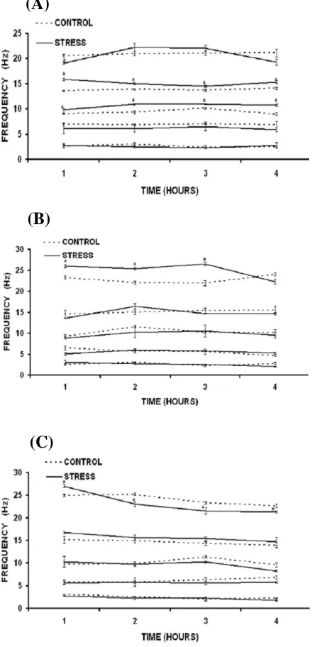
dominant components relative to their respective control values indicates that the 2 frequency components in EEG power spectrum show biphasic activity during SWS (Fig. 1a). In the first hour of EEG recording, frequency of 2 components were decreased (P<0.05), followed by marginal
insignificant increase in second and third hours and finally became closure to the control value in fourth hour of EEG recording. Under same conditions, frequency components of 1 and bands were found significantly increased (P<0.05) in all four
hour of recording. Aside these changes, 2 frequency components in EEG power spectrum were also observed increased (P<0.05) up to third
hour of post stress EEG recording in REM sleep (Fig. 1b) following acute exposure to high environmental heat; while changes in other frequency components were insignificant. In AWA condition, after initial increase (P<0.05) in 2 frequency components in first hour of EEG recording, persistent decrease (P<0.05) was
observed up to four hours (Fig. 1c). Such fluctuations were not evident in other four frequencies. Most of the changes in EEG power spectrum due to high environmental heat were observed totally reversed following four hours of cooling at room temperature.
EEG frequency analysis following chronic heat stress. After 21 days of chronic heat stress at 38C,
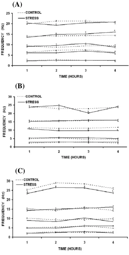
the 2 frequency components were found to be decreased significantly (P<0.05) in second hour of
EEG recording in SWS (Fig. 2a). Then it comes closer to the control value. The 1 frequency components were also observed increased significantly (P<0.05) from second hour of
recording that continued up to fourth hour. The changes in other frequency bands were found to be insignificant. In REM sleep conditions (Fig. 2b), following the chronic heat stress, 2 frequency components were observed significantly decreased (P<0.05) in third hour of EEG recording. These
changes were not evident in the first, second and fourth hour of recording. The frequency components of band were observed decreased
significantly (P<0.05) during all four hours of EEG
recording in REM sleep phase. In other two frequency components, the changes were analyzed to be insignificant. After chronic exposure of heat,
2 frequency components were found to decrease significantly (P<0.05) in all four hours of EEG
recording in AWA condition (Fig. 2c). However, the other bands of frequencies were observed to be unchanged.
Fig. 1. Data shown in mean ± S.E (n = 5, for both stressed and control groups). Figures show the EEG frequencies of all five bands (β2,β1,α, θ and ∂)in (A), SWS; (B), REM sleep and
(C), AWA state, following acute heat stress (*P<0.05 compared to respective control groups).
(A)
(B)
(C)
Fig. 2. Data shown in mean ± S.E (n = 5, for both stressed and control groups). Figures show the EEG frequencies of all five bands (β2,β1, α, θ and ∂) in (A), SWS; (B), REM sleep and
(C), AWA state, following chronic heat stress (*P<0.05 compared to respective control groups).
DISCUSSION
Results from the observations of body temperature as stress markers following heat stress, either acute or chronic, provide evidences of the stressful conditions. Acute heat stress, similar to the findings of Sharma et al.[5], significantly increased
the body temperature of all groups of rats. However, following the 21 days of chronic exposure of the high environmental heat, the body temperature of the rats were found to set at the higher temperature
similar to the results obtained by Dey [2]. The change in set point of the body temperature following chronic heat exposure indicates behavioral and physiological adaptations [2]. By considering the changes in body temperature in rats following acute or chronic heat exposure as an indication of stressful event, in the present study, an effort has been made to monitor and analyze EEG variations following acute as well as chronic heat stress on brain electrical activities with the help of EEG power spectra.
Most of the previous reports related with the EEG changes in hot environment are mainly concerned with the effects of hyperthermia, and very few researchers have studied the changes in EEG activities following the stressful events. The animal model of heat stress mentioned in this paper has already been established to produce clinical symptoms of heat induced injuries (acute conditions) and the acclimatization of animal’s physiological system to the new environmental conditions [5], which is rarely addressed. At the same time, it has been established that the changes produced by acute heat stress are reversible [11], the recordings of EEG signal was carried just after the removal of the stressful conditions. Conversely, to avoid the EEG changes, if any, produced owing the one-hour chronic exposure on the 21stday, the recording was performed on day 22 following the same recording protocol.
Dubois et al. [11], has reported an initial increase
in EEG frequency with increase in body temperature either by spontaneous or artificially induced fever. If the elevation of body temperature maintained long enough or above 41-42C, a major transient reduction in EEG activity was observed. They also shown that as cooling was resumed, these changes in EEG frequencies were usually totally reversible. Occasionally, the changed EEG frequencies did not return to the control level and that may occur due to CNS damage, which attributed to anorexia, dehydration, metabolic imbalance, energy failure or cellular change following heat stress [5, 11]. Similar to this report, our findings show changes in 2 frequency components after acute heat stress that returned to the control level in four hours of cooling at room temperature except in AWA state. However, 1and
frequency components in SWS did not returned to the control level, which may reflect the neuronal and non-neuronal changes in the brain due to acute heat stress [4, 5, 11]. On the other hand, 21 days of chronic heat exposure significantly decreases the 2 frequency in AWA state in all four hours of EEG
(A)
(B)
(C)
recording. The 2frequency components were also observed decreased in second hour SWS and third hour in REM sleep in EEG recording. Although, no report has been known to the author on the study of brain cortical electrical activity with the similar model of chronic heat stress, it has been supposed that the changed EEG activity has been recorded due to adaptations of animal’s physiological systems to the new ambient environmental conditions similarly as suggested by Sarbadhikari and his co-workers [17] in their study on chronic exercise stress.
Great attention has been made to study the heat-afflicted injuries in hypothalamus, as it is the main site of thermoregulation. As many thermosensitive neurons of the preoptic area of hypothalamus showed alterations in their firing rate with changes in sleep-wake states, their involvement in regulation of both sleep and body temperature has been suggested [14, 15]. The altered firing of the thermosensitive neurons of the hypothalamus may have changed the EEG activities in animals following exposure to high environmental heat. Reports from various laboratories also indicate that the neuroglial cells are highly activated by heat stress [4]. Literatures strongly support that glial cells control the ion environment and neurotransmitter metabolism in the brain [5] and helps in the genesis of cortical EEG waves [19, 20]. Thus the contribution of glial cells in altering the EEG frequency following high heat stress cannot also be ruled out.
Several reports have been published on the effects of high environmental heat on neurotransmitter systems. Sharma and his co-workers [5, 21] with their works on similar animal model of heat stress have suggested the involvement of serotonin in increased permeability in blood-brain barrier as well as in heat adaptations. Furthermore, it has been suggested that the EEG activation in different sleep-wake states are dependent on different neurons and neurotransmitter systems, which work differently in different states of sleep-wakefulness [22]. Although, the changes in cortical electrical activities or EEG signals observed in heat stressed rats are highly dependent on the neurotransmitters released and their metabolism in brain and the body; it is very difficult to explain the functional correlation of each neurotransmitter with each frequency components of EEG power spectra in different sleep-wake states. Therefore, the understanding of alteration in neurotransmitters following exposure to high environmental heat and its selective vulnerability in CNS, if any, is still not
clear and thus need detailed investigation.
In the present study, it has been found that the analyses of EEG power spectra in sleep-wake stages in generalization are observed sensitive to hot environment and found dependent upon the different sleep-wake stages. The results revealed that both acute and chronic heat stress changes the EEG frequency components in rats. It has also been shown that the higher frequency component (2) of the EEG was more sensitive than other frequency bands in both acute and chronic heat stress conditions in all three sleep-wake cycle. Further, the results demonstrate that the spectral analysis of long term EEG recordings can be used for obtaining useful results in analyzing heat induced changes in electrophysiological activities of cerebral cortex.
ACKNOWLEDGEMENTS
The authors are grateful to the Coordinator, School of Biomedical Engineering, Institute of Technology, Banaras Hindu University, Varanasi (India) for providing laboratory facilities for carrying out this study. Authors are also very thankful to Prof. B. M. Karan, Head of the Department, Electrical and Electronics Engineering and Prof. B. N. Das, Head of the department, Biomedical Instrumentation for their help and providing technical assistance to prepare this paper in the present form.
REFERENCES
1. Sminia, P., Van Der Zee, J., Wondergem, J. and Haveman, J. (1994) Effect of hyperthermia on the
central nervous system: a review. Int. J.
Hyperthermia 10: 1-30.
2. Dey, P.K. (2000) Involvement of endogenous opiates in heat stress. Biomedicine 20: 143-148. 3. Westman, J. and Sharma, H.S. (1998) Heat shock
protein response in the central nervous system
following hyperthermia. In: Progress in Brain
Research(Sharma, H.S. and Westman, J. eds.), Vol. 115, Elsevier, Amsterdam. pp. 209-239.
4. Cervó s-Navarro, J., Sharma, H.S., Westman, J. and Bongcam-Rudloff, E. (1998) Glial reaction in the central nervous system following heat stress. In: Progress in Brain Research (Sharma, H.S. and Westman, J. eds.), Vol. 115, Elsevier, Amsterdam pp. 241-274.
5. Sharma, H.S., Westman, J. and Nyberg, F. (1998) Pathophysiology of brain edema and cell changes following hyperthermic brain injury. In: Progress in
Brain Research (Sharma, H.S. and Westman, J. eds.), Vol. 115, Elsevier, Amsterdam. pp. 351-412. 6. Morimoto, T., Nagao, H., Sano, N., Takahashi, M.
and Matsuda, H. (1991) Electroencephalographic study of rat hyperthermic seizures. Epilepsia 32: 289-293.
7. Yang, R.C., Yang, S.L., Chen, S.W., Lai, S.L., Chen, S.S. and Chiang, C.S. (1996) Previous heat shock treatment attenuates bicuculline-induced convulsions in rats. Exp. Brain Res. 108: 18-22.
8. Sinha, R.K. and Ray, A.K. (2003) Effect of p-CPA pretreatment on EEG power spectra in experimental open brain injury in rats. Iran. Biomed. J. 7: 119-126.
9. Sinha, R.K. (2003) Artificial neural network detects changes in electro-encephalogram power spectrum of different sleep-wake states in an animal model of heat stress. Med. Biol. Eng. Comput. 41: 595-600.
10. Sinha, R.K. (2004) Electro-encephalogram
disturbances in different sleep-wake states following exposure to high environmental heat. Med. Biol. Eng. Comput. 42 (In press).
11. Dubois, M., Sato, S., Lees, D.E., Bull, J.M., Smith, R., White, B.G., Moore, H. and Macnamara, T.E. (1980) Electroencephalographic changes during
whole body hyperthermia in humans.
Electroencephalogr. Clin. Neurophysiol. 50: 486-495.
12. Nielsen, B., Hyldig, T., Bidstrup, F., Gonzalez-Alonso, J. and Christoffersen, G.R. (2001) Brain activity and fatigue during prolonged exercise in the heat. Pflugers Arch. 442: 41-48.
13. Glotzbach, S.F. and Heller, H.C. (1984) Changes in the thermal characteristics of hypothalamic neurons during sleep and wakefulness. Brain Res. 309: 17-26.
14. Alam, M.N., McGinty, D. and Szymusiak, R. (1996) Preoptic/anterior hypothalamic neurons: Thermo-sensitive in wakefulness and non rapid eye movement sleep. Brain Res. 29: 76-82.
15. Thomas, T.C. and Kumar, V.M. (2002) Effect of ambient temperature on brain temperature and sleep-wakefulness in medial preoptic area lesioned rats. Ind. J. Physiol. Pharmacol. 46: 287-297.
16. Cai, Z. (1991) The function of sleep: further analysis. Physiol. Behav. 50: 53-60.
17. Sarbadhikari, S.N., Dey, S. and Ray, A.K. (1996) Chronic exercise alters EEG power spectra in an
animal model of depression. Ind. J. Physiol.
Pharmacol. 40: 47-57.
18. Jervis, B.W., Coelho, M. and Morgan, G.W. (1989) Spectral analysis of EEG responses. Med. Biol. Eng. Comput. 27: 230-238.
19. Caspars, H., Speckmann, E.J. and Lehmankuehler, A. (1984) Electrogenesis of slow potentials of the brain. In: Self-regulation of the Brain and Behavior (Elbert, T., Rockstroh, B., Luetzenberger, W. and Birbaumer, N. eds.), Springer, New York.
20. Speckmann, E.J. and Elger, C.E. (1987) Introduction to the neurophysiological basis of the EEG and DC
potentials. In: Electroencephalography. Basic
Principles, Clinical Applications and Related Fields (Niedermeyer, E. and Lopes da Silva, F. eds.), Urban and Schwarzenberg, Baltimore.
21. Dey, P.K. (1999) Modification of functional responsiveness of central serotonin receptor subtypes in rats following exposure to chronic heat stress. Biomedicine 19:57-65.
22. Montplaisir, J., Dominique, P., Serge, G., Hélène, G. and Anne, D. (1998) Sleep disturbances and EEG slowing in Alzheimer’s disease. Sleep Res. Online 1: 147-151.