*Corresponding Author;
Comparing Invasive and Non-Invasive of Isolated Shigella flexneri
by Electron Microscopy of Cell Culture, SDS-PAGE and
Congo Red Method
Mojdeh Hakemi Vala
*1, Jamileh Nowroozi
1, Farideh Ghazi
1, Parvaneh Nabavi Tabatabai
2and Saeed Haghighi
31
Dept. of Medical Microbiology, Medical School, Iran University of Medical Sciences; 2Dept. of Electron Microscopy, Medical School, Iran University of Medical Sciences; 3 Dept. of Microbiology, Pasture Institute of Iran, Tehran, Iran
Received 12 November 2005; revised 24 June 2006; accepted 9 August 2006
ABSTRACT
Background: The aim of this study was to compare invasive and non-invasive strains of Shigella flexneri
isolated from Tehran by a 120 kDa protein band by SDS-PAGE, electron microscopy of cell culture and
Congo red dye methods. Methods: S. flexneri strains were isolated by standard bacterial methods from fecal
specimens of children attending to the 3 children’s hospitals. Phenotype analysis for screening virulent of
strains of S. flexneri was done on a plate of tryptic soy agar contained 0.003% Congo red dye. Whole
membrane protein preparations were used to examine the protein profiles of the inner and outer membrane of these Gram-negative bacteria. The protein mixture was electrophoresed through a polyacrylamide gel. The gel was stained with Coomassie brilliant blue R250 and destained with ethanol and acetic acid. HeLa cell culture was done by two-step preparations: one for light microscopy and the other for electron microscopy.
Results: Some of S. flexneri (46%) were Congo red positive colonies. S. flexneri with negative Congo red phenotype could not enter the HeLa cell culture. A 120 kDa protein band was found in 46% of these bacteria which could enter into HeLa cell culture. Pseudopod structures which facilitate bacterial cell-to-cell spread
were readily identified by electron microscopy. Discussion: Since the existence of 120-kDa protein band was
corresponded to enter of S. flexneri into the HeLa cell culture and correlated with Congo red dye positive, for
identification of invasive and non-invasive S. flexneri strains, the use of a 120-kDa protein band by
SDS-PAGE or a simple, rapid and very cheap Congo red dye method is recommended. Because, there are some
deaths due to Shigella sp. in our country, notification on the isolation of these bacteria in both children
hospitals laboratories and private clinical laboratories is important. Iran. Biomed. J. 11 (1): 47-52, 2007
Keywords: Shigella flexneri, HeLa cell culture, Electron microscopy, Congo red dye
INTRODUCTION
mong pathogenic microorganisms invasive bacteria have the ability to penetrate
mammalian epithelial cells both in vivo and
in vitro [1]. Shigella flexneri, which is responsible
for a dysenteric syndrome in human, belongs to the invasive group of pathogens [2-4]. It is known that the ability of S. flexneri to penetrate epithelial cells is encoded by a 20-kilo base portion of a 220-kilo base plasmid [1, 4, 5]. This is unexpected for a non-motile microorganism which has been related to icsA inter cellular spread, vir G, a gene encoding a
120-KDa outer membrane protein which allows interaction with microfilaments [1, 6]. The entry of
S. flexneri into epithelial cells is achieved through
internalization of the bacterium into a membrane bound vacuole derived from the host cell plasma
membrane [1]. Shigella species remain within
human intestinal epithelial cells where they cause the destruction of enterocytes and induce an
inflammatory response [2]. S. flexneri requires both
adhesive and invasive phenotype to efficiently colonize follicle-associated epithelium (FAE) [7].
Recent studies on the enter invasive pathogen, S.
flexneri, have shown that, in addition to allowing
A
48 Hakemi Vala et al. Iran. Biomed. J., January 2007
intracellular growth [8], lysis of the phagocyte vacuole also allows bacteria to spread intracellular and infect adjacent cells [8, 3]. Cell cultures are commonly used to assess the ability of intracellular bacteria to invade susceptible eukaryotic cells [9]. In the years of 1980, there were a lot of studies on
identification and the intracellular existence of S.
flexneri. But in recent years, because of health
improvement in conditions in developed countries, search for isolation of this bacteria decreased but in developing countries, such as Iran and Taiwan, many children still lose their life for infection of S.
flexneri [10, 11]. So, the aim of this study was to
compare the invasive and non-invasive properties of
isolated S. flexneri by electron microscopy (EM) of
cell culture, SDS-PAGE and Congo red.
MATERIALS AND METHODS
Bacterial strains. S. flexneri strains were isolated
from fecal specimens of children attending to the 3 children’s hospitals (Markaze-Tebbi Kodakan, Aliasghar and Mofid Hospitals) from January 2001
to December 2003. In this study, 350 Shigella sp.
were isolated and identified by standard methods.
After that, 100 S. flexneri strains were randomly
chosen and stored in peptone and glycerol at -70°C.
Bacterial suspensions. Bacteria were harvested in
tryptic soy broth in the exponential phase, washed in PBS [NaCl (8.8 gL-1); Na2HPO4. 2H2O (2.250 gL
-1
); NaH2PO4. H2O(0.257 gL
-1
): pH 7.4] and suspended at the appropriate density of 2 × 10⁶ cfu ml-1 in MEM.
Congo red binding. Phenotype analysis for
screening virulent of strains of S. flexneri was done by Congo red dye. A colony of fresh culture of isolated bacteria was inoculated on a plate contained TSA (tryptic soy agar) and Congo red solution at final concentration of 0.003% to detect red pigmentation colony [12, 13].
Protein preparation and SDS-PAGE. Whole
membrane preparations were used to examine the protein profiles of the inner and outer membrane of Gram-negative bacteria. In this process, pellet of the cells were suspended in 30 mM Tris-HCl (pH 8.1) and resuspended in 20% sucrose/30 mM Tris-HCl (pH 8.1) plus lysozyme. Then, 3 mM EDTA (pH
7.3) was added. Terminal pellets were suspended in l
× LUG buffer [Tris-HCl (pH 6.8), 50 ml; SDS 25
ml, 0.25 M; glycerol, 2g; beta-mercaptoethanol, 5 ml; bromophenol blue, 2 ml; of 1%, distilled water, 100 ml] [14]. Following certain preparative steps, the protein mixture was electrophoresed through a polyacrylamide 8% gel. The gel was stained with Coomassie brilliant blue R250 and continuously destained with mixture of ethanol and acetic acid. The 120-kDa band was compared with protein
ladder (protein ladder, Page Rulertm # Smo661,
Fermentas, Lituani).
HeLa cell cultures and EM. To proceed for
experiment, two-step preparations were done: one for light microscopy and the other, for EM. HeLa cell was obtained from the Public Health Medical School of Tehran University (Tehran, Iran). New cultures were prepared in tissue culture (trays) wells
consisting of cover slip (22 × 22 mm) and
concentration of 2 × 10⁵ cell/ml using 0.5 ml MEM
(Minimal Essential Medium) with 5% FCS. Then, bacterial suspensions with MOI (multiplicity of infection) 10 bacteria/cells were added to each well. Process of bacterial suspension preparation was as explained below:
After gentle mixing, the trays were inoculated in
the CO2 incubator at 37°C for 1 h and then washed
three times and fresh tissue culture medium (MEM) with 5% FCS containing gentamicin at a final concentration of 40 mg/ml was added to each well and were incubated in the CO2 incubator at 37°C for
another 2 h. The cover slips were fixed with
methanol at 4°C overnight and then stained with
Giemsa stain and washed with Giemsa buffer for light microscopy examination.
For EM investigation, HeLa cell monolayer was seeded in 100 ml culture flasks and after washing for three times, the bacterial suspensions were added. All flasks scraped off and centrifuged. Fixation with glutaraldehyde 2% was done on pellets of cells for 2 hours. Continuously, a centrifuge step was done and melted agar 2% was added to each cell pellet, mixed well and post fixed for 1 h with Osmium tetroxide, and then, 4 concentrations of aceton were added for dehydration and samples were embedded in resin spur (R1032, Agar Scientific, UK). Gold sections were taken and stained with saturated uranyl acetate and lead citrate [15]. A gride of cell culture
without infection with S. flexneri was used as
control.
http://IBJ.pasteur.ac.ir
RESULTS
Congo red binding. All 100 S. flexneri isolated
were tested for binding to Congo red dye. Then, isolated bacteria were identified as positive invasive phenotype with red colonies and non-invasive phenotype identified with white colonies. In this
study, 46 (46%) isolated S. flexneri were Congo red
positive colonies on TSA contained 0.003% Congo red dye. All the Congo red positives (100%) strains
produced β haemolysin on blood agar.
SDS-PAGE. From 100 isolated S. flexneri, a
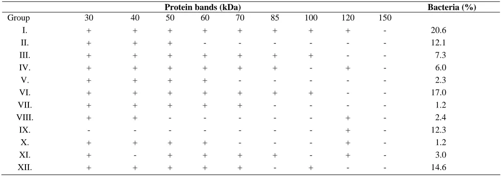
120-kDa band was detected in 46 isolates (46%). The range of protein bands was from 30 kDa to 150 kDa. The protein profiles of some strains are shown in Table 1 and Figure 1. On the bases of protein bands, these were 12 distinct groups. Protein bands with the same size were present in several strains, for example, most of strains (20.7%) were in group I.
HeLa cell culture and EM. As it was described
above, all isolates were tested for binding to Congo red dye; so, invasive and non-invasive strains were separated. Bacterial suspensions from S. flexneri
2a strain (Reference Strain, RS) with Congo red
positive and probably IcsA positive, S. flexneri
strains with Congo red positive and probably IcsA positive and S. flexneri strains with Congo red
negative and probably IcsA negative isolated from
patients were used to infect HeLa cell line. HeLa cell cultures which were infected with either RS or
S. flexneri strains with Congo red positive and
probably IcsA positiveat MOI 10:1, showed marked
Fig. 1. SDS-PAGE of some strains of S. flexneri. Lane1, ladder; lane 2, S. flexneri 2a standard had a 120-kDa band; lane 3, negative sample had not a 120-kDa band; lanes 4-6, positive samples; all had a120-kDa band.
loss of confluence and viable cells were heavily infected with bacteria and showed morphology with indistinct membrane (46%).
According to pictures, invasive shigella strains
penetrated into these cells (Fig. 2). HeLa cells
infected with S. flexneri strains with Congo red
positive and probably IcsA positive, exhibited structures similar to those of non-infected cells.
However, S. flexneri strains with Congo red positive
and probably IcsA positive adhered and occasionally entered to HeLa cells. Furthermore, pseudopod structures used to facilitate bacterial cell-to-cell spread were readily identified.
Table 1. Protein profiles of S. flexneri isolated.
Protein bands (kDa) Bacteria (%)
Group 30 40 50 60 70 85 100 120 150
I. + + + + + + + + - 20.6
II. + + + - - - 12.1
III. + + + + + + + - - 7.3
IV. + + + + + + - + - 6.0
V. + + + + - - - 2.3
VI. + + + + + + + - - 17.0
VII. + + + + + - - - - 1.2
VIII. + + - - - + - 2.4
IX. - - - + - 12.3
X. + + + + - - - + - 1.2
XI. + - + + + + - + - 3.0
XII. + + + + + - + - - 14.6
+, exist; -, not exist .
50 Hakemi Vala et al. Iran. Biomed. J., January 2007
Fig. 2. Penetration of invasive S. flexneri (vacuole formation) which had a 120-kDa band (presumptive IcsA protein) and Congo red positive.
Electron microscopy. S. flexneri 2a (RS) with
Congo red positiveand probably icsA positive and
patient’s isolated S. flexneri strains with Congo red
positive and patient’s isolated S. flexneri strains
with Congo red negativeandprobably icsA negative
which infected HeLa cell cultures were used for EM investigation. Internalization, existence of bacteria into cells and cell disruption of both Congo red
positive and probably IcsA positive S. flexneri
strains and S. flexneri 2a (RS) were detected.
Existence of pseudopod filaments were confirmed by EM (Fig. 3).
Fig. 3. Pseudopod filaments formation in infected HeLa cell with S. flexneri strain which had a 120-kDa band and Congo red positive.
DISCUSSION
In this study, 350 Shigella spp. from fecal of
patients were isolated and 142 (40.57%) were S.
flexneri. One hundred of S. flexneri strains which
recovered from stool specimens of children’s were chosen randomly for presumptive determination of IcsA protein (120 kDa band) and Congo red binding, then, infect HeLa cell line as noted above. Forty six (46%) of S. flexneri strains were Congo red positive colonies on TSA plate contained 0.003% Congo red dye. Relation between the virulence of S. flexneri 2a and its ability to absorb Congo red was examined in Mounier study [16]. This property is correlated to invasiveness phenotype of S. flexneri in cell culture [17]. Present results showed, all Congo red positive isolates had adhesion and invasion properties to HeLa cells. These data were agreed with those of other studies [1, 9, 18-22]. Francis and Thomas showed that,
infected Caco-2 or HeLa cell cultures by L.
morocytogenes at high MOI, had extensive invasion,
intra-cellular multiplication and finally cell lysis [10, 21].
As it was noted above, in 46 (46%) of isolated S.
flexneri strains , a 120 kDa band was detected in
SDS- PAGE method , that might be correlated to IcsA protein. Presence of a 120 kDa band and phenotype Congo red positive were detected in 46 (46%) of isolates in the same time. In this study, both S. flexneri 2a RS and S. flexneri strains with Congo red positive and probably IcsA Positive were isolated from patients, showed similar internalization, cell existence and cell disruption. In addition, pseudopod filaments were confirmed with EM.
In contrast, most of S. flexneri strains probably
IcsA negative could not enter HeLa cells culture.
Moreover, a few of these bacteria could adhere and enter to the HeLa cells, but cell disruption was not detected. These bacteria produce necrosis later, but can not be detected after 3 h incubation. Unlike the
Hly (Haemolysin) positive strains of L.
monocytogenes, none of Hly negative bacteria had
spread inter-cellular after 2 h of incubation and only a few number of them had spread after 4 h of incubation [18].
In other study, interaction of Salmonella
typhimurium, Listeria monocytogenes with murin’s
M cells were compared. Tissue infected with the lower dose of organisms did not show significant M
http://IBJ.pasteur.ac.ir
cell disruption at the various times examined and had the same appearance as the FAE. In contrast, at the higher dose, the interactions between S. flexneri strains and the epithelium of peyer’s patches were
similar to those observed for L. monocytogenes.
Destroyed regions revealed, membrane blebs and a denuded epithelial surface that closely resembled to
those observed for Listeria. These data demonstrated
that L. monocytogenes and S. flexneri possess the
ability to induce massive destruction of FAE when
inoculated into intestinal loops at inoculums of 4 ×
109 cfu per ml [5].
In this study, the 120 kDa protein band which detected by SDS-PAGE has been correlated with IcsA protein (1), the detection of this protein band may be a useful tool in epidemiological studies for searching prevalence sources.
Results showed, the existence of the 120 kDa
protein band was corresponded to enter of S. flexneri
into the HeLa cell culture, demonstrated by EM and
also, this band protein was found in all S. flexneri
strains with Congo red dye positive. In the other hand, our results detected by different methods (SDS-PAGE, EM and Congo red) could confirm
each other. Moreover, the ability of S. flexneri to
enter the host cells, spread to adjacent cells and intra cellular growth could damage and lyse the host cells, which indicated invasiveness property (8). So, for
identification of invasive and non-invasive S.
flexneri strains, detection of the 120 kDa protein
band by SDS-PAGE or a simple, rapid and very cheap Congo red dye method is recommended. Because, there are some deaths particularly in
children due to Shigella spp in our country [12]
notification on the isolation of this bacteria in both children hospitals laboratories and private clinical laboratories is important.
ACKNOWLEDGEMENTS
We gratefully acknowledge from Dr. H. Monavari in Hazrate Rasol Hospital Laboratory (Tehran, Iran) who helped us for cell culture. Moreover, we thank Miss Nafisi, Mrs. Abedini from Microbiology Laboratory of Mofid and Tebbi Kodakan Hospitals respectivly.
REFERENCES
1. Clerc, P.L., Berthon, B., Claret, M. and Sansonetti P.J. (1989) Internalization of Shigella flexneri into HeLa cells occur without an increase in cytosolic
Ca2+ concentration. Infect. Immune. 57 (9): 2919-2922.
2. Behrana, J.V., Harty J.T and Jones B.D. (1998) Interaction of invasive pathogens Salmonella typhimurium, Listeria monocytogenes, and Shigella flexneri with M cells and murine peyer’s patches. Infect. Immune. 66(8): 3758-3766.
3. La Brec, E.H., Schneider, H., Magnani, T.J. and Formal S.B. (1964) Epithelial cell penetration as an essential step in the pathogenesis of bacillary dysentery. J. Bacteriol. 88: 1503-1518.
4. Wing, H.J., Yan, A.W., Goldman, S.R. and Goldberg, M.B. (2004) Regulation of IcsP, the outer membrane protease of the shigella actin tail assembly protein IcsA, by virulence plasmid regulators virF and virB. J. Bacteriol. 186: 699-705.
5. Clark, M.A., Jepson, M.A., Simmons, N.L and Hirst, B.H. (1994) Differential surface characteristics of M cells from mouse intestinal peyer’s and Caecal patches. Histochem. J. 271-280.
6. Wing, H.J., Goldman, S.R., Ally, S. and Goldberg, M.B. (2005) Modulation of a protease contributes to the virulence defect of Shigella flexneri strains carrying a mutation in the virK locus. Infect. Immun. 73 (2): 1217-1220.
7. Sansonetti, P.J., Arondel, J., Cantey, J.R., Provost, M.C. and Huerre, M. (1996) Infection of rabbit peyer’s patches by Shigella flexneri: effect of adhesive or invasive bacterial phenotypes on follicle-associated epithelium. Infect. Immun. 2752-2764. 8. Suzuki, T. and Sasakawa, C. (2001) Molecular basis
of intracellular spreading of shigella. Infect. Immun. 69 (10): 5959- 5966.
9. Clerc, P. and Sansonetti, P.J. (1987) Entry of Shigella flexneri into HeLa cells: evidence for directed phagocytosis involving actin polymerization and myosin accumulation. Infect. Immun., 55: 2681-2688.
10. Chen, J.H., Chiou, C.S., Chen, P.C., Liao, T.L., Li, J.M. and Hsu, W.B. (2003) Molecular epidemiology of shigella in a Taiwan during 1996 to 2000. J. Clin. Microbiol. 41 (6): 3078-3088.
11. Ranjbar, R., Soltan Dalal, M.M., Pourshafie, M.R., Aslani, M.M., Majdzadeh, R. and Khoramzade, M.R. (2004) Serogroup distribution of shigella in Tehran, Iran. J. Pub. Health 33: 32-35.
12. Sun, A.N., Camelli, A. and Porthy, D.A. (1990) Isolation of Listeria monocytogenes small-plaque mutants defective for intracellular growth and cell-to-cell spread. Infect. Immune. 58 (11): 3770-3778. 13. Goldberg, M.B. and Therist J.A. (1995) Shigella
flexneri surface protein IcsA is sufficient to direct action-based motility. Proc. Natl. Acad. Sci. USA. 92: 6572-6576.
14. Morona, R., VanDen Bosch, L. and Manning P.A. (1995) Molecular genetic and topologic
52 Hakemi et al. Iran. Biomed. J., January 2007
characterization of O-antigen chain length regulation in Shigella flexneri. J. Bacteriol. 177:1059-1068. 15. Hoppert, M. and Holzenburg, A. (1998) Electron
microscopy in microbiology. 1st ed. BIOS. Scientific Publishers Ltd.
16. Monier, J., Ryter, A. and Sansonetti, P.J. (1990) Intracellular and cell-to-cell spread of Listeria monocytogenes involves interactions with F-actin in the enterocyte like cell line Caco-2. Infect. Immun. 65 (2): 1048-1058.
17. Maurelli, A.T., Blackman, B. and Curtiss III, R. (1987) Loss of pigmentation in S. flexneri 2a is correlated with loss of virulence and virulence associated plasmid. Infect. Immun. 44 (1): 397-401. 18. Sechi, A.S., Wehland, J. and Small, V. (1997) The
isolated comet tail pseudopodium of Listeria monocytogenes, A tail of 2 actin filament population, long and axial and short and random. J. Cell Biol. 137: 3867-3871.
19. Daskaleros, D. and Payne, M. (1987) Congo red binding phenotype is associated with hemin binding and increased infectivity of Shigella flexneri in the HeLa cell model. Infect. Immun. 55 (6): 1393-1398. 20. Bernardini, M., Mounier, J. and Sansonetti, P.J.
(1989) Identification of icsA, a plasmid locus of
Shigella flexneri that governs bacterial intra and intercellular spread through interaction with F-actin.
Proc. Natl. Acad. Sci. USA. 86: 3867-3871.
21. Francis, M.S. and Thomas, C.J. (1996) Effect of multiplicity of infection on Listeria monocytogenes
pathogenicity for HeLa and Caco-2 cell line. J. Med. Microbiol. 45: 323-330.
22. Mounier, J., Ryter, A.T., Coquis-Rondon, M. and Sansonetti, P.J. (1990) Intracellular and cell-to-cell spread of Listeria monocytogenes involves interaction with F-actin in the enterocyte like cell line Caco-2. Infect. Immun. 58 (4): 1048-1058.