Personalised medicine in interstitial lung
diseases
Maria A. Kokosi, George A. Margaritopoulos and Athol U. Wells
Number 6 in the Series
“
Personalised medicine in respiratory diseases
”
Edited by Renaud Louis and Nicolas Roche
Affiliation: Interstitial Lung Disease Unit, Royal Brompton and Harefield NHS Foundation Trust, London, UK. Correspondence: Maria A. Kokosi, Interstitial Lung Disease Unit, Royal Brompton and Harefield NHS Foundation Trust, Sydney Street, London, SW3 6NP, UK. E-mail: M.Kokosi@rbht.nhs.uk
@ERSpublications
Personalised medicine provides a potential road map for guiding clinical care in interstitial lung diseaseshttp://ow.ly/xZop30iQOoQ
Cite this article as:Kokosi MA, Margaritopoulos GA, Wells AU. Personalised medicine in interstitial lung diseases.Eur Respir Rev2018; 27: 170117 [https://doi.org/10.1183/16000617.0117-2017].
ABSTRACT Interstitial lung diseases in general, and idiopathic pulmonary fibrosis in particular, are complex disorders with multiple pathogenetic pathways, various disease behaviour profiles and different responses to treatment, all facets that make personalised medicine a highly attractive concept. Personalised medicine is aimed at describing distinct disease subsets taking into account individual lifestyle, environmental exposures, genetic profiles and molecular pathways. The cornerstone of personalised medicine is the identification of biomarkers that can be used to inform diagnosis, prognosis and treatment stratification. At present, no data exist validating a personalised approach in individual diseases. However, the importance of the goal amply justifies the characterisation of genotype and pathway signatures with a view to refining prognostic evaluation and trial design, with the ultimate aim of selecting treatments according to profiles in individual patients.
Introduction
Interstitial lung diseases (ILDs) are a group of heterogeneous disorders, either idiopathic or associated with an overreaction of the immune system. Until recently, no effective therapy existed in patients with a progressive fibrotic phenotype, in part due to limited knowledge of pathogenesis. However, during the last decade, new concepts have developed on aetiological factors, pathogenetic mechanisms (inflammation versusactive fibrosis) and genetic susceptibility. There has been an exponential increase in publications on the pathogenesis of ILDs and this applies especially to idiopathic pulmonary fibrosis (IPF), the most prevalent and progressive ILD. This period has seen the advent of antifibrotic therapies with proven treatment effects in IPF, which, among ILDs, continues to be the main focus of ongoing research.
Despite recent advances, there are many unanswered questions regarding the prediction of clinical behaviour and responsiveness to treatment in IPF. Initially, IPF was considered as a single disease with a
Copyright ©ERS 2018. ERR articles are open access and distributed under the terms of the Creative Commons Attribution Non-Commercial Licence 4.0.
Previous articles in this series: No. 1:Chung KF. Personalised medicine in asthma: time for action.Eur Respir Rev 2017; 26: 170064. No. 2: Bonsignore MR, Suarez Giron MC, Marrone O, et al. Personalised medicine in sleep respiratory disorders: focus on obstructive sleep apnoea diagnosis and treatment.Eur Respir Rev2017; 26: 170069.No. 3:Mascaux C, Tomasini P, Greillier L,et al. Personalised medicine for nonsmall cell lung cancer.Eur Respir Rev2017; 26: 170066. No. 4: Noell G, Faner R, Agusti A. From systems biology to P4 medicine: applications in respiratory medicine. Eur Respir Rev2018; 27: 170110.No. 5: Wouters EFM, Wouters BBREF, Augustin IL,et al. Personalised pulmonary rehabilitation in COPD.Eur Respir Rev2018; 27: 170125.
Received: Oct 15 2017 | Accepted after revision: March 05 2018
characteristic pattern of disease behaviour. However, our understanding of the pathogenesis of IPF has evolved significantly [1]. The currently favoured model of disease is an epithelial-driven and fibroblast-activated process in which inflammation has an ancillary role that is not yet fully elucidated [2]. More specifically, accelerated parenchymal senescence determined by either telomere dysfunction or genetic defects, together with the concurrent noxious effect of smoking, severely compromise the regenerative potential of parenchymal epithelial stem cells, triggering a cascade of molecular signals and events (scarring, bronchiolar proliferation, abnormal remodelling) leading eventually to epithelial– mesenchymal transition and irreversible lung remodelling [3]. In the non-IPF group of ILDs, inflammation plays the key role, although the two pathogenetic processes have several molecular pathways in common.
The involvement of multiple pathogenetic pathways may account for the heterogeneity of the clinical behaviour of IPF. Some patients experience a slow decline, whereas others decline rapidly and die within a few months from the time of diagnosis. In another patient subset, there are episodes of acute deterioration (“acute exacerbations”) that are associated with a very high short-term mortality [4]. The accurate prediction of the course of IPF is one of the greatest challenges in this field, with major implications for the accurate design of clinical trials.
In 2014, two antifibrotic compounds were licensed for the treatment of IPF. Both pirfenidone and nintedanib slow the rate of progression of disease by 50%, as judged by serial forced vital capacity (FVC) trends [5, 6]. Both drugs are highly pleiotropic, acting on many disease pathways, and this may account for their efficacy across the broad group of IPF patients. By contrast, drugs targeting a single pathway have not proven to be efficacious. The possibility exists that nonpleiotropic agents are beneficial in a small subset of IPF patients, accounting for nonsignificant trends in some trials. However, if so, there is currently no means of identifying key patient subgroups that may respond to targeted therapies.
The desirability of matching individual treatments to the upregulation of key disease pathways in individuals, a need common to all ILDs, underpins growing interest in the concept of personalised medicine (also called precision or stratified medicine). Personalised medicine refers to the medical approach that emphasises the customisation of healthcare, with all decisions and practices tailored to individual patients [7], based on behavioural and environmental factors and genetic and molecular profiles. It is hoped that tailored therapy will improve treatment outcomes, reduce side-effects, prevent unnecessary exposure to ineffective therapies and save money through more efficient use of healthcare resources. The cornerstone of personalised medicine is the identification of biomarkers that can be used to inform diagnosis, prognosis, treatment stratification and/or therapeutic response.
Personalised medicine has already been successfully applied in the field of respiratory medicine. In asthma, the interleukin (IL)-5 monoclonal antibody mepolizumab is more efficacious in reducing severe asthma exacerbations in patients with elevated sputum eosinophils [8, 9]. Personalised medicine has had its greatest impact in oncology. In metastatic breast cancer, the expression of human epidermal growth factor receptor 2 (EGFR2) is associated with benefits from the EGFR2 monoclonal antibody trastuzumab [10]. In nonsmall cell lung cancer, tyrosine kinase inhibitors such as erlotinib, gefitinib and afatinib have increased efficacy in patients with mutations in EGFR [11, 12].
In this review, we summarise current data on behavioural and environmental factors as well as the most promising molecular and genetic biomarkers in ILDs, with specific focus on IPF, and we divide personalised medicine into predictive medicine and preventive/participatory medicine.
Predictive medicine
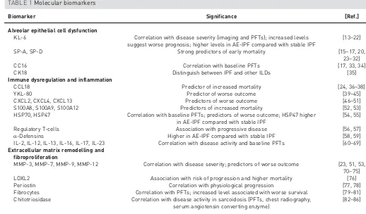
Molecular biomarkersA plethora of molecular biomarkers has been identified (table 1), but we will only focus on the most studied markers. Measurement of those proteins can potentially distinguish separate populations of individuals who have activation of different disease pathways and distinct prognosis.
post-treatment, suggesting a decrease in disease activity despite stability in respiratory physiology [21]. Serum KL-6 levels depend on mucin-1 (MUC1) gene polymorphism, which explains the differences in levels between ethnicities [13, 22].
Serum levels of SP-A and SP-D are elevated in IPF, SSc with associated ILD (SSc-ILD), hypersensitivity pneumonitis and PAP [15–17, 23–28]. Increased serum surfactant protein (SP)-A and SP-D levels are a strong independent predictor of early mortality in IPF patients in two separate studies [27, 28]. A prediction model using both SP-A and SP-D was superior to a model with one marker alone in an IPF cohort [27]. In SSc-ILD, SP-D levels are more sensitive than SP-A in detecting ILD, as defined by high-resolution computed tomography [29]. SP-D levels correlate negatively with diffusing capacity of the lung for carbon monoxide (DLCO) and FVC [30]. Polymorphism in the SP-D gene (SFTPD) affects the levels of SP-D [32].
The C-C chemokine ligand CCL18 is profibrotic and is elevated in serum, bronchoalveolar lavage (BAL) and lung tissue of IPF and SSc-ILD patients [36, 37]. Baseline CCL18 predicted the 6-month FVC change and correlated with survival in a cohort of 72 IPF patients. Increased mortality was observed above a certain cut-off value [37]. Cut-off values in both diseases have a potential role in predicting long-term outcome [37, 38].
Elevated serum chitinase-like protein (YKL-40) levels are elevated in patients with IPF [39, 40] and increased levels have been associated with worse survival [40]. YKL-40 is also increased in serum of SSc-ILD patients, and correlates with airway obstruction, reduced DLCO and poor prognosis [41]. Increased YKL-40 levels were found in patients with a variety of ILDs compared with healthy controls, with highest levels in those with more severe fibrosis and worse prognosis [42]. Increased levels have also been observed in patients with sarcoidosis [43], PAP [44] and hypersensitivity pneumonitis [45]. In hypersensitivity pneumonitis, higher baseline YKL-40 levels were associated with worse survival [45].
Matrix metalloproteinase (MMP)-7 represents one of the most extensively studied biomarkers in the pathogenesis of IPF. Higher plasma MMP-7 levels correlate with disease severity as measured by FVC and DLCO[70], and predict survival in IPF [23]. Furthermore, MMP-7 is a marker of poor prognosis even after TABLE 1Molecular biomarkers
Biomarker Significance [Ref.]
Alveolar epithelial cell dysfunction
KL-6 Correlation with disease severity (imaging and PFTs); increased levels suggest worse prognosis; higher levels in AE-IPF compared with stable IPF
[13–22]
SP-A, SP-D Strong predictors of early mortality [15–17, 20,
23–32]
CC16 Correlation with baseline PFTs [17, 33, 34]
CK18 Distinguish between IPF and other ILDs [35]
Immune dysregulation and inflammation
CCL18 Predictor of increased mortality [24, 36–38]
YKL-80 Predictor of worse outcome [39–45]
CXCL2, CXCL4, CXCL13 Predictors of worse outcome [46–51]
S100A8, S100A9, S100A12 Predictors of increased mortality [52, 53]
HSP70, HSP47 Correlation with baseline PFTs; predictors of worse outcome; HSP47 higher in AE-IPF compared with stable IPF
[54, 55]
Regulatory T-cells Association with progressive disease [56, 57]
α-Defensins Higher in AE-IPF compared with stable IPF [58, 59]
IL-2, IL-12, IL-13, IL-16, IL-17, IL-23 Correlation with disease activity and baseline PFTs [60–69] Extracellular matrix remodelling and
fibroproliferation
MMP-3, MMP-7, MMP-9, MMP-12 Correlation with disease severity; predictors of worse outcome [23, 51, 53, 70–75]
LOXL2 Association with risk of progression and higher mortality [76]
Periostin Correlation with physiological progression [77, 78]
Fibrocytes Correlation with PFTs; increased level associated with worse survival [79–81] Chitotriosidase Correlation with disease activity in sarcoidosis (PFTs, chest radiography,
serum angiotensin converting enzyme)
[82–86]
adjustment for disease severity [53]. MMP-7 plasma levels in IPF have been associated with two single nucleotide polymorphisms in the gene’s promoter region, thus providing a potential genetic contribution to MMP-7 upregulation [53]. MMP-3 is highly upregulated in bronchiolised areas in IPF and peripheral blood MMP-3 levels are predictive of a more rapid decline, independently of disease severity [51]. Recently, analysis from the PROFILE study data has shown that concentrations of protein fragments generated by MMP activity are increased in the serum of IPF patients compared with healthy controls. The rate of change between baseline and 3 months of six neoepitopes was strongly predictive of overall survival, and the increased risk was proportional to the magnitude of change in neoepitope concentrations [71].
Periostin is an extracellular matrix and intracellular protein localised in fibroblasts, and upregulated in fibroblastic foci in IPF. IL-13 leads to production of periostin by bronchial epithelial cells. Periostin promotes extracellular matrix deposition, mesenchymal cell proliferation and fibrosis. Lung tissue and serum periostin levels are elevated in IPF and correlate with physiological progression [77, 78].
Serum chitotriosidase is a true chitinase mainly expressed in differentiated and polarised macrophages, and deregulated in granulomatous and fibrotic ILDs. Several studies have shown that this biomarker is associated with disease activity in sarcoidosis as judged by radiographic stage, FVC,DLCO, presence or not of extrapulmonary disease, serum angiotensin converting enzyme and serum IL-2 [82–86]. Although no rigorous comparative analysis has been performed between sarcoidosis biomarkers, one study suggested that chitinase levels were more strongly associated than serum angiotensin converting enzyme levels with active sarcoidosis [86].
Genetic biomarkers
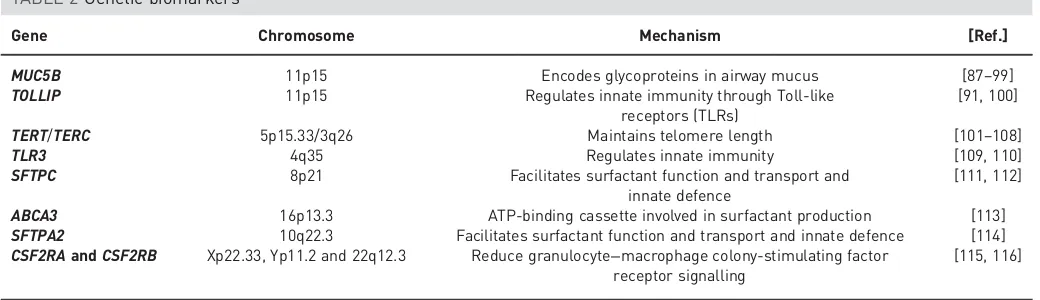
Genetic biomarkers are summarised in table 2.
In 2011, a single nucleotide polymorphism was identified in the promoter region of the mucin 5B (MUC5B) gene (rs35705950), on the short arm of chromosome 11, which was highly associated with both familial pulmonary fibrosis and sporadic IPF, with odds ratios of 6.3 and 8.3, respectively [87]. This genetic variant is present in 31–42% of patients with IPF [88–92]. The associations between MUC5B rs35705950 and short telomere length with extent of fibrosis, histopathological features of usual interstitial pneumonia and reduced survival in patients with chronic hypersensitivity pneumonitis were recently described [93]. No association has been found with SSc-ILD or sarcoidosis [89]. Increasing copies of the MUC5B promoter polymorphism were associated with progresssion of “interstitial lung abnormalities” (ILAs) [94]. TheMUC5Bvariant is associated with better survival in two separate cohorts of IPF patients, independently of disease severity [97]. In sporadic IPF, survival was not influenced by MUC5B alleles; in familial pulmonary fibrosis, MUC5B minor allele predicted better survival [98] and was associated with a lower pulmonary bacterial burden and, in turn, a better survival [99].
A recent genome-wide association study reported the association of three Toll interacting protein (TOLLIP) gene single nucleotide polymorphisms with pulmonary fibrosis, implicating innate immunity processes in IPF pathogenesis. Interestingly, the minor allele rs5743890_G was protective against the development of IPF, but was associated with increased mortality [91]. A different single nucleotide polymorphism rs3750920 within TOLLIP was associated with a differential response to N-acetylcysteine (NAC) treatment in a retrospective analysis of subjects in the PANTHER trial. While NAC treatment was associated with a significant reduction in a composite end-point risk (defined as death, transplant, hospitalisation or⩾10% FVC decline) in patients with a rs3750920 TT genotype, a nonsignificant increase in end-point risk was seen in patients with a CC genotype [100].
TABLE 2Genetic biomarkers
Gene Chromosome Mechanism [Ref.]
MUC5B 11p15 Encodes glycoproteins in airway mucus [87–99] TOLLIP 11p15 Regulates innate immunity through Toll-like
receptors (TLRs)
[91, 100]
TERT/TERC 5p15.33/3q26 Maintains telomere length [101–108]
TLR3 4q35 Regulates innate immunity [109, 110]
SFTPC 8p21 Facilitates surfactant function and transport and innate defence
[111, 112]
ABCA3 16p13.3 ATP-binding cassette involved in surfactant production [113] SFTPA2 10q22.3 Facilitates surfactant function and transport and innate defence [114] CSF2RAandCSF2RB Xp22.33, Yp11.2 and 22q12.3 Reduce granulocyte−macrophage colony-stimulating factor
receptor signalling
Mutations in both telomerase reverse transcriptase (TERT) and telomerase RNA component (TERC) have been reported in 8–15% of familial pulmonary fibrosis cases, creating a pathogenic link between short telomeres and pulmonary fibrosis [102–104]. Short telomeres are common in sporadic IPF, with∼25% of patients demonstrating blood leukocyte telomere lengths below the 10th percentile [105]. Blood leukocyte telomere lengths were predictive of transplant-free survival in IPF, independent of conventional markers, but interestingly not in other ILDs [107], and independently associated with worse survival in another IPF cohort [108].
Recently, a specific variant in the Toll-like receptor-3 gene (L412F) was found to be associated with early mortality and accelerated lung function decline in IPF [110]. Mutations in the SP-C gene (SFTPC) are associated with early-onset NSIP, desquamative interstitial pneumonia and PAP [111]. In adults, they are mostly associated with a usual interstitial pneumonia pattern, but NSIP, desquamative interstitial pneumonia or organising pneumonia may also be observed [112]. Abnormalities that severely reduce granulocyte–macrophage colony-stimulating factor receptor signalling, including loss of heterozygosity and a function-altering point mutation in CSF2RAand homozygousCSF2RBmutations, have been identified as the cause of hereditary PAP [115, 116].
The so-called 8.1 ancestral haplotype (human leukocyte antigen (HLA) A_0101: Cw_0701: B_0801: DRB1_0301: DQA1_0501: DQB1_0201), which is common in Europeans, has been associated with the development of sarcoidosis. HLA-B8 and HLA-B7 seem to increase the risk of disease, and a common haplotype in Swedish patients HLA-A*03, B*07, DRB1*15 increases the risk of chronic disease [117]. The strongest associations have been reported with the HLA class II genes, whereHLA-DRB1*01andDRB1*04 are protective, but DRB1*03, DRB1*11, DRB1*12, DRB1*14 and DRB1*15 represent risk factors for sarcoidosis. Interestingly, in African-Americans, HLA-DQB1 and not DRB1 alleles were suggested to be primarily associated with sarcoidosis [118].
HLA genes have also been identified as the strongest genetic risk factor for SSc-ILD.HLA-DRB5*01:05is a novel risk factor for developing ILD in patients with SSc, being significantly more frequent in SSc patients with ILD than in those without or in healthy controls [119].
Tumour necrosis factor (TNF)-α plays an important role in the pathogenesis of sarcoidosis. TNF-α inhibitors have shown promising results in the treatment of refractory sarcoidosis [120, 121]. The presence of the TNF-α-308A variant allele may identify patients with a favourable response to therapy [122, 123]. Sarcoidosis patients without the TNF-α-308A variant allele (GG genotype) had a three-fold higher response to TNF inhibitors [123].
Decreased peripheral blood mononuclear cell expression of genes involved in the “costimulatory signal during T-cell activation” pathway (including those for CD28, inducible T-cell costimulator (ICOS), lymphocyte-specific protein tyrosine kinase (LCK) and IL-2-inducible T-cell kinase (ITK)) was associated with a shorter transplant-free survival in an IPF cohort. The addition of the gene expression levels of these four genes increased the predictive capacity of a prognostic model compared with one containing solely clinical data (age, sex and FVC %) [124].
A gene set that included downregulation of genes involved in T-cell immune responses was predictive of poor prognosis in IPF, even after adjusting for the composite physiological index [125].
Epigenetic biomarkers (microRNAs)
There have been a number of epigenomic studies of microRNA (miRNA) profiles, mostly in IPF. Downregulated miRNAs in IPF lungs include let-7d, miR-29 and the miR-200 family, while upregulated miRNAs include miR-155, miR-21, miR-199 and miR-154 [126, 127].
A panel of miRNAs (miR-302c, miR-423, miR-210, miR-376c and miR-185) in lung biopsies from patients with IPF could differentiate rapid from slow progressors [128]. Selected miRNAs were validated in independent cohorts and appeared to differentiate slow versus rapid progressors. Compared with slow progressors, and in turn with controls, rapid progressors displayed higher circulating miR-21, miR199a-5p and miR-200c, and lower levels of miR-31, let-7a and let-7d [129]. miR-29 is involved in regulation of fibroblast activation. A beneficial effect of intravenously administered miR-29 in the mouse model of pulmonary fibrosis has been demonstrated [130]. miR-29a is strongly downregulated in SSc fibroblasts and skin sections compared with healthy controls [131]. let-7d miRNA is consistently downregulated in IPF lungs and is believed to play a crucial role in inhibiting epithelial–mesenchymal differentiation [132]. Inhibition of the let-7d family resulted in upregulation of mesenchymal and downregulation of epithelial markers in alveolar epithelial cell lines [133].
Preventive/participatory medicine
smoking cessation campaigns should include this information. The role of diet and exercise could potentially contribute to disease development and progression, but further studies are required.
Factors originating from the external or internal environment also seem to contribute to the pathogenesis of IPF, but they have not been sufficiently explored. More recent work has focused on the interaction between the lung microbiome and the innate immune system, mostly in IPF, highlighting the role of host– environment interaction in disease pathogenesis.
BAL from IPF patients was characterised by a higher bacterial burden and a loss of bacterial species diversity compared with controls. Patients with the highest BAL bacterial burden had the worse survival, independent of disease severity, although no specific microbe was associated with the rate of progression [99]. SpecificStreptococcus orStaphylococcusspp. were associated with more rapid disease progression in patients with IPF [134]. More recently, integrated analysis of the host transcriptome and microbial signatures in IPF patients demonstrated an apparent host response to the presence of an altered or more abundant microbiome. These responses remained elevated in longitudinal follow-up, suggesting that the bacterial communities of the lower airways may act as persistent stimuli for repetitive alveolar injury in IPF [135]. In a pilot study of patients with acute exacerbations of IPF and matched control patients with stable IPF, acute exacerbations of IPF were associated with an increased BAL bacterial burden compared with stable IPF. The bacterial communities of the stable IPF subjects were similar to those found in the airways of healthy individuals as well as subjects with asthma, chronic obstructive pulmonary disease and IPF. Following an acute exacerbation of IPF there was a notable change in the microbiota, with an increase inCampylobacter sp. and Stenotrophomonas sp., coupled with a significant decrease in Veillonella sp. The apparent translocation of bacteria usually confined to the gastrointestinal tract suggests a potential role for aspiration in the development of acute exacerbations [136]. These data suggest that the lung microbiome may serve as a preventive or therapeutic target as well as prognostic biomarker in IPF and potentially other ILD.
The unforeseeable and often rapidly progressive nature of IPF means that patients often have to deal with a number of challenges at different points during the disease course. Therefore, efficient communication between the healthcare professional and the patient is of major importance in helping the patient and their family cope and feel supported in the disease process. Another problem for IPF patients at the stages of diagnosis and treatment is education. Sufficient information should be provided regarding the prognosis and available treatments. Participatory medicine, a patient-centred approach, where the patient has the key role in decision making after sufficient education and communication, should be part of personalised medicine, and the focus of future work and research.
Implications of personalised pathways in appraising historical IPF data
At present, the only treatment data providing direct support for a personalised approach in IPF consist of the analyses showing a differential treatment effect according toTOLLIPgenotype status [100]. It must be stressed that this study was underpowered and that the findings need to be confirmed. The findings have face and content validity, and can be viewed as establishing“proof of concept”. However, the stimulus to harnessing personalised medicine in IPF and other ILDs comes primarily from data in other chronic lung diseases. For this reason, the main body of this review consists of a detailed compilation of candidate biomarkers that might lead to a personalised approach in future. Given these data, findings in historical studies can now be reappraised.
Several IPF treatment trials have been performed based on positive results in pre-clinical studies and early-phase clinical trials that suggested the possibility of benefit. Most did not fulfil expectations. However, increased awareness of personalised medicine has raised the question of whether negative findings reflected the necessarily indiscriminate recruitment of IPF patients, without reference to key pathway signatures.
The first attempt to apply personalised medicine prospectively in an IPF treatment trial was unsuccessful. Lysyl oxidase-like 2 (LOXL2) is a regulator of collagen cross-linking. Higher serum levels in a subgroup of IPF patients were associated with rapidly progressive disease [76]. A recent phase II study of simtuzumab, an anti-LOXL2 monoclonal antibody, was terminated for lack of efficacy (with no difference in progression-free survival between treatment arms) [140]. However, the study design serves as a template for future attempts to apply personalised medicine to IPF interventions.
Variations between trial cohorts in the prevalence of key pathogenetic pathways may also explain the existence of major differences in treatment effects in different trials of the same therapeutic agent. In most trials, pirfenidone has had a major beneficial effect on serial FVC trends. In the one negative study, the placebo arm exhibited a FVC decline that was considerably lower than generally observed in other IPF placebo arms [141]. Again, it can be argued that the study may have been negative because of underrepresentation of patients with key pathways associated with progressive disease. This may be a greater problem in those trials with a numerical imbalance between treatment and placebo arms (e.g.a 2:1 recruitment strategy), with smaller placebo populations cohorts at greater risk of outlying findings.
This concept may also explain major divergences in the reported efficacy of antioxidant therapy in IPF. In the first positive IPF trial, prednisolone, azathioprine and NAC (compared with prednisolone, azathioprine and placebo) was associated with a beneficial effect on FVC and DLCO trends [142]. Although triple therapy was subsequently discredited due to an increased number of deaths and hospitalisations [143], no conclusion could be reached on pulmonary function trends because of the need for early cessation of the triple-therapy arm. Subsequently, the same investigators found no difference in FVC trends between the NAC monotherapy and placebo arms [144]. Although the study was, at first sight, definitively negative, subsequent analyses of the PANTHER cohort (discussed earlier) suggested that a major imbalance in outcomes existed according toTOLLIPstatus [100]. If this latter finding is confirmed, conflicting findings in historical studies of antioxidant therapy may be seen in a new light. In essence, an inability to apply personalised medicine may have resulted in misleading negative findings in a large IPF cohort, with an averaging of beneficial and adverse treatment effects.
The above examples illustrate the major difficulties that may arise in this complex therapeutic field, if interventions are not evaluated in relation to key pathogenetic pathways or if the wrong pathways are explored. In this regard, it is essential that pathways be chosen not only from pre-clinical data, but also from the validation of biomarker signals in key IPF subgroups. To this point, pathogenetic pathway identification has largely been driven by analyses of selected biomarkers across the whole spectrum of IPF, examined against pulmonary function trends (quantified as continuous variables) and survival (variably adjusted for baseline severity). A potentially fruitful complementary approach is to pre-define criteria for the rapidity of IPF disease progression and to determine which among a great many candidate pathways distinguish most accurately between rapid and slow progressors. For this purpose, the design of the PROFILE study can be seen as an important exemplar.
Another major opportunity lies in long-term studies of ILAs, the subject of several recent large cohort analyses. Much work remains to be done in refining the description of ILAs, which in different cohorts include varying proportions of nondependent ground-glass or reticular abnormalities, diffuse centrilobular nodularity, nonemphysematous cysts, honeycombing and traction bronchiectasis [94, 145–151]. There is a need for an international group to produce guidance to standardise future ILA studies in such a way as to allow underpowered findings in key subgroups to be amalgamated in a definitive evidence base. With reference to IPF, 80% of ILAs in the Framington cohort were found to be subpleural and reticular [94]. Importantly, ILAs and IPF have very similar demographic profiles and a common association with MUC5Brs35705950T positivity [145].
The major difficulty is that the prevalence of ILA in elderly cohorts of 5–7% greatly exceeds the prevalence of IPF. The opportunity lies in the identification of key pathogenetic pathways, operating in the subgroup who ultimately develop IPF [152]. In this regard, the higher plasma galectin-3 levels associated with the presence of ILAs, and with lower lung volumes and DLCO levels, provide proof of concept [153]. ILAs provide a unique opportunity to identify key biomarkers associated with progression to early IPF. In principle, this may allow pivotal upstream pathogenetic pathways to be isolated and to be studied selectively in future attempts to develop a personalised approach in established disease.
Conclusions
and pathway signatures with a view to refining prognostic evaluation and trial design, with the ultimate aim of selecting treatments according to profiles in individual patients. Some historical trials may have been unsuccessful because interventions that were beneficial in some IPF patients were buried within whole-cohort analyses. It must be acknowledged, based on the very large body of nondefinitive data summarised in the current review, that the way ahead will be difficult. However, the successful application of personalised medicine in other chronic lung diseases should continue to inspire IPF researchers in attempting to transform the field.
Conflict of interest: A.U. Wells reports personal fees from InterMune/Roche and Boehringer Ingelheim, outside the submitted work.
References
1 Margaritopoulos GA, Romagnoli M, Poletti V,et al.Recent advances in the pathogenesis and clinical evaluation of pulmonary fibrosis.Eur Respir Rev2012; 21: 48–56.
2 Selman M, King TE, Pardo A. Idiopathic pulmonary fibrosis: prevailing and evolving hypotheses about its pathogenesis and implications for therapy.Ann Intern Med2001; 134: 136–151.
3 Maher TM, Wells AU, Laurent GJ. Idiopathic pulmonary fibrosis: multiple causes and multiple mechanisms?Eur Respir J2007; 30: 835–839.
4 Collard HR, Ryerson CJ, Corte TJ,et al.Acute exacerbation of idiopathic pulmonary fibrosis. An international working group report.Am J Respir Crit Care Med2016; 194: 265–275.
5 King TE Jr, Bradford WZ, Castro-Bernardini S,et al.A phase 3 trial of pirfenidone in patients with idiopathic pulmonary fibrosis.N Engl J Med2014; 370: 2083–2092.
6 Richeldi L, du Bois RM, Raghu G,et al. Efficacy and safety of nintedanib in idiopathic pulmonary fibrosis. N Engl J Med2014; 370: 2071–2082.
7 Collins FS, Varmus H. A new initiative on precision medicine.N Engl J Med2015; 372: 793–795.
8 Haldar P, Brightling CE, Hargadon B,et al. Mepolizumab and exacerbations of refractory eosinophilic asthma. N Engl J Med2009; 360: 973–984.
9 Nair P, Pizzichini MM, Kjarsgaard M, et al. Mepolizumab for prednisone-dependent asthma with sputum eosinophilia.N Engl J Med2009; 360: 985–993.
10 Aggarwal C. Targeted therapy for lung cancer: present and future.Ann Palliat Med2014; 3: 229–235.
11 Mok TS, Wu YL, Thongprasert S, et al. Gefitinib or carboplatin–paclitaxel in pulmonary adenocarcinoma. N Engl J Med2009; 361: 947–957.
12 Rosell R, Carcereny E, Gervais R, et al. Erlotinib versus standard chemotherapy as first-line treatment for European patients with advanced EGFR mutation-positive non-small-cell lung cancer (EURTAC): a multicentre, open-label, randomised phase 3 trial.Lancet Oncol2012; 13: 239–246.
13 Horimasu Y, Hattori N, Ishikawa N,et al. Different MUC1 gene polymorphisms in German and Japanese ethnicities affect serum KL-6 levels.Respir Med2012; 106: 1756–1764.
14 Satoh H, Kurishima K, Ishikawa H,et al.Increased levels of KL-6 and subsequent mortality in patients with interstitial lung diseases.J Intern Med2006; 260: 429–434.
15 Hant FN, Ludwicka-Bradley A, Wang HJ, et al. Surfactant protein D and KL-6 as serum biomarkers of interstitial lung disease in patients with scleroderma.J Rheumatol2009; 36: 773–780.
16 Janssen R, Sato H, Grutters JC,et al.Study of Clara cell 16, KL-6, and surfactant protein-D in serum as disease markers in pulmonary sarcoidosis.Chest2003; 124: 2119–2125.
17 Bonella F, Ohshimo S, Miaotian C, et al. Serum KL-6 is a predictor of outcome in pulmonary alveolar proteinosis.Orphanet J Rare Dis2013; 8: 53.
18 Collard HR, Calfee CS, Wolters PJ, et al. Plasma biomarker profiles in acute exacerbation of idiopathic pulmonary fibrosis.Am J Physiol Lung Cell Mol Physiol2010; 299: L3–L7.
19 Ohshimo S, Ishikawa N, Horimasu Y,et al. Baseline KL-6 predicts increased risk for acute exacerbation of idiopathic pulmonary fibrosis.Respir Med2014; 108: 1031–1039.
20 Yanaba K, Hasegawa M, Hamaguchi Y,et al.Longitudinal analysis of serum KL-6 levels in patients with systemic sclerosis: association with the activity of pulmonary fibrosis.Clin Exp Rheumatol2003; 21: 429–436.
21 Okada M, Suzuki K, Matsumoto M,et al.Intermittent intravenous cyclophosphamide pulse therapy for the treatment of active interstitial lung disease associated with collagen vascular diseases.Mod Rheumatol2007; 17: 131–136.
22 Janssen R, Kriut A, Grutters JC,et al.The mucin-1 568 adenosine to guanine polymorphism influences serum Krebs von den Lungen-6 levels.Am J Respir Crit Care Med2006; 34: 496–499.
23 Song JW, Do KH, Jang SJ, et al.Blood biomarkers MMP-7 and SP-A: predictors of outcome in idiopathic pulmonary fibrosis.Chest2013; 143: 1422–1429.
24 Elhaj M, Charles J, Pedroza C,et al. Can serum surfactant protein D or CC-chemokine ligand 18 predict outcome of interstitial lung disease in patients with early systemic sclerosis?J Rheumatol2013; 40: 1114–1120. 25 Ihn H, Asano Y, Kubo M,et al. Clinical significance of serum surfactant protein D (SP-D) in patients with
polymyositis/dermatomyositis: correlation with interstitial lung disease.Rheumatology2002; 41: 1268–1272. 26 Lin FC, Chen YC, Chang SC. Clinical importance of bronchoalveolar lavage fluid and blood cytokines, surfactant
protein D, and Krebs von Lungren 6 antigen in idiopathic pulmonary alveolar proteinosis.Mayo Clin Proc2008; 83: 1344–1349.
27 Kinder BW, Brown KK, McCormack FX,et al.Serum surfactant protein-A is a strong predictor of early mortality in idiopathic pulmonary fibrosis.Chest2009; 135: 1557–1563.
28 Barlo NP, van Moorsel CH, Ruven HJ,et al.Surfactant protein-D predicts survival in patients with idiopathic pulmonary fibrosis.Sarcoidosis Vasc Diffuse Lung Dis2009; 26: 155–161.
30 Yanaba K, Hasegawa M, Takehara K, et al. Comparative study of serum surfactant protein-D and KL-6 concentrations in patients with systemic sclerosis as markers for monitoring the activity of pulmonary fibrosis. J Rheumatol2004; 31: 1112–1120.
31 Asano Y, Ihn H, Yamane K,et al.Clinical significance of surfactant protein D as a serum marker for evaluating pulmonary fibrosis in patients with systemic sclerosis.Arthritis Rheum2001; 44: 1363–1369.
32 Horimasu Y, Hattori N, Ishikawa N,et al. Differences in serum SP-D levels between German and Japanese subjects are associated withSFTPDgene polymorphisms.BMC Med Genet2014; 15: 4.
33 Hermans C, Petrek M, Kolek V,et al.Serum Clara cell protein (CC16), a marker of the integrity of the air-blood barrier in sarcoidosis.Eur Respir J2001; 18: 507–514.
34 Hasegawa M, Fujimoto M, Hamaguchi Y,et al.Use of serum Clara cell 16-kDa (CC16) levels as a potential indicator of active pulmonary fibrosis in systemic sclerosis.J Rheumatol2011; 38: 877–884.
35 Cha SI, Ryerson CJ, Lee JS,et al.Cleaved cytokeratin-18 is a mechanistically informative biomarker in idiopathic pulmonary fibrosis.Respir Res2012; 13: 105.
36 Prasse A, Pechkovsky DV, Toews GB,et al.CCL18 as an indicator of pulmonary fibrotic activity in idiopathic interstitial pneumonias and systemic sclerosis.Arthritis Rheum2007; 56: 1685–1693.
37 Prasse A, Probst C, Bargagli E, et al. Serum CC-chemokine ligand 18 concentration predicts outcome in idiopathic pulmonary fibrosis.Am J Respir Crit Care Med2009; 179: 717–723.
38 Tiev KP, Hua-Huy T, Kettaneh A,et al.Serum CC chemokine ligand-18 predicts lung disease worsening in systemic sclerosis.Eur Respir J2011; 38: 1355–1360.
39 Furuhashi K, Suda T, Nakamura Y,et al.Increased expression of YKL-40, a chitinase-like protein, in serum and lung of patients with idiopathic pulmonary fibrosis.Respir Med2010; 104: 1204–1210.
40 Korthagen NM, van Moorsel CH, Barlo NP,et al.Serum and BALF YKL-40 levels are predictors of survival in idiopathic pulmonary fibrosis.Respir Med2011; 105: 106–113.
41 Nordenbaek C, Johansen JS, Halberg P,et al.High serum levels of YKL-40 in patients with systemic sclerosis are associated with pulmonary involvement.Scand J Rheumatol2005; 34: 293–297.
42 Korthagen NM, van Moorsel CH, Zanen P,et al.Evaluation of circulating YKL-40 levels in idiopathic interstitial pneumonias.Lung2014; 192: 975–980.
43 Johansen JS. Studies on serum YKL-40 as a biomarker in diseases with inflammation, tissue remodelling, fibroses and cancer.Dan Med Bull2006; 53: 172–209.
44 Bonella F, Long X, He X, et al.Serum YKL-40 is a reliable biomarker for pulmonary alveolar proteinosis. Respirology2017; 22: 1371–1378.
45 Long X, He X, Ohshimo S,et al. Serum YKL-40 as predictor of outcome in hypersensitivity pneumonitis.Eur Respir J2017; 49: 1501924.
46 Antonelli A, Ferri C, Fallahi P,et al.CXCL10 (alpha) and CCL2 (beta) chemokines in systemic sclerosis–a longitudinal study.Rheumatology2008; 47: 45–49.
47 Su R, Nguyen ML, Agarwal MR, et al. Interferon-inducible chemokines reflect severity and progression in sarcoidosis.Respir Res2013; 14: 121.
48 Romagnani P, Maggi L, Mazzinghi B,et al.CXCR3-mediated opposite effects of CXCL10 and CXCL4 on TH1 or TH2 cytokine production.J Allergy Clin Immunol2005; 116: 1372–1379.
49 van Bon L, Affandi AJ, Broen J,et al.Proteome-wide analysis and CXCL4 as a biomarker in systemic sclerosis. N Engl J Med2014; 370: 433–443.
50 Vuga LJ, Tedrow JR, Pandit KV, et al.C-X-C motif chemokine 13 (CXCL13) is a prognostic biomarker of idiopathic pulmonary fibrosis.Am J Respir Crit Care Med2014; 189: 966–974.
51 DePianto DJ, Chandriani S, Abbas AR,et al.Heterogeneous gene expression signatures correspond to distinct lung pathologies and biomarkers of disease severity in idiopathic pulmonary fibrosis.Thorax2015; 70: 48–56. 52 van Bon L, Cossu M, Loof A, et al. Proteomic analysis of plasma identifies the Toll-like receptor agonists
S100A8/A9 as a novel possible marker for systemic sclerosis phenotype.Ann Rheum Dis2014; 73: 1585–1589. 53 Richards TJ, Kaminski N, Baribaud F,et al.Peripheral blood proteins predict mortality in idiopathic pulmonary
fibrosis.Am J Respir Crit Care Med2012; 185: 67–76.
54 Kahloon RA, Xue J, Bhargava A,et al.Patients with idiopathic pulmonary fibrosis with antibodies to heat shock protein 70 have poor prognoses.Am J Respir Crit Care Med2013; 187: 768–775.
55 Kakugawa T, Yokota S, Ishimatsu Y,et al.Serum heat shock protein 47 levels are elevated in acute exacerbation of idiopathic pulmonary fibrosis.Cell Stress Chaperones2013; 18: 581–590.
56 Kotsianidis I, Nakou E, Bouchliou I,et al. Global impairment of CD4+CD25+FOXP3+ regulatory T cells in idiopathic pulmonary fibrosis.Am J Respir Crit Care Med2009; 179: 1121–1130.
57 Reilkoff RA, Peng H, Murray LA, et al. Semaphorin 7a+ regulatory T cells are associated with progressive idiopathic pulmonary fibrosis and are implicated in transforming growth factor-beta1-induced pulmonary fibrosis.Am J Respir Crit Care Med2013; 187: 180–188.
58 Yang IV, Luna LG, Cotter J,et al. The peripheral blood transcriptome identifies the presence and extent of disease in idiopathic pulmonary fibrosis.PLoS One2012; 7: e37708.
59 Konishi K, Gibson KF, Lindell KO,et al.Gene expression profiles of acute exacerbations of idiopathic pulmonary fibrosis.Am J Respir Crit Care Med2009; 180: 167–175.
60 Antoniou KM, Tzouvelekis A, Alexandrakis MG,et al.Upregulation of Th1 cytokine profile (IL-12, IL-18) in bronchoalveolar lavage fluid in patients with pulmonary sarcoidosis.J Interferon Cytokine Res2006; 26: 400–405. 61 Mroz RM, Korniluk M, Stasiak-Barmuta A, et al. Increased levels of interleukin-12 and interleukin-18 in
bronchoalveolar lavage fluid of patients with pulmonary sarcoidosis.J Physiol Pharmacol 2008; 59: Suppl. 6, 507–513.
62 Liu DH, Cui W, Chen Q,et al.Can circulating interleukin-18 differentiate between sarcoidosis and idiopathic pulmonary fibrosis?Scand J Clin Lab Invest2011; 71: 593–597.
63 Antoniou KM, Alexandrakis MG, Sfiridaki K,et al.Th1 cytokine pattern (IL- 12 and IL-18) in bronchoalveolar lavage fluid (BALF) before and after treatment with interferon gamma-1b (IFN-gamma-1b) or colchicine in patients with idiopathic pulmonary fibrosis (IPF/UIP).Sarcoidosis Vasc Diffuse Lung Dis2004; 21: 105–110. 64 Komura K, Fujimoto M, Hasegawa M,et al.Increased serum interleukin 23 in patients with systemic sclerosis.
65 Wuttge DM, Wildt M, Geborek P,et al.Serum IL-15 in patients with early systemic sclerosis: a potential novel marker of lung disease.Arthritis Res Ther2007; 9: R85.
66 Keicho N, Kitamura K, Takaku F,et al. Serum concentration of soluble interleukin-2 receptor as a sensitive parameter of disease activity in sarcoidosis.Chest1990; 98: 1125–1129.
67 Bargagli E, Bianchi N, Margollicci M, et al. Chitotriosidase and soluble IL-2 receptor: comparison of two markers of sarcoidosis severity.Scand J Clin Lab Invest2008; 68: 479–483.
68 Grutters JC, Fellrath JM, Mulder L,et al.Serum soluble interleukin-2 receptor measurement in patients with sarcoidosis: a clinical evaluation.Chest2003; 124: 186–195.
69 Ziegenhagen MW, Rothe ME, Schlaak M,et al.Bronchoalveolar and serological parameters reflecting the severity of sarcoidosis.Eur Respir J2003; 21: 407–413.
70 Rosas IO, Richards TJ, Konishi K, et al. MMP1 and MMP7 as potential peripheral blood biomarkers in idiopathic pulmonary fibrosis.PLoS Med2008; 5: e93.
71 Jenkins RG, Simpson JK, Saini G,et al.Longitudinal change in collagen degradation biomarkers in idiopathic pulmonary fibrosis: an analysis from the prospective, multicentre PROFILE study.Lancet Respir Med2015; 3: 462–472.
72 Kim WU, Min SY, Cho ML, et al. Elevated matrix metalloproteinase-9 in patients with systemic sclerosis. Arthritis Res Ther2005; 7: R71–R79.
73 Moinzadeh P, Krieg T, Hellmich M,et al.Elevated MMP-7 levels in patients with systemic sclerosis: correlation with pulmonary involvement.Exp Dermatol2011; 20: 770–773.
74 Oka S, Furukawa H, Shimada K,et al.Serum biomarker analysis of collagen disease patients with acute-onset diffuse interstitial lung disease.BMC Immunol2013; 14: 9.
75 Manetti M, Ibba-Manneschi L, Fatini C, et al. Association of a functional polymorphism in the matrix metalloproteinase-12 promoter region with systemic sclerosis in an Italian population.J Rheumatol2010; 37: 1852–1857.
76 Chien JW, Richards TJ, Gibson KF,et al. Serum lysyl oxidase-like 2 levels and idiopathic pulmonary fibrosis disease progression.Eur Respir J2014; 43: 1430–1438.
77 Okamoto M, Hoshino T, Kitasato Y,et al. Periostin, a matrix protein, is a novel biomarker for idiopathic interstitial pneumonias.Eur Respir J2011; 37: 1119–1127.
78 Tajiri M, Okamoto M, Fujimoto K,et al.Serum level of periostin can predict long-term outcome of idiopathic pulmonary fibrosis.Respir Investig2015; 53: 73–81.
79 Bellini A, Mattoli S. The role of the fibrocyte, a bone marrow-derived mesenchymal progenitor, in reactive and reparative fibroses.Lab Invest2007; 87: 858–870.
80 Fujiwara A, Kobayashi H, Masuya M, et al. Correlation between circulating fibrocytes, and activity and progression of interstitial lung diseases.Respirology2012; 17: 693–698.
81 Moeller A, Gilpin SE, Ask K, et al. Circulating fibrocytes are an indicator of poor prognosis in idiopathic pulmonary fibrosis.Am J Respir Crit Care Med2009; 179: 588–594.
82 Tercelj M, Salobir B, Simcic S,et al.Chitotriosidase activity in sarcoidosis and some other pulmonary diseases. Scand J Clin Lab Invest2009; 69: 575–578.
83 Bargagli E, Margollicci M, Nikiforakis N,et al.Chitotriosidase activity in the serum of patients with sarcoidosis and pulmonary tuberculosis.Respiration2007; 74: 548–552.
84 Grosso S, Margollicci MA, Bargagli E,et al.Serum levels of chitotriosidase as a marker of disease activity and clinical stage in sarcoidosis.Scand J Clin Lab Invest2004; 64: 57–62.
85 Bargagli E, Rottoli P. Serum chitotriosidase activity in sarcoidosis patients.Rheumatol Int2007; 27: 1187. 86 Boot RG, Hollak CE, Verhoek M, et al. Plasma chitotriosidase and CCL18 as surrogate markers for
granulomatous macrophages in sarcoidosis.Clin Chim Acta2010; 411: 31–36.
87 Seibold MA, Wise AL, Speer MC,et al. A commonMUC5Bpromoter polymorphism and pulmonary fibrosis. N Engl J Med2011; 364: 1503–1512.
88 Zhang Y, Noth I, Garcia JG,et al. A variant in the promoter ofMUC5Band idiopathic pulmonary fibrosis. N Engl J Med2011; 364: 1576–1577.
89 Stock CJ, Sato H, Fonseca C,et al.Mucin 5B promoter polymorphism is associated with idiopathic pulmonary fibrosis but not with development of lung fibrosis in systemic sclerosis or sarcoidosis.Thorax2013; 68: 436. 90 Borie R, Quesnel C, Phin S,et al.Detection of alveolar fibrocytes in idiopathic pulmonary fibrosis and systemic
sclerosis.PLoS One2013; 8: e53736.
91 Noth I, Zhang Y, Ma SF,et al.Genetic variants associated with idiopathic pulmonary fibrosis susceptibility and mortality: a genome-wide association study.Lancet Respir Med2013; 1: 309–317.
92 Fingerlin TE, Murphy E, Zhang W,et al.Genome-wide association study identifies multiple susceptibility loci for pulmonary fibrosis.Nat Genet2013; 45: 613–620.
93 Ley B, Newton CA, Arnould I,et al.TheMUC5Bpromoter polymorphism and telomere length in patients with chronic hypersensitivity pneumonitis: an observational cohort-control study. Lancet Respir Med 2017; 5: 639–647.
94 Araki T, Putman RK, Hatabu H,et al.Development and progression of interstitial lung abnormalities in the Framingham Heart Study.Am J Respir Crit Care Med2016; 194: 1514–1522.
95 Seibold MA, Smith RW, Urbanek C, et al. The idiopathic pulmonary fibrosis honeycomb cyst contains a mucocilary pseudostratified epithelium.PLoS One2013; 8: e58658.
96 Conti C, Montero-Fernandez A, Nicholson AG,et al.Distribution of mucins MUC5B and MUC5AC in distal airways and honeycomb spaces: a comparison between UIP and other ILD patterns.Am J Respir Crit Care Med 2015; 191: A2161.
97 Peljto AL, Zhang Y, Fingerlin TE,et al.Association between theMUC5Bpromoter polymorphism and survival in patients with idiopathic pulmonary fibrosis.JAMA2013; 309: 2232–2239.
98 van der Vis JJ, Snetselaar R, Kazemier KM, et al. Effect of Muc5b promoter polymorphism on disease predisposition and survival in idiopathic interstitial pneumonias.Respirology2016; 21: 712–717.
100 Oldham JM, Ma SF, Martinez FJ,et al. TOLLIP,MUC5Band the response toN-acetylcysteine among individuals with idiopathic pulmonary fibrosis.Am J Respir Crit Care Med2015; 192: 1475–1482.
101 Selman M, Pardo A. Revealing the pathogenic and aging-related mechanisms of the enigmatic idiopathic pulmonary fibrosis. an integral model.Am J Respir Crit Care Med2014; 189: 1161–1172.
102 Armanios MY, Chen JJ, Cogan JD,et al.Telomerase mutations in families with idiopathic pulmonary fibrosis. N Engl J Med2007; 356: 1317–1326.
103 Tsakiri KD, Cronkhite JT, Kuan PJ,et al. Adult-onset pulmonary fibrosis caused by mutations in telomerase. Proc Natl Acad Sci USA2007; 104: 7552–7557.
104 Alder JK, Chen JJ, Lancaster L,et al.Short telomeres are a risk factor for idiopathic pulmonary fibrosis.Proc Natl Acad Sci USA2008; 105: 13051–13056.
105 Cronkhite JT, Xing C, Raghu G,et al.Telomere shortening in familial and sporadic pulmonary fibrosis.Am J Respir Crit Care Med2008; 178: 729–737.
106 Rode L, Bojesen SE, Weischer M,et al.Short telomere length, lung function and chronic obstructive pulmonary disease in 46,396 individuals.Thorax2013; 68: 429–435.
107 Stuart BD, Lee JS, Kozlitina J,et al.Effect of telomere length on survival in patients with idiopathic pulmonary fibrosis: an observational cohort study with independent validation.Lancet Respir Med2014; 2: 557–565. 108 Dai J, Cai H, Li H,et al.Association between telomere length and survival in patients with idiopathic pulmonary
fibrosis.Respirology2015; 20: 947–952.
109 Kawai T, Akira S. The role of pattern-recognition receptors in innate immunity: update on Toll-like receptors. Nat Immunol2010; 11: 373–384.
110 O’Dwyer DN, Armstrong ME, Trujillo G, et al. The Toll-like receptor 3 L412F polymorphism and disease progression in idiopathic pulmonary fibrosis.Am J Respir Crit Care Med2013; 188: 1442–1450.
111 Thomas AQ, Lane K, Phillips J III,et al.Heterozygosity for a surfactant protein C gene mutation associated with usual interstitial pneumonitis and cellular nonspecific interstitial pneumonitis in one kindred.Am J Respir Crit Care Med2002; 165: 1322–1328.
112 Borie R, Kannengiesser C, Nathan N,et al.Familial pulmonary fibrosis.Rev Mal Respir2015; 32: 413–434. 113 Young LR, Nogee LM, Barnett B,et al.Usual interstitial pneumonia in an adolescent withABCA3mutations.
Chest2008; 134: 192–195.
114 Wang Y, Kuan PJ, Xing C,et al.Genetic defects in surfactant protein A2 are associated with pulmonary fibrosis and lung cancer.Am J Hum Genet2009; 84: 52–59.
115 Suzuki T, Sakagami T, Young LR,et al.Hereditary pulmonary alveolar proteinosis: pathogenesis, presentation, diagnosis, and therapy.Am J Respir Crit Care Med2010; 182: 1292–1304.
116 Suzuki T, Maranda B, Sakagami T,et al.Hereditary pulmonary alveolar proteinosis caused by recessiveCSF2RB mutations.Eur Respir J2011; 37: 201–204.
117 Fischer A, Grunewald J, Spagnolo P, et al. Genetics of sarcoidosis.Semin Respir Crit Care Med 2014; 35: 296–306.
118 Spagnolo P, Grunewald J. Recent advances in the genetics of sarcoidosis.J Med Genet2013; 50: 290–297. 119 Odani T, Yasuda S, Ota Y,et al.Up-regulated expression of HLA-DRB5 transcripts and high frequency of the
HLA-DRB5*01:05allele in scleroderma patients with interstitial lung disease.Rheumatology2012; 51: 1765–1774. 120 Baughman RP, Drent M, Kavuru M,et al.Infliximab therapy in patients with chronic sarcoidosis and pulmonary
involvement.Am J Respir Crit Care Med2006; 174: 795–802.
121 Judson MA, Baughman RP, Costabel U,et al.Efficacy of infliximab in extrapulmonary sarcoidosis: results from a randomised trial.Eur Respir J2008; 31: 1189–1196.
122 Wijnen PA, Nelemans PJ, Verschakelen JA, et al. The role of tumor necrosis factor alpha G-308A polymorphisms in the course of pulmonary sarcoidosis.Tissue Antigens2010; 75: 262–268.
123 Wijnen PA, Cremers JP, Nelemans PJ, et al. Association of the TNF-alpha G-308A polymorphism with TNF-inhibitor response in sarcoidosis.Eur Respir J2014; 43: 1730–1739.
124 Herazo-Maya JD, Noth I, Duncan SR,et al.Peripheral blood mononuclear cell gene expression profiles predict poor outcome in idiopathic pulmonary fibrosis.Sci Transl Med2013; 5: 205ra136.
125 Huang Y, Ma SF, Vij R,et al.A functional genomic model for predicting prognosis in idiopathic pulmonary fibrosis.BMC Pulm Med2015; 15: 147.
126 Yang IV, Schwartz DA. Epigenetics of idiopathic pulmonary fibrosis.Transl Res2015; 165: 48–60.
127 Tzouvelekis A, Kaminski N. Epigenetics in idiopathic pulmonary fibrosis.Biochem Cell Biol2015; 93: 159–170. 128 Oak SR, Murray L, Herath A,et al.A micro RNA processing defect in rapidly progressing idiopathic pulmonary
fibrosis.PLoS One2011; 6: e21253.
129 Yang G, Yang L, Wang W, et al. Discovery and validation of extracellular/circulating microRNAs during idiopathic pulmonary fibrosis disease progression.Gene2015; 562: 138–144.
130 Xiao J, Meng XM, Huang XR,et al.miR-29 inhibits bleomycin-induced pulmonary fibrosis in mice.Mol Ther 2012; 20: 1251–1260.
131 Zhu H, Li Y, Qu S,et al.MicroRNA expression abnormalities in limited cutaneous scleroderma and diffuse cutaneous scleroderma.J Clin Immunol2012; 32: 514–522.
132 Pandit KV, Corcoran D, Yousef H,et al.Inhibition and role of let-7d in idiopathic pulmonary fibrosis. Am J Respir Crit Care Med2010; 182: 220–229.
133 Huleihel L, Ben-Yehudah A, Milosevic J,et al.Let-7d microRNA affects mesenchymal phenotypic properties of lung fibroblasts.Am J Physiol Lung Cell Mol Physiol2014; 306: L534–L542.
134 Han MK, Zhou Y, Murray S,et al.Lung microbiome and disease progression in idiopathic pulmonary fibrosis: an analysis of the COMET study.Lancet Respir Med2014; 2: 548–556.
135 Molyneaux PL, Willis-Owen SAG, Cox MJ,et al.Host–microbial interactions in idiopathic pulmonary fibrosis. Respir Res2017; 18: 29.
136 Molyneaux PL, Cox MJ, Wells AU,et al.Changes in the respiratory microbiome during acute exacerbations of idiopathic pulmonary fibrosis.Respir Res2017; 18: 29.
138 King TE, Brown KK, Raghu G, et al. BUILD-3: a randomized, controlled trial of bosentan in idiopathic pulmonary fibrosis.Am J Respir Crit Care Med2011; 184: 92–99.
139 Shulgina L, Cahn AP, Chilvers ER, et al. Treating idiopathic pulmonary fibrosis with the addition of co-trimoxazole: a randomised controlled trial.Thorax2013; 68: 155–162.
140 Raghu G, Brown KK, Collard HR,et al. Efficacy of simtuzumab versus placebo in patients with idiopathic pulmonary fibrosis: a randomised, double-blind, controlled, phase 2 trial.Lancet Respir Med2017; 5: 22–32. 141 Noble PW, Albera C, Bradford WZ, et al. Pirfenidone in patients with idiopathic pulmonary fibrosis
(CAPACITY): two randomised trials.Lancet2011; 377: 1760–1769.
142 Demedts M, Behr J, Buhl R,et al.High-dose acetylcysteine in idiopathic pulmonary fibrosis.N Engl J Med2005; 353: 2229–2242.
143 Idiopathic Pulmonary Fibrosis Clinical Research Network. Prednisone, azathioprine, and N-acetylcysteine for pulmonary fibrosis.N Engl J Med2012; 366: 1968.
144 Idiopathic Pulmonary Fibrosis Clinical Research Network. Randomized trial of acetylcysteine in idiopathic pulmonary fibrosis.N Engl J Med2014; 370: 2093–3101.
145 Hunninghake GM, Hatabu H, Okajima Y, et al. MUC5B promoter polymorphism and interstitial lung abnormalities.N Engl J Med2013; 368: 2192–2200.
146 Rosas IO, Ren P, Avila NA,et al.Early interstitial lung disease in familial pulmonary fibrosis.Am J Respir Crit Care Med2007; 176: 698–705.
147 Lederer DJ, Enright PL, Kawut SM,et al. Cigarette smoking is associated with subclinical parenchymal lung disease: the Multi-Ethnic Study of Atherosclerosis (MESA)-lung study.Am J Respir Crit Care Med2009; 180: 407–414.
148 Washko GR, Lynch DA, Matsuoka S,et al.Identification of early interstitial lung disease in smokers from the COPDGene study.Acad Radiol2010; 17: 48–53.
149 Jin GY, Lynch D, Chawla A,et al.Interstitial lung abnormalities in a CT lung cancer screening population: prevalence and progression rate.Radiology2013; 268: 563–571.
150 Putman RK, Hatabu H, Araki T,et al.Association between interstitial lung abnormalities and all-cause mortality. JAMA2016; 315: 672–681.
151 Sverzellati N, Guerci L, Randi G,et al.Interstitial lung diseases in a lung cancer screening trial.Eur Respir J 2011; 38: 392–400.
152 Wells AU, Kokosi MA. Subclinical interstitial lung abnormalities: toward the early detection of idiopathic pulmonary fibrosis?Am J Respir Crit Care Med2016; 194: 1445–1446.