ID: 19-0092; November 2019 DOI: 10.1530/EDM-19-0092
Polar vasculosis in advanced diabetes mellitus
Y Oda and others
Significant polar vasculosis in a patient with
a 30-year history of type 2 diabetes
Yasuhiro Oda1, Masayuki Yamanouchi1, Hiroki Mizuno1, Rikako Hiramatsu1,
Tatsuya Suwabe1, Junichi Hoshino1,2, Naoki Sawa1, Kenichi Ohashi3,4, Takeshi Fujii3 and Yoshifumi Ubara1,2
1Nephrology Center, 2Okinaka Memorial Institute for Medical Research, 3Department of Pathology, Toranomon Hospital, Tokyo, Japan, and 4Department of Pathology, Graduate School of Medicine, Yokohama City University, Yokohama, Japan
Summary
We report the renal histology of a 66-year-old man with hypertension, cardiovascular disease, and a 30-year history of type 2 diabetes mellitus with proliferative diabetic retinopathy, diabetic neuropathy, and diabetic foot status post toe amputation.Urinaryproteinexcretionwas1.4 g/gCr,serumcreatininelevel0.86 mg/dL,estimatedglomerularfiltration rate69 mL/min/1.73 m2, and HbA1c 13–15%, despite using insulin. Light microscopy showed global glomerulosclerosis in 37%oftheglomeruli,buttheremainingglomeruliwereintact.Significantpolarvasculosiswaspresent,whilearteriolar sclerosis was mild. Electron microscopy revealed a thickened glomerular basement membrane, which is compatible with the early stage of diabetic glomerulopathy. The presented case was unique because glomerular changes seen typically in diabetes were not seen in the patient, despite the long-standing history of diabetes and diabetic comorbidities, while prominent polar vasculosis was found. Polar vascular formation helps preserve the glomeruli by allowing hyperosmotic blood bypass the glomeruli; this decreases intraglomerular pressure and minimizes glomerular endothelial damage.
Correspondence should be addressed to Y Oda or Y Ubara
yasuhirooda3@gmail.com or ubara@toranomon.gr.jp
Learning points:
• A 66-year-old man with a 30-year history of type 2 diabetes mellitus with poor glycemic control underwent renal biopsy, which showed scarce glomerular changes typically seen in diabetic kidney disease and instead revealed significantpolarvasculosis.
• Past studies demonstrated that the increased small vessels around the vascular hilus in diabetic patients originatedfromtheafferentarteriolesanddrainedintotheperitubularcapillaries.
• Polarvascularformationmaypreserveglomerularfunctionbyallowingthebloodflowtobypasstheglomeruliand decreasing the intraglomerular pressure, which minimizes endothelial damage of the glomerular tufts.
Background
Diabetic kidney disease (DKD) is one of the major microangiopathies of diabetes mellitus. Past studies demonstrate that the glomeruli in early-stage DKD with albuminuria or normoalbuminuria may have typical structural changes, including mesangial expansion and nodular sclerosis (1). These changes become more prevalent as the duration of diabetes gets longer; one study found
Case presentation
A 66-year-old man with hypertension and a 30-year history of T2DM was referred to our hospital for evaluation of overt proteinuria. He was diagnosed with T2DM at the age of 36 years. Although insulin therapy was started when he was 50 years old, the patient’s HbA1c had always been elevated at 13–15% due to poor adherence to insulin injection.
The patient was suffering from polyvascular diseases secondary to diabetic angiopathy. Photocoagulation for proliferative diabetic retinopathy was performed twice. Angina pectoris occurred at the age of 63 years; coronary angiography revealed 90% stenosis of the proximal segment of the right coronary artery, and hence, stent implantation was performed. Multiple percutaneous interventions were done upon follow-up coronary angiographies. He also had peripheral artery disease status post toe amputations due to diabetic foot ulcers. Pain and numbness in the legs were suggestive of diabetic neuropathy. His other past medical history included hypertension, dyslipidemia, and obstructive sleep apnea syndrome. He had been prescribed with aspirin 100 mg, clopidogrel 75 mg, lansoprazole 15 mg, nifedipine 20 mg,
rosuvastatin 15 mg, methormine 1500 mg, insulin
glulisine 40 units, and insulin degludec 26 units per day, but he had poor adherence to medication regimen. He had smoked three cigarettes per day for 5 years in the past. Both parents of the patient had a history of myocardial infarction.
Investigation
On physical examination, height was found to be 165 cm, weight was 78.3 kg, and the BMI was 28.8. Blood pressure was 118/59 mmHg. Mild pitting edema was present in the lower extremities. Urine protein excretion was 1.43 g/ gCr, and urinary sediment contained 5–10 erythrocytes per high-power field with no casts. Laboratory findings were as follows: erythrocyte count 4.97 × 10⁶/μL (reference range (RR): 4.35 × 10⁶–5.55 × 10⁶); hemoglobin: 14.1 g/ dL (RR: 13.7–16.8); hematocrit: 43.1% (RR: 40.7–50.1);
leukocyte count: 6700/μL (RR: 3300–8600); platelet
count: 242 × 103/μL (RR: 158 × 103–348 × 103); total protein concentration: 7.6 g/dL (RR: 6.6–8.1); albumin: 4.3 g/ dL (RR: 4.1–5.1); urea nitrogen: 17 mg/dL (RR: 8–20);
creatinine: 0.86 mg/dL (RR: 0.65–1.07); potassium:
4.1 mEq/L (RR: 3.6–4.8); calcium: 10.1 mg/dL (RR: 8.8– 10.1); inorganic phosphorus: 3.3 mg/dL (RR: 2.7–4.6); glucose: 227 mg/dL (RR: 73–109); HbA1c: 13.6% (RR:
4.6–6.2); IgA: 432 mg/dL (RR: 93–393); CH50: 72 U/mL (RR: 25–48); C3: 147 mg/dL (RR: 86–160); PR3-ANCA: negative; MPO-ANCA: negative; anti-nuclear antibody: negative; and anti-double-stranded-DNA antibody: negative. His estimated glomerular filtration rate (eGFR) was 69 mL/min/1.73 m2 according to the Modification of Diet in Renal Disease (MDRD) equation for estimating glomerular filtration rate with variables adjusted to Japanese population (3). His fasting serum insulin level was 13 μU/mL (2–10), and fasting serum C-peptide level 3.09 ng/mL (0.7–3.5), both of which showed preserved
β-cell function. Anti-glutamic acid decarboxylase antibody was not detected.
Based on his history, DKD was assumed to be the most likely cause of overt proteinuria. Renal biopsy was performed to determine the etiology.
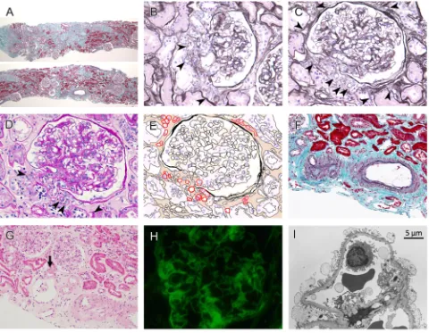
Light microscopic examination of the renal biopsy specimen revealed global sclerosis in 7 out of 19 glomeruli (37%). Tubulointerstitial fibrosis and atrophy occupied approximately 30–40% of the renal cortex (Fig. 1A). However, the residual glomeruli were intact and lacked structural changes typically seen in DKD: mesangial expansion, segmental mesangiolysis, nodular sclerosis, microaneurysm, and exudative lesions were absent (Fig. 1B, C and D). The most prominent feature of the pathology was polar vasculosis, which is the term for neovascularization (arteriolar proliferation) at the glomerular vascular pole (Fig. 1B, C and D). A schematic drawing of the glomeruli is shown in Fig. 1E. Arteriolar hyalinosis was mild (Fig. 1F), and a few arterioles had droplet-like deposition of hyaline (Fig. 1G). Immunofluorescence microscopy demonstrated linear IgG deposition along the glomerular basement
membrane (GBM) (Fig. 1H). Electron microscopy
showed GBM thickening with 480 nm in width, but no mesangial expansion (Fig. 1I). A GBM width of more than 430 nm is diagnostic of DKD (4). These findings were compatible with class I diabetic nephropathy according to the pathologic classification of diabetic nephropathy suggested by the Renal Pathology Society (4).
Treatment
Nutrition therapy and glycemic control were maintained, but adherence to insulin injection remained poor.
Outcome and follow-up
protein excretion remained stable between 0.3 and 0.9 g/ gCr, and hematuria was persistently negative.
Discussion
This report describes a case of a 66-year-old man with a 30-year history of T2DM with poor glycemic control, overt proteinuria, and polyvascular disease, whose renal biopsy did not reveal any glomerular changes typically seen in DKD but revealed remarkable proliferation of arterioles adjacent to the glomerular pole (polar vasculosis).
Historically, diabetic nephropathy was often diagnosed clinically without pathological evaluation, especially if
a patient with diabetes manifests persistent proteinuria, diabetic retinopathy, and no findings contradictory to diabetic nephropathy. Parving and his colleagues studied the renal pathology of 35 non-insulin-dependent diabetic patients with persistent albuminuria (>300 mg/24 h) and reported that all 15 patients with retinopathy had mesangial expansion or nodular sclerosis, which denote diabetic nephropathy (5). Considering the rarity of concomitant non-diabetic diseases that would affect treatment regimens, renal biopsy was often performed in a limited group of diabetic patients who presented with atypical features in the course of diabetes (6). The presented case followed a typical clinical course of a diabetic patient, but the renal
Figure 1
pathology unexpectedly showed scarce glomerular changes that are often seen in DKD.
Attempts have been made to study and categorize the pathology of DKD by performing renal biopsies extensively (7, 8, 9, 10). Referring to prior studies, Renal Pathology Society has proposed a pathologic classification for diabetic nephropathy, where GBM thickening, mesangial expansion, nodular sclerosis, and advanced glomerular sclerosis are categorized to classes I, II, III, and IV, respectively (4). The notion that the latter findings appear as the nephropathy progresses may not apply to all cases with DKD, considering that the presented case had urine protein excretion of 1.43 g/gCr, which could not be pathologically attributed to any kidney disease other than DKD. In contrast to the presented case, Klessens et al. showed that morphologic changes of the kidney were seen in diabetic patients even without albuminuria or other clinical manifestation of kidney disease (11). These findings suggest that the pathology of DKD does not follow a uniform pattern and is rather diverse, just as its clinical course is (12).
The mechanism of how the patent glomeruli did not undergo morphological changes is of particular interest, considering that poor glycemic control caused interstitial fibrosis and global glomerulosclerosis in 37% of the glomeruli. One hypothesis is that the polar vasculosis seen adjacent to the remaining glomeruli relieved glomerular hypertension by allowing the blood flow to bypass the glomeruli. Min and Yamanaka first studied neovascularization at the vascular pole region of glomeruli in detail with three-dimensional analysis of the
increased vasculature (13). They found increased small vessels around the vascular hilus in diabetic patients concomitant with varying severity of nodule, diffuse, and exudative lesions of the glomeruli. These extra vessels were not seen in control cases with hypertension or membrano-proliferative glomerulonephritis, thus were estimated to be caused by diabetes. They noted that the small vessels were seen adjacent to both the glomeruli with severe changes and those with slight changes. Three-dimensional analysis revealed that most of the small vessels originated from the afferent arterioles and drained into the peritubular capillaries. According to these findings, the authors assumed that the small vessels served as shunts and facilitated efferent blood flow from the glomeruli (13). A schematic picture of the shunt is shown in Fig. 3. To the contrary, Stout et al. observed that nearly all extra vessels were connected to the efferent arterioles and drained into the peritubular capillaries (14). Regardless of this opposite finding, Stout et al. also assumed that the extra vessels might help preserve glomerular function by decreasing the intraglomerular pressure, since the extra vessels contained muscle that is identical to the efferent arterioles and did not block the glomerular outflow by anatomically hindering other arterioles in the vascular pole (14). In patients with diabetes, hyperglycemia and overhydration lead to glomerular hyperfiltration of hyperosmotic fluid, which causes typical diabetic glomerulopathy via glomerular endothelial damage. If hyperosmotic blood bypasses the glomeruli by flowing directly from the afferent arteriole to the peritubular capillaries, glomerular damage may be hypothetically minimized.
Several studies have discussed the etiology of the development of the extra vessels. Østerby and
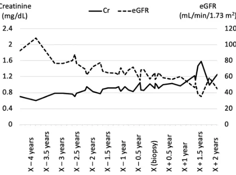
Figure 2
Serumcreatininelevelandestimatedglomerularfiltrationrate.Thesolid line indicates serum creatinine level, and the broken line refers to estimatedglomerularfiltrationrate(eGFR).Serumcreatininelevelshows agradualincrease,whileeGFRdeclinesaccordingly.
A
B
Figure 3
her colleagues observed a strong correlation between frequency of neovascularization, mesangial expansion, and the severity of diabetic glomerulopathy (low glomerular filtration rate) and concluded that the extra vessels appeared as a compensatory consequence of
the developing structural glomerular changes (15).
Kanesaki et al. supported this idea by showing a positive correlation between vascular endothelial growth factor mRNA expression, mesangial matrix expansion, and the frequency of extra small vessels (16). Stout et al. also observed a positive association between the number of glomeruli with extra vessels and the severity of diffuse and/or nodular lesions (14). However, they also noted that the largest number of extra vessels were found in older patients with a long-standing history of T2DM with only mild-to-moderate, presumably slowly progressing, diffuse and nodular lesions of the glomeruli. In contrast to this, several cases of T2DM with only midrange number of extra vessels showed moderate-to-severe diffuse and nodular lesions, focal mesangiolysis, and fibrin cap lesions, which all indicate more rapid progression of the DKD. They assumed that long-standing modest increases in glomerular inflow may have compensated through the formation of extra vessels, thus slowing the development of severe diffuse and/or nodular lesions (14). This may explain the etiology of the DKD in our presented case, where renal function was preserved at the time of renal biopsy and morphologic changes of the patent glomeruli were scarce, while remarkable polar vasculosis was present. It is of note that the blood pressure of the presented case was well controlled with an angiotensin receptor blocker and a calcium blocker.
In summary, we evaluated the renal histology of a 66-year-old man with a 30-year history of T2DM with poor glycemic control, overt proteinuria, and typical diabetic complications including retinopathy and neuropathy. Although patients with these clinical features have been known to show typical diabetic glomerulopathy including nodular lesions, the renal histology of the presented case showed GBM thickening, scarce glomerular changes, and prominent polar vasculosis. Polar vascular formation may preserve glomerular function by decreasing the intraglomerular pressure, which sheds light on further investigation into the etiology and treatment of DKD.
Declaration of interest
The authors declare that there is no conflict of interest that could be perceived as prejudicing the impartiality of the research reported.
Funding
Thisresearchdidnotreceiveanyspecificgrantfromanyfundingagencyin thepublic,commercialornot-for-profitsector.
Patient consent
Informed and voluntary consent for publication was obtained from the patient described in the article.
Author contribution statement
Y Oda acquired, analyzed, and interpreted data, and wrote the manuscript. M Yamanouchi, H Mizuno, R Hiramatsu, T Suwabe, J Hoshino and N Sawa followed the patient and were major contributors in revising the manuscript. K Ohashi and T Fujii performed the histopathology investigations. Y Ubara followed the patient, was a major contributor in revising the manuscript, and supervised the work.
References
1 Ekinci EI, Jerums G, Skene A, Crammer P, Power D, Cheong KY, Panagiotopoulos S, McNeil K, Baker ST, Fioretto P, et al. Renal structure in normoalbuminuric and albuminuric patients with type 2 diabetes and impaired renal function. Diabetes Care 2013 36 3620–3626. (https://doi.org/10.2337/dc12-2572)
2 Hong D, Zheng T, Jia-qing S, Jian W, Zhi-hong L & Lei-shi L. Nodular glomerular lesion: a later stage of diabetic nephropathy?
Diabetes Research and Clinical Practice 2007 78 189–195. (https://doi. org/10.1016/j.diabres.2007.03.024)
3 Matsuo S, Imai E, Horio M, Yasuda Y, Tomita K, Nitta K, Yamagata K, Tomino Y, Yokoyama H, Hishida A, et al. Revised equations for estimated GFR from serum creatinine in Japan. American Journal of Kidney Diseases 2009 53 982–992. (https://doi.org/10.1053/j. ajkd.2008.12.034)
4 Tervaert TW, Mooyaart AL, Amann K, Cohen AH, Cook HT, Drachenberg CB, Ferrario F, Fogo AB, Haas M, de Heer E, et al. Pathologic classification of diabetic nephropathy. Journal of the American Society of Nephrology 2010 21 556–563. (https://doi. org/10.1681/ASN.2010010010)
5 Parving HH, Gall MA, Skøtt P, Jørgensen HE, Løkkegaard H, Jørgensen F, Nielsen B & Larsen S. Prevalence and causes of albuminuria in non-insulin-dependent diabetic patients. Kidney International 1992 41 758–762. (https://doi.org/10.1038/ki.1992.118)
6 Olsen S & Mogensen CE. How often is NIDDM complicated with non-diabetic renal disease? An analysis of renal biopsies and the literature. Diabetologia 1996 39 1638–1645. (https://doi.org/10.1007/ s001250050628)
7 Mazzucco G, Bertani T, Fortunato M, Bernardi M, Leutner M, Boldorini R & Monga G. Different patterns of renal damage in type 2 diabetes mellitus: a multicentric study on 393 biopsies. American Journal of Kidney Diseases 2002 39 713–720. (https://doi.org/10.1053/ ajkd.2002.31988)
8 Gambara V, Mecca G, Remuzzi G & Bertani T. Heterogeneous nature of renal lesions in type II diabetes. Journal of the American Society of Nephrology 1993 3 1458–1466.
9 Fioretto P, Mauer M, Brocco E, Velussi M, Frigato F, Muollo B, Sambataro M, Abaterusso C, Baggio B, Crepaldi G, et al. Patterns of renal injury in NIDDM patients with microalbuminuria. Diabetologia
1996 39 1569–1576. (https://doi.org/10.1007/s001250050616)
diabetes mellitus: relationship with retinopathy. Nephrology Dialysis Transplantation 1998 13 2547–2552. (https://doi.org/10.1093/ ndt/13.10.2547)
11 Klessens CQ, Woutman TD, Veraar KA, Zandbergen M, Valk EJ, Rotmans JI, Wolterbeek R, Bruijn JA & Bajema IM. An autopsy study suggests that diabetic nephropathy is underdiagnosed.
Kidney International 2016 90 149–156. (https://doi.org/10.1016/j. kint.2016.01.023)
12 Retnakaran R, Cull CA, Thorne KI, Adler AI, Holman RR & UKPDS Study Group. Risk factors for renal dysfunction in type 2 diabetes: U.K. Prospective Diabetes Study 74. Diabetes 2006 55 1832–1839.
(https://doi.org/10.2337/db05-1620)
13 Min W & Yamanaka N. Three-dimensional analysis of increased vasculature around the glomerular vascular pole in diabetic nephropathy. Virchows Archiv: A, Pathological Anatomy and
Histopathology 1993 423 201–207. (https://doi.org/10.1007/ bf01614771)
14 Stout LC & Whorton EB. Pathogenesis of extra efferent vessel development in diabetic glomeruli. Human Pathology 2007 38 1167–1177. (https://doi.org/10.1016/j.humpath.2007.01.019)
15 Østerby R, Asplund J, Bangstad HJ, Nyberg G, Rudberg S, Viberti GC & Walker JD. Neovascularization at the vascular pole region in diabetic glomerulopathy. Nephrology Dialysis Transplantation 1999 14 348–352. (https://doi.org/10.1093/ ndt/14.2.348)
16 Kanesaki Y, Suzuki D, Uehara G, Toyoda M, Katoh T, Sakai H & Watanabe T. Vascular endothelial growth factor gene expression is correlated with glomerular neovascularization in human diabetic nephropathy. American Journal of Kidney Diseases 2005 45 288–294.
(https://doi.org/10.1053/j.ajkd.2004.09.020)