Tanaffos (2008) 7(4), 37-43
©2008 NRITLD, National Research Institute of Tuberculosis and Lung Disease, Iran
Relationship of Pleural and Serum Cholesterol and
Lipoprotein Levels in Exudative and
Transudative Effusions
Ebrahim Razi 1, Gholam Abbas Moosavi 2, Eemail Fakharian 3, Mohammad Abedi 1
1 Department of Internal Medicine, Shahid Beheshti Hospital, 2 Department of Biostatistics, 3 Department of Surgery, Kashan University of Medical Sciences, KASHAN-IRAN.
ABSTRACT
Background: This study aimed to assess whether total cholesterol (CHOL), low-density lipoprotein cholesterol (LDL), and high-density lipoprotein (HDL) are sensitive markers for discriminating between transudative and exudative pleural effusions (PE).
Materials and Methods: In this study CHOL, LDL, HDL, TG, protein and LDH were analyzed in PE and serums of 119
patients with pleural effusion out of which 49 had transudative and 70 had exudative pleural effusion. Sensitivity, specificity, and area under the curve (AUC) of CHOL, LDL and HDL were measured by receiver operating characteristic curve (ROC).
Results: Pleural fluid CHOL, LDL and HDL levels were significantly lower in the transudate group compared to the exudate (29.6 ±16.3 mg/dl versus 65.24 ± 25.9 mg/dl, p<0.001; 17 ±14.8 mg/dl versus 43.94 ± 21.6 mg/dl, p<0.001; and 9.2 ± 4.8 mg/dl versus14.9 ± 6.3 mg/dl, p<0.001, respectively). Sixty-seven percent of cases with pleural transudates were secondary to heart failure, while 41% and 39% of those with pleural exudates were of parapneumonic effusion and neoplastic origin, respectively. Pleural fluid CHOL, LDL and HDL levels were significantly higher in malignant pleural effusion (71.5±18.6, 48.1±17.4 and 16.1±6.6mg/dl, respectively), and in parapneumonic effusion (70.7±28.5, 49.4±22.4 and 14.7±6.4 mg/dl, respectively) than in heart failure (30.6±11.9, 17.5±10.4 and 9.6 ±5.4 mg/dl, respectively). The optimum cut-off value for pleural fluid CHOL level of ≥38 mg/dL had a sensitivity of 87% and 80% specificity, for LDL pleural fluid level a cut-off value of ≥ 22.5 mg/dl had a sensitivity of 87% and 78% specificity, and for HDL pleural fluid a cut-off value of ≥ 10.5 mg/dl had a sensitivity of 70% and 69% specificity. AUC values were 0.906, 0.883, and 0.783, for CHOL, LDL, and HDL of pleural fluid, respectively.
Conclusion: We conclude that pleural fluid CHOL, LDL and HDL are significantly increased in exudative effusions compared to the transudative ones. Measurement of CHOL, LDL and HDL concentrations in pleural effusions is useful in distinguishing exudates from transudates. (Tanaffos 2008; 7(4): 37-43)
Key words: Cholesterol, Exudate, Transudate, Lipoproteins, Pleural effusion
Correspondence to: Razi E
Address: Department of Internal Medicine, Kashan University of Medical Sciences, Kashan-Iran
Email address:ebrahimrazi@yahoo.com
INTRODUCTION
Pleural effusions, classified into transudates and exudates, develop in different thoracic or systemic diseases (1, 2).
When encountering this finding, it is necessary to determine its etiology in order to treat the disease. The distinction between exudative and transudative pleural effusions is the basis for defining the etiology. In aclassic article by Light et al, presence of one or more of the following test results was defined as diagnostic clues for pleural exudates: pleural fluid to serum proteinratio > 0.5, pleural fluid LDH > 200 IU, and a pleuralfluid to serum LDH ratio > 0.6 (3).
This has been reported as the best method for discriminating exudates from transudates (4), although others (5)have modified the cut-off points used by Light et al (3). A considerable number of other biochemical markers have beencompared with Light’s criteria. These include measuring pleural fluid cholesterol, albumin gradient, and serum/pleuralfluid bilirubin ratio (6-10).
For better assessment of the role of PE cholesterol level in discrimination of transudates from exudates, a deeper understanding of the relationship between the CHOL level in serum and PE would be useful.
Cholesterol and other lipids are carried in body fluids by lipoproteins. The plasma lipoproteins are classified into five major groups based on their relative densities: chylomicrons, very low-density lipoprotein (VLDL), intermediate-density lipoprotein (IDL), low-density lipoprotein (LDL), and high-density lipoprotein (HDL). LDL is the major cholesterol-carrying lipoprotein in the plasma which consist nearly 70% of total plasma cholesterol. It is composed of approximately 75% lipid and 25% protein. HDL contains about 50% lipid and 50% protein (11).
There are a few studies focusing on the relationship between serum and pleural fluid lipoproteins. Pfalzer et al. reported that in
transudative pleural effusion the level of LDL represented 16% of serum levels, whereas inflammatory and malignant pleural effusions exhibited high levels of LDL, with 67% and 69% of serum levels, respectively (12). In another study Vaz et al. reported that cholesterol levels in pleural effusion were related to serum cholesterol levels and the permeability of the pleura (13).
The purpose of this study was to determine the sensitivity and specificity of CHOL, LDL and HDL measurements in distinguishing exudative from transudative pleural effusions.
MATERIALS AND METHODS
Statistical analysis:
Results were presented as the mean ±SD. Differences in quantitative variables between groups were assessed by using an unpaired t test.
Receiver operating characteristic (ROC) curves and areas under the ROC curves (AUC) with 95% confidence intervals were calculated for each criterion in order to evaluate the optimum cut-off points. In addition to using the cut-off points derived from ROC curves, the use of each criterion for identifying exudates was evaluated by calculating its sensitivity and specificity in distinguishing the pleural exudates from transudates. The optimum cut-off level was determined by selecting points of test values that provided the greatest sum of sensitivity and specificity. A p-value <0.05 was considered significant.
RESULTS
One hundred-nineteen cases including 52 (43.7%) males and 67 (56.3%) females were enrolled in this study. The mean age was 63.5±15 years (range 18-89 years). Pleural fluid was exudative in 70 cases (58.8%), and transudative in 49 (41.2%). Causes for exudative pleural effusion were thoracic malignancy in 27, parapneumonic effusion in 30, pulmonary embolism in 4, tuberculosis in 2, and cardiac failure in 7 patients. The causes for transudative pleural effusion were cardiac failure in 33, chronic liver disease in 7, and renal failure in 9 cases. Patients with unknown or more than one diagnosis were excluded from the study. The average levels of biochemical parameters in pleural fluid were higher among the exudative group than the transudative patients. The means and standard errors of cholesterol, LDL, and HDL in pleural effusion were evaluated in transudative and exudative effusions (Table 1). For each test, the differences in mean values between the transudate and the exudate groups were statistically significant (t-test, p<0.001).
Table 1 Pleural fluid biochemical parameters in patients with exudative and transudative pleural effusion*.
Pleural effusion Biochemical Parameters
Exudative (70 cases)
Transudative
(49 cases) P value
PE Cholesterol, mg/dl 65.24 ± 25.9 29.6 ±16.3 <0.001 Serum cholesterol, mg/dl 151.5 ± 45.6 143.5 ± 48 0.363 PE LDL, mg/dl 43.94 ± 21.6 17 ±14.8 <0.001 Serum LDL, mg/dl 89.64± 33.6 90.9± 35.6 0.845 PE HDL, mg/dl 14.9 ± 6.3 9.2 ± 4.8 <0.001 Serum HDL, mg/dl 35.77± 11.8 32.8± 10 0.146 PE VLDL, mg/dl 6.1 ± 3.6 3.7 ± 2.4 0.905 Serum VLDL, mg/dl 26.5± 17.3 23.4± 17.8 0.184 PE Triglyceride, mg/dl 32.6 ± 20.6 21.8 ± 21.4 0.158 Serum Triglyceride, mg/dl 130.2± 73.2 126.5± 117.5 0.241
*Data are presented as Mean ± SD. PE: pleural effusion
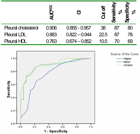
The cut-off value, sensitivity and specificity of pleural cholesterol, pleural LDL, and pleural HDL in discriminating exudative from transudative pleural effusions were obtained by using ROC curve (Fig. 1 and Table 2).
Table 2. The value of pleural cholesterol, LDL, and HDL in the differential diagnosis of exudative and transudative pleural effusion.
AU
C
RO
C
CI
Cut of
f
Sensitivity
%
Specificit
y
%
Pleural cholesterol 0.906 0.855 - 0.957 38 87 80
Pleural LDL 0.883 0.822 – 0.944 22.5 87 78
Pleural HDL 0.763 0.674 – 0.852 10.5 70 69
The area under the ROC curve for pleural fluid cholesterol was 0.906 (95% CI = 0.855–0.957). The optimal cut-off value for pleural fluid cholesterol was determined to be greater than 38 mg/dl, with a sensitivity of 87.1%, and a specificity of 79.6%. The area under the ROC curve for pleural fluid LDL was 0.883 (95% CI = 0.822–0.944); at a cut-off limit of greater than 22.5 mg/dl, the sensitivity was 87.1%, and the specificity was 77.6%. The area under the ROC curve for pleural fluid HDL was 0.763 (95% CI = 0.674–0.852); at a cut-off limit of greater than 10.5 mg/dl, the sensitivity was 70 %, and the specificity was 69.4%.
In comparison of both pleural cholesterol and pleural LDL, pleural HDL had lower sensitivity
(70%) and lower specificity (69%).
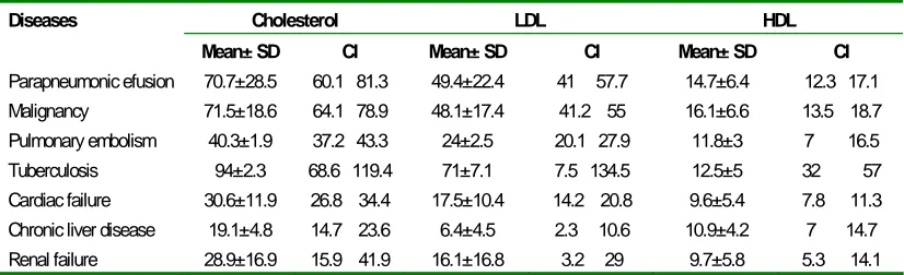
Table 3 demonstrates the mean± SD values and confidence interval of CHOL, LDL, and HDL lipoproteins according to various etiologies of exudative and transudative pleural effusions. As shown in Table 3, levels of CHOL, LDL, and HDL lipoproteins were significantly higher in parapneumonic and malignant pleural effusions than cardiac failure or other causes of transudative pleural effusion.
The areas under the ROC curve for discriminating exudative from transudative pleural effusions for cholesterol, LDL and HDL of pleural effusion were 0.909, 0.883, and 0.763; respectively.
Table 3. Lipoproteins levels by diagnostic groups.
Cholesterol LDL HDL
Diseases
Mean± SD CI Mean± SD CI Mean± SD CI
Parapneumonic efusion 70.7±28.5 60.1 81.3 49.4±22.4 41 57.7 14.7±6.4 12.3 17.1
Malignancy 71.5±18.6 64.1 78.9 48.1±17.4 41.2 55 16.1±6.6 13.5 18.7
Pulmonary embolism 40.3±1.9 37.2 43.3 24±2.5 20.1 27.9 11.8±3 7 16.5
Tuberculosis 94±2.3 68.6 119.4 71±7.1 7.5 134.5 12.5±5 32 57
Cardiac failure 30.6±11.9 26.8 34.4 17.5±10.4 14.2 20.8 9.6±5.4 7.8 11.3
Chronic liver disease 19.1±4.8 14.7 23.6 6.4±4.5 2.3 10.6 10.9±4.2 7 14.7
Renal failure 28.9±16.9 15.9 41.9 16.1±16.8 3.2 29 9.7±5.8 5.3 14.1
Values are expressed as mean±SD (95% confidence interval).
DISCUSSION
This study showed that measurement of CHOL, LDL and HDL concentrations in pleural effusions was useful in distinguishing exudates from transudates. There are elevated levels of pleural fluid cholesterol, LDL, and HDL lipoproteins in patients with parapneumonic and malignant pleural effusions compared to those with heart failure.
The known lipoproteins are chylomicron, VLDL, LDL, HDL and IDL.
Approximately 90% of blood cholesterol is
associated with LDL and HDL. LDL is synthesized in the vessels while HDL is synthesized in both liver and vessels. These lipids have an important role in cellular metabolism.
in the LDL (52% of LDL weight is due to cholesterol) and HDL (19% of HDL weight is due to cholesterol).It is believed that PE cholesterol is also related to LDL and HDL (13).
Lipoproteins are bound to some specific cell membrane receptors. Liver takes up some lipoproteins, then they are released into intestines via gallbladder. It is expected that pleural fluid cholesterol be composed of LDL and HDL. Pleural cholesterol is thought to be derived from degenerating cells and vascular leakage from increased permeability. Measurement of pleural cholesterol has been used to improve the accuracy of differentiating transudative and exudative effusions.
For distinguishing between pleural transudates and exudates the most analyzed parameters have been the PE cholesterol level and the PE to serum cholesterol ratio. A pleural cholesterol level of greater than 45 mg/dL is not by itself a definitive criterion for an exudate, but does figure in the two and three-test rules as noted above (10). In other studies, different cut-off values were reported (5, 10, 14). Hamm et al. reported that cholesterol level was superior to Light’s criteria in distinguishing transudates from exudates (7). The application of a cut-off of 60 mg/dL yielded a sensitivity and specificity of 90 and 100%, respectively. Hamm et al. concluded that measurement of pleural fluid cholesterol level is a simple and cost-effective aid in differentiating exudative from transudative pleural effusions (7).
Several recent studies evaluated the diagnostic utility of measuring lipoproteins in pleural fluid.
Vaz et al. demonstrated that cholesterol levels in PE were related to serum cholesterol levels and to the permeability of the pleura (13). They found that the percentage of cholesterol associated with LDL and HDL in the PE was much lower than that associated with LDL and HDL in serum, suggesting that lipoproteins are modified once they enter the pleural
space (13).
Pfalzer et al. reported that LDL levels in effusions correlated with serum levels in exudates but did not correlate with those in transudates (12). In his study, transudates had a low cholesterol content of 35 ± 12 mg/dl (mean ± SD) because of low levels of low-density lipoprotein (LDL) cholesterol--representing 16% of serum levels--whereas inflammatory exudates (cholesterol 92 ± 26 mg/dl) and malignant exudates (cholesterol 86 ± 6 mg/dl) exhibited high levels of LDL, with 67% and 69% of serum levels, respectively. Pfalzer et al. concluded that:
1) LDL levels in effusions correlated with serum levels in exudates but did not correlate with those in transudates.
2) Alterations of lipoproteins occur in chronic inflammation and in malignancy with possible de novo synthesis of apolipoprotein E by tumor cells. 3) LDL levels in effusions correlated with serum levels in exudates but did not correlate with those in transudates.
4) A strongly abnormal HDL level with accumulation of cholesterol was found in a long-standing tuberculous effusion (12). Regarding the origin of cholesterol in pleural effusion, Hamm et al. reported that, in inflammatory effusions of recent onset most cholesterol was bound to LDL with corresponding apoprotein B levels (15). The chronic tuberculous exudate showed a shift of cholesterol binding towards HDL. In the chyliform effusion most cholesterol was found in the HDL region (15).
In the present study, we found that the mean LDL level was significantly lower in transudates than exudates. The mean level of HDL was also lower in transudates. This can be explained by increased pleural permeability in exudates, which allows transporting of large molecules like LDL.
It means that pleural permeability for proteins is higher than lipids, which might be due to the larger size of lipids versus proteins (16). Some studies indicated that lipid metabolization is the reason for its reduced levels in pleural fluid (12, 13, 17). Other studies related to the investigation of these mechanisms are reviewed. According to a study by Raymond et al. on alterations of LDL, inflamed pleural space was a condition where leukocytes and protease were strongly activated and LDL level was elevated (18). Rerabek et al. reported that the cholesterol content in pleural exudates simply reflected serum levels (19). In a study conducted by Vaz et al. other factors such as permeability of the pleura may also be involved in the pleural fluid lipid levels (13).
In the current study, sensitivity, specificity and AUCRoc of cholesterol and LDL for discrimination of exudative and transudative pleural effusions were 87.1% versus 87.1%, 79.6% versus 77.6% and 0.906 versus 0.883, respectively, which are higher than those of HDL: 70%, 69.4% and 0.763, respectively. These findings indicated that cholesterol and LDL lipoproteins could substitute HDL for distinction of exudative and transudate pleural effusions. Measurement of HDL and LDL in PE and calculating HDL/LDL ratio can be suggested to aid differentiation between pleural exudates and transudates with the advantage of not requiring serum levels (20). Koukturk et al. concluded that the mean HDL level was 21.37±11.87 in exudates and17.79±7.64 in transudates (p=0.17). The mean LDL level was 58.69±29.8 in exudates and 12.32±13.08 in transudates (p=0.001). In our study, the mean HDL level was 17±14.8 in exudates and 9.2±4.8 in transudates (p<0.001). The mean LDL level was 43.94 ± 21.6 in exudates and 14.9 ±6.3 in transudates (p<0.001). Koukturk et al. concluded that the value of pleural HDL/LDL ratio that best
differentiated between transudates and exudates was 0.6, with the sensitivity of 89%, and specificity of 79% (20).
Transudative effusions from patients with heart insufficiency and nephrotic syndrome had significantly lower levels of cholesterol, LDL, and HDL. As shown in Table 3, the mean values of cholesterol, LDL, and HDL in cardiac failure were 28.9, 16.1 and 9.7 mg/dl, respectively; but in renal failure these rates were 30.6, 17.5, and 9.6 mg/dl, respectively. In our study the highest level of LDL was seen in parapneumonic effusion (49.4±22.4), and level of HDL in malignant pleural effusion was more than that in other causes of pleural effusion (Table 3). Similar to our study, Jenss et al. reported that higher than average values were found for HDL-cholesterol in patients with malignant diseases and for LDL-cholesterol in patients with pneumonia (21).
Our analyses yielded two important observations. First, transudative fluids were apparently characterized by low levels of cholesterol and its lipoproteins. Second, CHF-related pleural effusions could be differentiated from parapneumonic and malignant pleural effusions by the low levels of cholesterol, LDL, and HDL.
In conclusion, diagnostic usefulness of cholesterol and LDL in discrimination of exudative and transudative pleural effusions was higher than HDL. The value of cholesterol for this differentiation was: 38 mg/dl ( 87% sensitivity and 80% specificity), and for LDL was 22.5 mg/dl (87% sensitivity, and 78% specificity).
REFERENCES
1. Black LF. The pleural space and pleural fluid. Mayo Clin
Proc 1972; 47 (7): 493- 506.
2. Sahn SA. State of the art. The pleura. Am Rev Respir Dis
3. Light RW, Macgregor MI, Luchsinger PC, Ball WC Jr.
Pleural effusions: the diagnostic separation of transudates
and exudates. Ann Intern Med 1972; 77 (4): 507- 13.
4. Vives M, Porcel JM, Vicente de Vera M, Ribelles E, Rubio
M. A study of Light's criteria and possible modifications for
distinguishing exudative from transudative pleural
effusions. Chest 1996; 109 (6): 1503- 7.
5. Romero S, Candela A, Martín C, Hernández L, Trigo C, Gil
J. Evaluation of different criteria for the separation of pleural transudates from exudates. Chest 1993; 104 (2): 399- 404.
6. Gil Suay V, Martínez Moragón E, Cases Viedma E, Perpiñá
Tordera M, León Fábregas M, Sanchis Aldás J. Pleural cholesterol in differentiating transudates and exudates. A prospective study of 232 cases. Respiration 1995; 62 (2): 57- 63.
7. Hamm H, Brohan U, Bohmer R, Missmahl HP. Cholesterol
in pleural effusions. A diagnostic aid. Chest 1987; 92 (2): 296- 302.
8. Ortega L, Heredia JL, Armengol R, Mir I, Romanillas T,
Armengol J. The differential diagnosis between pleural exudates and transudates: the value of cholesterol. Med Clin (Barc) 1991; 96 (10): 367- 70.
9. Roth BJ, O'Meara TF, Cragun WH. The serum-effusion
albumin gradient in the evaluation of pleural effusions. Chest 1990; 98 (3): 546- 9.
10. Heffner JE, Brown LK, Barbieri CA. Diagnostic value of
tests that discriminate between exudative and transudative pleural effusions. Primary Study Investigators. Chest 1997; 111 (4): 970- 80.
11. Mahley RW, Weisgraber KW, Farese RV Jr. Disorders of
lipid metabolism. In: Williams textbook of endocrinology,
10th edition (Larsen PR, Kronenberg HM, Melmed S,
Polonsky KS, eds) WB Saunders Philadelphia, 2003; 1642-1705.
12. Pfalzer B, Hamm H, Beisiegel U, Ostendorf P. Lipoproteins
and apolipoproteins in human pleural effusions. J Lab Clin Med 1992; 120 (3): 483- 93.
13. Vaz MA, Teixeira LR, Vargas FS, Carmo AO, Antonangelo
L, Onishi R, et al. Relationship between pleural fluid and serum cholesterol levels. Chest 2001; 119 (1): 204- 10.
14. Garcia-Pachon E, Padilla-Navas I, Sanchez JF, Jimenez B,
Custardoy J. Pleural fluid to serum cholinesterase ratio for the separation of transudates and exudates. Chest 1996;
110 (1): 97-101.
15. Hamm H, Pfalzer B, Fabel H. Lipoprotein analysis in a
chyliform pleural effusion: implications for pathogenesis and diagnosis. Respiration 1991; 58 (5-6): 294- 300.
16. Silverman LM, Christenson RH. Amino Acids and Proteins.
In: Burtis CA, Ashwood ER (eds): Tietz Fundamentals of
Clinical Chemistry, 4th ed, WB Saunders, Philadelphia,
1996; 240-82.
17. Wiener-Kronish JP, Albertine KH, Licko V, Staub NC.
Protein egress and entry rates in pleural fluid and plasma in sheep. J Appl Physiol 1984; 56 (2): 459- 63.
18. Raymond TL, Reynolds SA. Lipoproteins of the
extravascular space: alterations in low density lipoproteins of interstitial inflammatory fluid. J Lipid Res 1983; 24 (2): 113-9.
19. 1: Rerabek JE.Low density lipoproteins and
immunoglobulins in human pleural effusions. Clin Chim Acta 1977; 76 (3): 363- 9.
20. Köktürk O, Ulukavak Ciftci T, Firat H, Firat S. HDL/LDL
ratio: a useful parameter for separation of pleural
transudates from exudates.
Tuberk Toraks 2005; 53(1):
34-9.
21. Jenss U, Töwe D, Diwok K. Determination of individual