I J H S
Original Article
I nternational Journal of Health Studies
Differential Expression of CD16 and CD56 on Natural Killer (NK) Cell Subsets in
Multiple Sclerosis and Neuromyelitis Optica
Mehdi Khaksari1, Sayyed Hamid Zarkesh-Esfahani2, Masoud Etemadifar3, Mohsen Masjedi4, Roqayeh Aliyari5, Majid Rahmati6*
1 Dept.of Physiology, School of Medicine, Shahroud University of Medical Sciences, Shahroud, Iran. 2 Dept. of Immunology, School of Medicine, Isfahan University of Medical Sciences, Isfahan, Iran. 3 Dept. of Neurology, School of Medicine, Isfahan University of Medical Sciences, Isfahan, Iran. 4 Dept. of Immunology, School of Medicine, Isfahan University of Medical Sciences, Isfahan, Iran.
5 Dept. of Biostatistics and Epidemiology, School of Public Health, Shahroud University of Medical Sciences, Shahroud, Iran. 6 Dept. of Medical Biotechnology, School of Medicine, Shahroud University of Medical Sciences, Shahroud, Iran.
Received: 8 December 2014 Accepted: 11 March 2015
Abstract
Background: Multiple sclerosis (MS) and Neuromyelitis optica (NMO) are inflammatory and demyelinating diseases of the central nervous system (CNS). NK cells are supposed to play an important role in the pathophysiology of MS, but their role in the NMO remains unknown. The aim of this study was to compare the prevalence of different subpopulations of NK cells in the patients with MS and NMO and healthy individuals.
Methods: Treatment Naive MS and NMO patients, age, and sex matched controls were included in this study. PBMCs were isolated from peripheral blood and different phenotypes of circulating NK cells were compared with the flow cytometry analysis.
Results: There were no significant differences in the mean percentages of circulating NK cells expressing the CD56 bright molecule in patients with MS and NMO. However, the mean percentages of circulating NK cells expressing the CD56bright molecule were significantly lower in all patients groups, compared to controls. In addition, the mean percentages of circulating NK cells expressing the CD16dim molecule was significantly higher in the patients with MS, compared to controls/any other groups. The mean percentages of circulating NK cells expressing the CD56dim molecule were significantly higher in the patients with MS than the controls. There were significant differences in the mean percentages of circulating NK cells expressing the CD16bright molecule between the patients with MS, and NMO/controls.
Conclusion: The results indicate that evaluation of NK cell subsets has an implication for the biomarker discovery and therapeutic targets in given diseases.
Keywords: Multiple sclerosis, Neuromyelitis optica, Devic disease, Natural killer (NK) cell.
*Corresponding to: M Rahmati, Email: rahmatima@hotmail.com Please cite this paper as: Khaksari M, Zarkesh-Esfahani SH, Etemadifar M, Masjedi M, Aliyari R, Rahmati M. Differential expression of CD16 and CD56 on natural killer (NK) cell subsets in multiple sclerosis and neuromyelitis optica. Int J Health Stud 2015;1(1):1-7.
I
ntroduction:
Multiple sclerosis (MS) is an inflammatory disease that affects the central nervous system. Two major forms of MS have been reported: Relapsing-remitting (RR)-MS, which is the most frequent (85-90%), and primary progressive (PP)-MS. Indeed, most patients with MS enjoy clinically stable periods of variable duration (remission), although interrupted unpredictably by occurrence of relapse. Furthermore, most RR-MS patients later develop secondary progressive (SP)-MS. About 10-15% of patients present with insidious disease onset and steady progression and therefore termed (PP)-MS. It is not clear what
factors are responsible for the different courses. Regarding the pathogenesis of MS, it has been shown that auto reactive T cells targeting myelin components play a crucial role in mediating the inflammatory process, particularly in the early stages of RR-MS.
The etiology of MS remains unclear, though studies have demonstrated that there is a strong genetic component determining susceptibility to disease, the low concordance rates of monozygotic twins indicates a contribution of non-genetic factors to MS etiology. Hence, even in a genetically susceptible individual, additional environmental factors may be necessary for the pathogenic reaction against self-tissue- but the non-genetic trigger is still unknown. Studies using animal models suggest that the innate response is involved in determining whether or not an autoimmune reaction will occur.1
In 1894, Devic and Gault described the sine qua non clinical characteristics of NMO: Optic neuritis and acute transverse myelitis. The patients described by Devic and Gault had monophasic or relapsing courses of NMO.2 Various antecedents or coexisting infections, vaccinations, and systemic autoimmune diseases have been linked to NMO but cause of the disease is unknown.3-5 The definition of NMO developed from the recognition that attacks of optic neuritis are more commonly unilateral than bilateral and that attacks of optic neuritis and myelitis usually occur sequentially rather than simultaneously.6
The interval separating disease defining attacks of optic neuritis and myelitis can be years or decades.2,6 Ocular pain with loss of vision, and myelitis with severe symmetric paraplegia, sensory loss below the lesion, and bladder dysfunction are typical features of neuromyelitis optica. Cervical myelitis can extend into the brain stem, resulting in nausea, hiccoughs, or acute neurogenic respiratory failure,6-8 which is exceedingly rare in MS. Other symptoms typical of spinal cord demyelination that are seen in both NMO and MS include paroxysmal tonic spasms (recurrent, stereotypic painful spasms of the limbs and trunk that last 20-45 seconds) and Lhermitte’s symptom (spinal or limb dysesthesias caused by neck flexion).6,7
minority (approximately 10%) of cells is CD56bright and CD16dim/neg. CD56 is an isoform of the human neural-cell adhesion molecule with unknown function on the human NK cells, though it might mediate interactions between NK cells and other cells. While the functional significance of the CD56 molecule is unknown on NK cells, its bright or dim expression correlates with the expression of several other surface markers that confer unique functional properties to the CD56bright and CD56dim subsets. These NK cell subsets show important differences in their cytotoxic potential, capacity for cytokine production and responses to cytokine activation.11,12 The CD56bright NK cells are the primary population of NK cells that produce immunoregulatory cytokines, including interferon (IFN)-, tumor necrosis factor (TNF)-α, TNF-β, granulocyte monocyte-colony stimulating factor (GM-CSF), interleukin (IL)-10 and IL-13 following monokine stimulation.11,13
CD56dimCD16+ NK cells constitute about 90% of total blood NK cells, efficiently kill target cells and secrete only low levels of IFN-γ. In contrast, CD56brightCD16Neg NK cells constitute 5-10% of total blood NK cells but are enriched in secondary lymphoid tissues14 and sites of autoimmune inflammation.15
In contrast to the CD56dim NK cell subset, CD56bright cells produce a large amount of cytokines upon stimulation, but acquire cytolytic activity only after prolonged activation.16 Therefore, NK cells could mediate tissue damage and regulate autoimmune T-cell responses through cytokine secretion and cytotoxicity in secondary lymphoid organs and in the inflamed CNS.
Because of lack of mouse strains that are selectively deficient in NK cells, the study of NK cell function in vivo has been challenging in the past.17 MS animal models provide evidence for both disease-accelerating and disease-protective effects of NK cells.18-20
It has been suggested that NK cells could be pathogenic by shaping Th1-polarized adaptive immune responses, activating
CNS-infiltrating DCs and/or via direct recognition and lysis of glial and neuronal cells.20,21 However, most studies in the experimental autoimmune encephalomyelitis (EAE) model reported that NK cells protect us from autoimmune mediated tissue damage, presumably by editing initiator and effector cell populations.18, 22-24 Such apparently controversial findings might be explained, at least in part, by differences in timing of myelin immunization and NK cell activation/depletion in these animal models. Furthermore, NK cells could also assist T-cell polarization and effector function during the initiation of autoimmune responses against neuroantigens but might acquire a more suppressive function during progression of the established disease. Alternatively, distinct subsets of NK cells could mediate divergent effects on EAE initiation and progression. The observation by de Jager25 and others26 in humans, reduced frequencies of NK cells in MS patients together with NK cell expansion during effective immunotherapy,
27-29
suggests that these innate lymphocytes exert beneficial functions. However, the mechanisms that could mediate such immunoregulatory NK cell functions in multiple sclerosis are poorly understood.
The aim of this study is to compare the mean percentages of peripheral blood NK cell subsets expressing the CD16 and CD56 molecules in patients with MS, Devic disease (NMO), and healthy control subjects.
Materials and Methods
Twenty eight patients with RRMS, according to the McDonald
30
criteria and EDSS(expanded disability status scale) 31 less than five, took part in the study. Moreover based on Wingerchuk 32 criteria twelve seropositive NMO-IgG Devic patients and fifteen seronegative NMO-IgG Devic patients, according to the same criteria were classified into the two separated groups. Untreated patients, less than fifty years old and less than ten years disease history were included in the study. Also thirty healthy donors, whose age, sex and environmental conditions were matched with the patients, were included in the study as the control group. None of the controls was on any immunomodulatory or immunosuppressive therapy and none of them had a history of neurological symptoms that would indicate an acute or chronic neurological disease. This group did not have any acute or chronic diseases.
This study was approved by the ethical committee of Isfahan University of Medical Sciences. Informed consent forms were obtained from all patients who contributed in this study. PBMCs were freshly isolated by density gradient centrifugation using Ficoll and stored in RPMI 1640 containing 50% fetal bovine serum (FBS) and 10% dimethyl sulfoxide (DMSO) at –80°C for later use. After controlling that freezing and thawing does not alter the viability of the cells of interest (the viability was always greater than 80%, and determined by trypan blue), the full series of samples from each patient was recovered from the freezer and immunofluorescence (IF) stained.
The following antibodies were used: RPE/Cy5-conjugated mouse antihuman CD3 monoclonal antibody (mAb), FITC-conjugated mouse antihuman CD16 mAb, RPE-FITC-conjugated mouse antihuman CD56 mAb and PE/FITC/Cy5 conjugated mouse IgG1 Negative Controls, all from Serotec UK.
NK cells were examined in a FACSCalibur flow cytometer with cell Quest software (Becton Dickinson) using three- color staining. Cells in Phosphate buffered saline (PBS) were incubated for 20 min, at 4C in the dark with specific or control antibodies. After staining, the cells were washed (1500 rpm, 10 min) and resuspended in PBS. Flow cytometer data were presented as percentage of positive cells calculated by gating against samples labeled with control isotype –matched monoclonal antibody.
In this study, flow cytometry techniques were performed on a BD FACSCalibur flow cytometer (Becton Dickinson, CA, USA) fitted with an air-cooled 488 nm argon laser. This flow cytometer allowed the detection of up to three different fluorescent parameters. The NK cells expressing CD16 and CD56 molecules lower than 103 molecules on the surface of each cell were considered as Dim cells and the expression of these molecules on the surface of each cell more than 103 has been considered as bright cells (Figure 5).
a phycobiliprotein-based fluorochrome, derived from algae, which can be conjugated to antibodies for use in immunophenotyping, and PE-Cy5 (PC-5). PC-5 is a tandem dye consisting of an indotricarbocyanine dye coupled to PE.
Results
The expression of CD3, CD16 and CD56 were studied on the peripheral blood mononuclear cells using three color-flow cytometry methods. Antibodies consisted of CD3, anti-CD16, anti-CD56 and isotype controls. Patients with MS (n=28), NMO-IgG seropositive Devic (n=12), NMO-IgG seronegative Devic (n=15) and controls (n=30) were included in this study. The age and ratio of male/female in the patients with MS and Devic disease were different. The results showed that there were no significant differences in the age means among the groups (P=0.112, Tables 1&2).
One-way ANOVA test showed that the mean percentages of peripheral blood NK cells expressing the CD56bright molecule were significantly different among the groups (P<0.0001, Table 3 & Figure 1). Analysis with the Scheffe test showed that the mean percentages of peripheral blood NK cells expressing the CD56bright molecule was higher in the controls than the other groups (P<0.0001). However, there were no significant differences between the patients with MS, NMO+ and NMO- (P>0.05).
One-way ANOVA test showed that the mean percentages of peripheral blood NK cells expressing the CD16dim molecule were significantly different between the groups (P<0.0001, Table 4). In this regard, analysis with the Scheffe test showed that the mean percentages of peripheral blood NK cells expressing the CD16dim molecule was higher in the patients with MS than the controls (P<0.0001). However, there were no significant differences between the other groups.
One-way ANOVA test showed that the mean percentages of peripheral blood NK cells expressing the CD56dim moleculewere significantly different in the groups (P=0.001, Table 5 & Figure 2). Analysis with the Scheffe test showed that the mean percentages of peripheral blood NK cells expressing the CD56dim molecule was significantly higher in the patients with MS than the controls (P=0.001) and there were no significant differences in the other groups.
Table 2. Gender distribution in all of the cases
Group
MS NMO + NMO - HC Total
Female 18(30) 9(15) 11(18.4) 22(36.7) 60
Male 10(40) 3(12) 4(16) 8(32) 25
Total 28(32.9) 12(14.1) 15(17.6) 30(35.3) 85
*The values are reported as Number (Percentage), MS: Multiple Sclerosis, NMO+: NMO-IgG seropositive Devic patients, NMO-: NMO-IgG seronegative Devic patients, HC: Healthy Controls, Chi-Square=0.812 and P.V=0.8.
Table 3. Comparison of the mean percentages of peripheral blood NK cells expressing the CD56bright molecule in the groups
Group N Mean% SD Min Max P.V*
MS 28 0.69 0.29 0.13 1.38
<0.0001
NMO+ 12 0.52 0.27 0.12 1.08
NMO- 15 0.70 0.52 0.11 1.76
HC 30 1.35 0.40 0.37 2.24
*P-value calculated as one-way ANOVA test, SD: Standard Deviation, MS: Multiple Sclerosis, NMO+: NMO-IgG seropositive Devic patients, NMO-: NMO-IgG seronegative Devic patients, HC: Healthy Controls
Table 4. Comparison of the mean percentages of peripheral blood NK cells expressing the CD16dim molecule in the groups
Group N Mean% SD Min Max P.V*
MS 28 25.30 15.52 2.20 61.75
<0.0001 NMO
+ 12 17.00 19.17 2.06 63.00
NMO - 15 15.90 9.43 1.11 30.58
HC 30 9.79 5.10 2.74 20.71
*P-value calculated as one-way ANOVA test, SD: Standard Deviation, MS: Multiple Sclerosis, NMO+: NMO-IgG seropositive Devic patients, NMO-: NMO-IgG seronegative Devic patients, HC: Healthy Controls
Figure 1. Comparison of the mean percentages of peripheral blood NK cells expressing the CD56bright molecule in the groups ± SD
Figure 2. Comparison of the mean percentages of peripheral blood NK cells expressing the CD16dim molecule in the groups ± SD
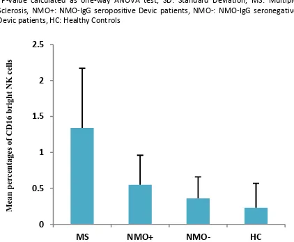
One-way ANOVA test showed that the mean percentages of peripheral blood NK cells expressing the CD16bright molecule were significantly different between the groups (P<0.0001, Table 6 & Figure 3). Analysis with the Scheffe test also showed that there were significant differences in the mean percentages of peripheral blood NK cells expressing the CD16dim molecule in patients with MS, compared to the Table 1. Comparison of the age means among different groups
Group N Mean% SD Min Max P.V*
MS 28 34.25 8.48 20.00 49.00
0.112
NMO + 12 28.91 8.09 14.00 43.00
NMO - 15 32.53 8.57 21.00 48.00
HC 30 29.53 7.99 15.00 47.00
*P-value calculated as one-way ANOVA test, SD: Standard Deviation, MS: Multiple Sclerosis, NMO+: NMO-IgG seropositive Devic patients, NMO-: NMO-IgG seronegative Devic patients, HC: Healthy Controls
0 5 10 15 20 25 30 35 40
MS NMO+ NMO- HC
M
ean
p
er
ce
n
tage
s
of
CD
56
d
im
NK
ce
ll
s
0 0.2 0.4 0.6 0.8 1 1.2 1.4 1.6 1.8 2
MS NMO+ NMO- HC
M
ean
p
er
ce
n
tage
s
of
CD
56
b
righ
t
NK
ce
ll
controls, NMO+ and NMO- groups (P<0.05). However, there were no significant differences between the other groups.
Table 5. Comparison of the mean percentages of peripheral blood NK cells expressing the CD56dim molecule in the groups
Group N Mean% SD Min Max P.V*
MS 28 24.03 13.07 2.25 48.46
0.001
NMO + 12 18.11 14.20 4.06 46.56
NMO - 15 21.11 13.15 3.98 44.30
HC 30 11.51 5.65 2.39 23.73
*P-value calculated as one-way ANOVA test, SD: Standard Deviation, MS: Multiple Sclerosis, NMO+: NMO-IgG seropositive Devic patients, NMO-: NMO-IgG seronegative Devic patients, HC: Healthy Controls
Figure 3. Comparison of the mean percentages of peripheral blood NK cells expressing the CD56dim molecule in the groups ± SD
Discussion
This study aimed to compare the mean percentages of peripheral blood NK cells expressing the cell surface markers of CD16 and CD56 in patients with Ms, Devic disease, and healthy controls. The heterogeneity of idiopathic inflammatory demyelinating diseases of the CNS is one of the main factors that confound epidemiologic, genetic and therapeutic studies of MS. Although previously NMO was considered as a subtype of MS, now withcombination of clinical, neuroimaging, serological, and pathological characteristics can be differentiated from MS (33).
The target antigen of NMO-IgG has been recently identified as the mercurial-insensitive water channel protein, aquaporin-4 (AQP4) which is the dominant water channel within the CNS (34). Aquaporins (AQPs) are a family of widely distributed membrane-inserted water channel proteins providing a pathway for the osmotically-driven water transport through cell membranes. CNS AQPs also play a role in the osmoreception, K+ siphoning, CSF formation and are strongly implicated in the pathogenesis of cerebral edema following water intoxication or focal cerebral ischemia (35).
These findings reinforce the notion that NK cells, originally named after their ability to mediate spontaneous cytotoxicity towards certain tumor cell lines, regulate the autoimmune responses in MS.
Because of the lack of mouse strains that are selectively deficient in NK cells, the study of NK cell function in vivo has
been challenging in the past (17). Studies from a laboratory revealed an unexpected mechanism of action of daclizumab in MS. It increases the quantity and function of immunoregulatory CD56bright NK cells, which downregulate adaptive T-cell responses.27
Although the overall NK cell quantity or function has been described as diminished in patients with MS, 26 the quantity of immunoregulatory CD56bright NK cells has not been systematically studied. Studies from this laboratory first identified the expansion of these cells by daclizumab, based on their combined phenotype as the CD8αdim lymphocytes that are CD3−, CD4−, and CD19−.29
Recently, an unbiased large-scale immunophenotyping approach identified a diminished quantity of CD8dim cells as a
biomarker that distinguishes patients with MS and patients with the CIS(clinically isolated syndrome) from healthy control subjects.25
Furthermore, the number of CD56bright NK cells is increased by several other effective immunomodulatory therapies in MS such as, IFN-β29 and rituximab.36 These data suggest that CD56bright NK cells may be relevant immunoregulatory cells in MS. Because type 1 interferons are known to enhance NK cell function 37 and conversely, because NK cells induce IFN-β during the viral infection,38 the following important question emerged: Is synergism between IFN-β and daclizumab required for the therapeutic efficacy in MS?
Although intravenous daclizumab (1 mg/kg) administered every 4 weeks blocks more than 95% of CD25 on the T cells in the blood,29 it is unclear whether CD25 saturation is also achieved in tissues and whether a higher dosage of daclizumab may be more efficacious. In addition, it has described pilot biomarkers that potentially allow the early identification of patients who may not respond optimally to daclizumab monotherapy at the standard dosage. Instead, these patients may need a higher dosage of daclizumab or combination with IFN-β to achieve full therapeutic benefit. Autoimmune activity was regulated in MS by a delicate balance between the autoreactive proinflammatory immune cells and regulatory cells that keeps the disease-promoting cells in control.39 Recently, NK cells have been implicated in maintaining the remission status of MS patients.40,41 IFN-β has been proved successfully in treatment of MS that aims to minimizing the number of relapses and slowing down the disease progression.29 IFN-β affects the function of the immune system by decreasing MHC class II expression, T-cell proliferation, IFN-β production and the expression of adhesion molecules.42
Table 6. Comparison of the mean percentages of peripheral blood NK cells expressing the CD16bright molecule in groups
Group N Mean% SD Min Max P.V*
MS 28 1.34 0.83 0.00 3.12
<0.0001
NMO + 12 0.55 0.41 0.13 1.31
NMO - 15 0.36 0.30 0.00 0.82
HC 30 0.23 0.34 0.00 1.46
*P-value calculated as one-way ANOVA test, SD: Standard Deviation, MS: Multiple Sclerosis, NMO+: NMO-IgG seropositive Devic patients, NMO-: NMO-IgG seronegative Devic patients, HC: Healthy Control.
0 0.5 1 1.5 2 2.5
MS NMO+ NMO- HC
M
ean
p
er
ce
n
tage
s
of
CD
16
b
righ
t
NK
c
ell
A decrease in the number of NK cells has also been observed after IFN-β-therapy.29,43
Figure 4. Flow cytometer analysis of NK cells
NKT cells comprise a subgroup of NK cells expressing both CD56 and CD3 on their surface.44 To get more insight into the properties of different NK cell subtypes in the context of MS, a study analyzed how CD56bright and CD56dim NK cell populations in the peripheral blood of MS patients, respond to IFN-β therapy.
The proportion of CD56bright NK-cells increases in the peripheral blood of MS patients after IFN-β therapy. Importantly, an immunoregulatory function has been suggested for the CD56bright NK-cells, based on their ability to secrete cytokines and home to lymph nodes45 and inflamed tissues.46 Moreover, treatment of uveitis or MS with daclizumab, an IL-2 antagonist, resulted in amelioration of the autoimmune disease and expansion of CD56bright regulatory NK-cells.8,27,29
Bielekova et al. studied 22 MS patients before and after daclizumab therapy; before the treatment 3.3% of PBL were CD56bright.28 Interestingly, the patients were using IFN-β when
the baseline sample was drawn, which might explain the rather high baseline percentage of the CD56bright cells when compared to the normal healthy controls.10 CD56bright NK-cells produce the anti-inflammatory cytokine IL-10 efficiently and this might contribute to the effective treatment associated with the expansion of CD56bright NK cells both after IFN-β and
daclizumab treatment.28 CD56bright lymphocytes might also control the adaptive immune responses in a cell-contact-dependent manner by the negative immunoregulation of activated lymphocytes.27 Recent finding of enrichment the CD56bright NK-cells in the secondary lymphoid tissues, as compared to the peripheral blood of the same individual, is consistent with the idea that the CD56bright NK-cells interact with auto reactive T cells in the secondary lymphoid organs. IFN-β treatment might drive enrichment of CD56bright NK-cells both in the secondary lymphoid tissues and in the peripheral blood.
Astrocytes and microglia are susceptible to NK cells injury, and NK cells have been detected both in the MS plaques and in the CSF of MS patients.22 However, another study suggests that NK cells play a significant role in protecting against the autoimmune disease, as mice deficient in NK cells develop more serious EAE disease.22,49 There are data to suggest that in the EAE model, NK-cell protection comes through killing of encephalitogenic T cells.50 In conclusion, the study demonstrates that different subtypes of NK cells are differentially modulated following IFN-β therapy.
IFN-β therapy is associated with an increase in the number of (CD56bright) regulatory NK cells in the circulation and a decrease in the number of CD56dim NK cells. In addition to taking part in controlling autoimmune activity by interacting with autoreactive T cells in the secondary lymphoid tissues, regulatory NK-cells may also perform this function within the CNS.23 This can be part of the mechanism whereby IFN-β helps to keep MS disease activity in control. Further studies are needed to better understand the mechanisms controlling the NK-cell subtype balance during the autoimmune disease activity, as this might aid in developing new therapeutic strategies for MS.
Regarding the mean percentages of peripheral blood NK cells expressing the CD16 and CD56 molecules in the patients with MS, Devic disease and controls, the findings of the present study are consistent with other findings. Both diseases are demyelinating inflammatory diseases, but this study confirms their differences concerning the immunological processes.
NK cell functions in the patients with MS and other autoimmune diseases will potentially generate exciting insights into the reciprocal regulation between NK cells mediated innate immunity and adaptive immune responses, improve our capacity to target these cells as surrogate marker for the disease activity and treatment response and, perhaps, provide new prospects for the NK cell-directed therapies in MS.
Future research on the pathogenesis of NMO would be facilitated by the animal models that develop the characteristic vascular-centric lesions in the spinal cord and optic nerves either spontaneously or by passive transfer of AQP4 IgG, or by active immunization with AQP4.
Experiments in the EAE model have clearly demonstrated that endogenous conventional NK cells can suppress neuroinflammation and subsequent damage to CNS tissues in the course of autoimmune demyelinating disease. These findings are made even more compelling by the observation that administration of an immunomodulatory agent resulted in the expansion of a subset of NK cells in MS patients in association with clinical and radiological improvement. Nonetheless, a number of critical issues remain unresolved.
Despite the demonstration that, under certain conditions, NK cells are cytotoxic towards activated T cells in vitro (including encephalitogenic myelin-specific T cells), the mechanism of action of regulatory NK cells in autoimmune disease in vivo remains uncertain. It is also unclear whether regulatory functions are a global property of NK cells or, more likely, possessed by a discrete subset. Such a subset might distinguish a unique cytokine and chemokine repertoire that facilitates its immunomodulatory functions. There is already a suggestion in the literature that regulatory NK cells preferentially
express CX3CR1; by extrapolation, they might express a unique panel of adhesion molecules and chemokine receptors that facilitate trafficking and/or contact-dependent interactions necessary for their function. While CX3CL1 signaling in NK cells appears to be critical for their recruitment to the CNS (at least during EAE), it is thus far unproven that secretion of soluble CX3CL1 by neurons is responsible for NK cell migration across the blood-brain-barrier. Alternatively, it is possible that NK cells are altered by a CX3CL1 dependent event in the periphery that in turn potentiates their trafficking to, or retention in, the CNS and/or promotes the acquisition of immunoregulatory traits.
The prospect of exploiting regulatory NK cells for therapeutic purposes, while intriguing, holds a number of caveats. If a cell surface profile of these cells is defined, one could imagine expanding them in vitro for autologous infusion. However, while a few of the studies contained an experiment in which purified NK cells were transferred into mice to prevent EAE, none showed that NK transfers could be used to suppress established disease.
Furthermore, potential negative side effects of therapies designed to promote NK cell activity and/or increase NK cell frequency (such as cytokine release syndromes or cytotoxic injury) have not been rigorously explored. Future experimental therapies will more likely be based on the manipulation of cytokine/ chemokine pathways and cell-to-cell interactions involving regulatory NK cells in a manner that will boost their efficiency without stimulating the proinflammatory NK subsets to damage the healthy tissues. Before carrying out phase I of clinical trials, further researches are required to understand the biological properties of this interesting leukocyte.
Acknowledgement
The author is willing to thank Dr. Amir- Hadi Moghvi and Mrs. Manijeh Narimany for the kind support and helpful comments.
Conflict of interest
The authors declare that they have no conflict of interest.
References
1. Morandi B, Bramanti P, Bonaccorsi I, Montalto E, Oliveri D, Pezzino G, et al. Role of natural killer cells in the pathogenesis and progression of multiple sclerosis. Pharmacological Research 2008;57:1-5.
2. Wingerchuk DM, Lennon VA, Lucchinetti CF, Pittock SJ, Weinshenker BG. The spectrum of neuromyelitis optica. The Lancet Neurology 2007;6:805-15.
3. Cree BA, Goodin DS, Hauser SL, editors. Neuromyelitis optica. Seminars in neurology; 2002: [New York]: Thieme-Stratton Inc.,[c1981-.
4. Scolding N. Devic's disease and autoantibodies. The Lancet Neurology 2005;4:136-7.
5. Weinshenker BG. Neuromyelitis optica: what it is and what it might be. The Lancet 2003;361:889-90.
6. Wingerchuk DM, Hogancamp WF, O’Brien PC, Weinshenker BG. The clinical course of neuromyelitis optica (Devic’s syndrome). Neurology 1999;53:1107-41.
7. Wingerchuk DM, Weinshenker BG. Neuromyelitis optica clinical predictors of a relapsing course and survival. Neurology 2003;60:848-53.
8. Pittock S, Weinshenker B, Wijdicks E. Mechanical ventilation and tracheostomy in multiple sclerosis. Journal of Neurology, Neurosurgery & Psychiatry 2004;75:1331-3.
10. Cooper MA, Fehniger TA, Caligiuri MA. The biology of human natural killer-cell subsets. Trends in Immunology 2001;22:633-40.
11. Cooper MA, Fehniger TA, Turner SC, Chen KS, Ghaheri BA, Ghayur T, et al. Human natural killer cells: a unique innate immunoregulatory role for the CD56bright subset. Blood 2001;97:3146-51.
12. Farag SS, Caligiuri MA. Human natural killer cell development and biology. Blood Reviews 2006;20:123-37.
13. Perussia B, Chen Y, Loza MJ. Peripheral NK cell phenotypes: multiple changing of faces of an adapting, developing cell. Molecular Immunology 2005;42:385-95.
14. Ferlazzo G, Thomas D, Lin S-L, Goodman K, Morandi B, Muller WA, et al. The abundant NK cells in human secondary lymphoid tissues require activation to express killer cell Ig-like receptors and become cytolytic. The Journal of Immunology 2004;172:1455-62.
15. Dalbeth N, Gundle R, Davies RJ, Lee YG, McMichael AJ, Callan MF. CD56bright NK cells are enriched at inflammatory sites and can engage with monocytes in a reciprocal program of activation. The Journal of Immunology 2004;173:6418-26.
16. Strowig T, Brilot F, Münz C. Noncytotoxic functions of NK cells: direct pathogen restriction and assistance to adaptive immunity. The Journal of Immunology 2008;180:7785-91.
17. Walzer T, Bléry M, Chaix J, Fuseri N, Chasson L, Robbins SH, et al. Identification, activation, and selective in vivo ablation of mouse NK cells via NKp46. Proceedings of the National Academy of Sciences 2007;104:3384-9.
18. Zhang B-n, Yamamura T, Kondo T, Fujiwara M, Tabira T. Regulation of experimental autoimmune encephalomyelitis by natural killer (NK) cells. The Journal of Experimental Medicine 1997;186:1677-87.
19. Vollmer TL, Liu R, Price M, Rhodes S, La Cava A, Shi F-D. Differential effects of IL-21 during initiation and progression of autoimmunity against neuroantigen. The Journal of Immunology 2005;174:2696-701.
20. Winkler-Pickett R, Young HA, Cherry JM, Diehl J, Wine J, Back T, et al. In vivo regulation of experimental autoimmune encephalomyelitis by NK cells: alteration of primary adaptive responses. The Journal of Immunology 2008;180:4495-506.
21. Morse RH, Séguin R, McCrea EL, Antel JP. NK cell-mediated lysis of autologous human oligodendrocytes. Journal of Neuroimmunology 2001;116:107-15.
22. Matsumoto Y, Kohyama K, Aikawa Y, Shin T, Kawazoe Y, Suzuki Y, et al. Role of natural killer cells and TCRγ δ T cells in acute autoimmune encephalomyelitis. European Journal of Immunology 1998;28:1681-8.
23. Huang D, Shi F-D, Jung S, Pien GC, Wang J, Salazar-Mather TP, et al. The neuronal chemokine CX3CL1/fractalkine selectively recruits NK cells that modify experimental autoimmune encephalomyelitis within the central nervous system. The FASEB Journal 2006;20:896-905.
24. Lu L, Ikizawa K, Hu D, Werneck MB, Wucherpfennig KW, Cantor H. Regulation of activated CD4+ T cells by NK cells via the Qa-1–NKG2A inhibitory pathway. Immunity 2007;26:593-604.
25. De Jager PL, Rossin E, Pyne S, Tamayo P, Ottoboni L, Viglietta V, et al. Cytometric profiling in multiple sclerosis uncovers patient population structure and a reduction of CD8low cells. Brain 2008;131:1701-11.
26. Segal BM. The role of natural killer cells in curbing neuroinflammation. Journal of Neuroimmunology 2007;191:2-7.
27. Bielekova B, Catalfamo M, Reichert-Scrivner S, Packer A, Cerna M, Waldmann TA, et al. Regulatory CD56bright natural killer cells mediate immunomodulatory effects of IL-2Rα-targeted therapy (daclizumab) in multiple sclerosis. Proceedings of the National Academy of Sciences 2006;103:5941-6.
28. Li Z, Lim WK, Mahesh SP, Liu B, Nussenblatt RB. Cutting edge: in vivo blockade of human IL-2 receptor induces expansion of CD56bright regulatory NK cells in patients with active uveitis. The Journal of Immunology 2005;174:5187-91.
29. Saraste M, Irjala H, Airas L. Expansion of CD56Bright natural killer cells in the peripheral blood of multiple sclerosis patients treated with interferon-beta. Neurological Sciences 2007;28:121-6.
30. McDonald WI, Compston A, Edan G, Goodkin D, Hartung HP, Lublin FD, et al. Recommended diagnostic criteria for multiple sclerosis: guidelines from the international panel on the diagnosis of multiple sclerosis. Annals of Neurology 2001;50:121-7.
31. Kurtzke JF. Rating neurologic impairment in multiple sclerosis: an expanded disability status scale (EDSS). Neurology 1983;33:1444-52.
32. Wingerchuk D, Lennon V, Pittock S, Lucchinetti C, Weinshenker B. Revised diagnostic criteria for neuromyelitis optica. Neurology 2006;66:1485-9.
33. Kerr DA. The lumping and splitting of inflammatory CNS diseases. Neurology 2006;66:1466-7.
34. Lennon VA, Kryzer TJ, Pittock SJ, Verkman A, Hinson SR. IgG marker of optic-spinal multiple sclerosis binds to the aquaporin-4 water channel. The Journal of Experimental Medicine 2005;202:473-7.
35. Manley GT, Fujimura M, Ma T, Noshita N, Filiz F, Bollen AW, et al. Aquaporin-4 deletion in mice reduces brain edema after acute water intoxication and ischemic stroke. Nature Medicine 2000;6:159-63.
36. Reis EA, Athanazio DA, Lima I, e Silva NO, Andrade JCS, Jesus RN, et al. NK and NKT cell dynamics after rituximab therapy for systemic lupus erythematosus and rheumatoid arthritis. Rheumatology International 2009;29:469-75.
37. Biron CA. Interferons α and β as immune regulators-a new look. Immunity 2001;14:661-4.
38. Iversen A-C, Norris PS, Ware CF, Benedict CA. Human NK cells inhibit cytomegalovirus replication through a noncytolytic mechanism involving lymphotoxin-dependent induction of IFN-β. The Journal of Immunology 2005;175:7568-74.
39. Viglietta V, Baecher-Allan C, Weiner HL, Hafler DA. Loss of functional suppression by CD4+ CD25+ regulatory T cells in patients with multiple sclerosis. The Journal of Experimental Medicine 2004;199:971-9.
40. Takahashi K, Miyake S, Kondo T, Terao K, Hatakenaka M, Hashimoto S, et al. Natural killer type 2 bias in remission of multiple sclerosis. The Journal of Clinical Investigation 2001;107:R23-9.
41. Infante-Duarte C, Weber A, Krätzschmar J, Prozorovski T, Pikol S, Hamann I, et al. Frequency of blood CX3CR1-positive natural killer cells correlates with disease activity in multiple sclerosis patients. The FASEB Journal 2005;19:1902-4.
42. Yong VW. Differential mechanisms of action of interferon-β and glatiramer acetate in MS. Neurology 2002;59:802-8.
43. Perini P, Wadhwa M, Buttarello M, Meager A, Facchinetti A, Thorpe R, et al. Effect of IFNβ and anti-IFNβ antibodies on NK cells in multiple sclerosis patients. Journal of Neuroimmunology 2000;105:91-5.
44. Bendelac A, Savage PB, Teyton L. The biology of NKT cells. Annu Rev Immunol 2007;25:297-336.
45. Fehniger TA, Cooper MA, Nuovo GJ, Cella M, Facchetti F, Colonna M, et al. CD56bright natural killer cells are present in human lymph nodes and are activated by T cell–derived IL-2: a potential new link between adaptive and innate immunity. Blood 2003;101:3052-7.
46. Dalbeth N, Callan MF. A subset of natural killer cells is greatly expanded within inflamed joints. Arthritis & Rheumatism 2002;46:1763-72.
47. Flodström M, Shi FD, Sarvetnick N, Ljunggren HG. The natural killer cell– friend or foe in autoimmune disease? Scand J Immunol 2002;55:432-41.
48. Shi F-D, Takeda K, Akira S, Sarvetnick N, Ljunggren H-G. IL-18 directs autoreactive T cells and promotes autodestruction in the central nervous system via induction of IFN-γ by NK cells. The Journal of Immunology 2000;165:3099-104.
49. Smeltz RB, Wolf NA, Swanborg RH. Inhibition of autoimmune T cell responses in the DA rat by bone marrow-derived NK cells in vitro: implications for autoimmunity. The Journal of Immunology 1999;163:1390-7.