MOLECULAR BIOLOGY AND PHYSIOLOGY
Expression and Characterization of a UDP-Glucose Pyrophosphorylase Gene
in Cotton
Earl Taliercio* and Reiner Kloth
E. Taliercio, and R. Kloth, USDA-ARS, 141 Experiment Station Rd., Stoneville, MS 38756.
* Corresponding author: ETaliercio@msa-stoneville.ars.usda.gov
ABSTRACT
UDP-glucose is the primary substrate for cellulose synthase, the enzyme that produces the main chemical constituent of cotton fiber. Two enzymes, UDP-glucose pyrophosphorylase (UD-PGp, EC 2.7.7.9) and sucrose synthase (SuSy, EC 2.4.1.13) catalyze the synthesis of UDP-glucose. SuSy plays an important role in cellulose metabo-lism during rapid secondary cell wall biogenesis by providing UDP-glucose directly to cellulose synthase. The exact role of UDPGp is unclear, but UDPGp enzyme activity increases during the period of development when cotton fiber is synthe-sizing massive amounts of cellulose. The objective of this study was to begin elucidating the role of UDPGp by determining the temporal expression of UDPGp genes during fiber development and in other cotton tissues. The results demonstrate that cotton fibers, seeds, and leaves express the same UDPGp gene at various stages of development, and there is an increase in the steady state level of UDPGp mRNA concomitant with secondary cell wall biosynthesis. The UDPGp steady state mRNA level is reduced in leaves upon wounding, so it is unlikely this UDPGp gene plays a role in callose biosynthesis. The deduced open reading frame of this sequence is 48% identical to an UDPGp from Dictyostelium discoideum Raper. The 5′ end of an UDPGp gene expressed in fiber was isolated and the organization of two introns determined. Motifs potentially important in controlling gene expression were also identified.
M
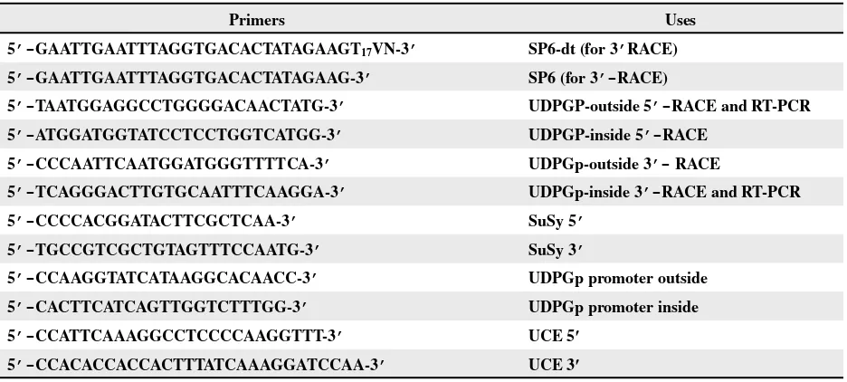
etabolic pathways leading to starch and cellulose biosynthesis have been well characterized in plants. UDP-glucose and ADP-glucose are known to play an important role in glucan biosynthesis. ADP-glucose provides theTable 1. Primers used in the isolation of UDPGp cDNA gene and in the evaluation of gene expression
Primers Uses
5’-GAATTGAATTTAGGTGACACTATAGAAGT17VN-3’ SP6-dt (for 3’RACE)
5’-GAATTGAATTTAGGTGACACTATAGAAG-3’ SP6 (for 3’-RACE)
5’-TAATGGAGGCCTGGGGACAACTATG-3’ UDPGP-outside 5’-RACE and RT-PCR
5’-ATGGATGGTATCCTCCTGGTCATGG-3’ UDPGP-inside 5’-RACE
5’-CCCAATTCAATGGATGGGTTTTCA-3’ UDPGp-outside 3’- RACE
5’-TCAGGGACTTGTGCAATTTCAAGGA-3’ UDPGp-inside 3’-RACE and RT-PCR
5’-CCCCACGGATACTTCGCTCAA-3’ SuSy 5’
5’-TGCCGTCGCTGTAGTTTCCAATG-3’ SuSy 3’
5’-CCAAGGTATCATAAGGCACAACC-3’ UDPGp promoter outside
5’-CACTTCATCAGTTGGTCTTTGG-3’ UDPGp promoter inside
5’-CCATTCAAAGGCCTCCCCAAGGTTT-3’ UCE 5′
5’-CCACACCACCACTTTATCAAAGGATCCAA-3’ UCE 3′
gene in cotton fiber and identify the transcriptional start site. Conserved motifs were identified in the promoter region of the UDPGp gene that defines regions for further evaluation.
MATERIALS AND METHODS
Plant material. Cotton seed refers to the devel-oping seed with seed coat and fiber removed. Mature leaves for the wounding experiments were detached from field grown plants and were either left intact or cut multiple times with a razor blade. Both treatments were stored in petri dishes with a piece of moist filter paper for 2 hr.
RNA isolation, cDNA synthesis, and PCR cloning. Total polyribosomal RNA was isolated as previously described from the indicated tissues of
Gossypium hirsutum L. (DES119; Delta and Pine Land Co.; Scott, MS) (Taliercio and Ray, 2002). Reverse transcription was performed on 1 µg of total polyribosomal RNA using the SMART cDNA Kit (Clontech; Palo Alto, CA) and Superscript II (Invitro-gen; Carlsbad, CA ) following the manufacturers’ in-structions with the following modifications. Briefly, 7µL (1 µg) of RNA was mixed with 1µL of a 10mM dNTP stock (Invitrogen), 1 µL of the SMART oligo (Clontech), and 1µL (25 pmoles) of oligo dT-SP6. The sequence of the SP6-dt primer and other primers are shown in Table 1. The mixture was heated to 80°C for 3 min in a PTC100 (MJ Research; Watertown, MA) thermal cycler and cooled to 37°C. A master mix containing 2X RT buffer, 20mM DTT, and 400 units Superscript II was heated to 37°C and an equal
volume was added to the RNA mix. The reaction was incubated at 37°C for 1hr, heated to 85°C for 3 min, and then cooled to 50°C. A master mix containing 1X RT buffer, 1mM dNTP, 10mM DTT, and 200U Su-perscriptII was heated to 50°C, and 5 µL was added to the first reaction. The reaction was incubated at 50°C for 20 min, 42°C for 20 min, and 37°C for 20 min. The reaction was heated to 85°C for 10 min, and 1 µL of RNAseH was added and incubated at 37°C for 20 min. RNAseH was inactivated by heating to 85°C for 10 min. The RT product was purified over a PCR purification column (Qiagen; Valencia, CA), eluted in 50µL buffer, and an equal volume of 10mM Tricine (pH 7) was added.
PCR product amplified from the RNA of various cotton tissues.The utility of UCE for normalizing fiber mRNA was based on results from Zhang et al. (2003) and was verified by RNA blot analysis (data not shown).
Isolation of the putative UDPGp promoter. The putative UDPGp promoter was isolated from an existing G. hirsutum (DES119) genomic phage library. The library was diluted 1/5 into 10mM Tris, pH 8, 1mM EDTA, 0.1% TritonX-100 to a final concentration of 5 X 10⁵ plaque forming units/µL. The diluted phage was disrupted by heating to 98°C for 4 min. Two µL of the phage DNA was used in a standard PCR reaction (50 µL) that included 1X PCR Advantage 2 buffer (Clontech; PaloAlto, CA), 200uM dNTP (Invitrogen), 10 pmoles 5′ primer, 10 pmoles 3′ primer, and 1µL Advantage 2 polymerase (Clontech). In the standard PCR, an initial 2 min denaturation at 95°C was followed by 30 cycles, where the denaturation temperature was 95°C for 15 s, the annealing temperature was 72°C for 15 s, and the extension temperature was 68°C for 10 min. To amplify the UDPGp promoter, nested primers were made to the 5’ end of the UDPGp cDNA sequence. The outside primer was used in an initial amplifica-tion followed by a nested PCR using 2 µL of the initial PCR and the inside primer (Table 1). These primers were paired with the standard T7 primers in the lambda vector to “walk” into new 5′ portions of the UDPGp gene. All sequencing was done at the MidSouth Area Genomic Center (USDA-ARS; Ston-eville, MS) using standard methods. All sequence analysis was done using Vector NTI (InforMax; Bethesda, MD). Transcription factor binding motifs were identified using the Plant Cis-Acting Regula-tory Element (Lescot et al., 2002) and the database of Plant Cis-Acting Regulatory DNA Elements (Higo et al., 1999). The DNA sequence of the putative pro-moter for cotton UDPGp was deposited in GenBank as accession number AY486082.
RESULTS
Expression of UDPGp in cotton. Steady state levels of UCE mRNA, UDPGp mRNA, and SuSy mRNA were determined for cotton fiber (7 DPA, 14 DPA, and 25 DPA), cotton seed (7 DPA, 14 DPA, and 25 DPA), 14-day-old roots, 14-day-old shoots, and mature leaves (wounded and unwounded). RT-PCR is a semiquantitive measure of mRNA abundance. Figure 1 shows PCR products for UCE,
Figure 1. RT-PCR quantification of relative transcript abundance. A. RT-PCR was performed on RNA from the indicated tissues (W: wounded and UW: unwounded). The same RT reaction mixture was used as a template for PCR with primers to amplify either UCE sequences, SuSy sequences, or UDPGp sequences and the densities of the bands were determined. The relative densities of the fiber samples were determined by dividing the UCE band density of each fiber sample by the UCE band density of the 14DPA fiber, because it was the highest value. The same method was used to determine the relative band density of seed and leaf samples. The relative densities are shown below the panel for the UCE amplification products. B. The bar chart shows the corrected abundance of the indicated transcripts. The corrected values were the densities of the SuSy and UDPGp bands divided by the relative densities
and divided by 106. No corrected abundances were applied
to the root and leaves, so the corrected values are the band
densities divided by 106.
relative density
0.7 1.0 0.8 1.0 0.8 0.8 N/A N/A 1.0 1.0
A
7 14 25 7 14 25 root shoot W UW
fiber seed leaves
UDPGp SuSy UCE AP D 7 AP D 41 AP D 52 AP D 7 AP D 41 AP D 52 to
or toohs dedn
uo w de dn uo wn u 0 10 20 30 40 50 60
fiber seed leaves
UDPGp, and SuSy amplified from various cotton tissues resolved on an agarose gel and stained with ethidium bromide. Selective sequencing of these products confirmed their identity (data not shown). Band densities were determined for all the PCR products by scanning the gel with a densitometer. The amount of UDPGp and SuSy products were normalized in comparison with the UCE products by assuming the amount of UCE product was consistent between similar types of tissues. There is almost a 3-fold increase in the level of UDPGp RNA in cotton fiber at 14 DPA compared with the amount in 7 DPA fiber that is maintained through the 25 DPA sample. SuSy mRNA was increased nearly 2-fold in the same tissues. The highest steady state level of both SuSy and UDPGp mRNA occurred in the 25 DPA seed, and the lowest level occurred in the 14 DPA seed. Both root and shoots had easily detectible levels of UDPGp mRNA and SuSy mRNA.
Comparisons of the 3’ends of UDPGp tran-scripts from various cotton tissues. The same cDNAs that were used in the semiquantitative PCR to determine the levels of UDPGp and SuSy mRNAs were also used to perform 5’-RACE and 3’-RACE to isolate the complete UDPGp mRNA. RACE was used to amplify the 3’ends of UDPGp mRNA from 14 DPA cotton fiber, 25 DPA cotton seed, and leaves. Figure 2 shows the alignments of these sequences. The nearly identical sequences at the 3′ noncoding region indicate that these mRNAs were derived from the same gene. The slightly shorter cotton seed se-quence probably represented internal priming in the five adenosine residues just 5’of the poly A tail.
Evaluation of the open reading frame. RACE was used to amplify the near full length 5′ end of the UDPGp mRNA from 14 DPA fiber. All 5’ RACE products sequenced were slightly shorter than the EST sequence (AI727382) available in GenBank. Combining the 3’RACE and EST sequences gen-erates the complete UDPGp open reading frame of 465 aa. Alignment of the deduced protein with a UDPGp from D.discoideum (AF150929) is shown in Figure 3. The deduced UDPGp protein from cotton was compared with the UDPGp from D. discoideum because the function of this protein has been experimentally determined by complementa-tion of an E.coli mutant. This UDPGp gene from
D.discoideum has been shown to encode a protein important in glycogen biosynthesis in the slime mold (Bishop et al., 2002). The proteins from E. coli and
D. discoideum are 48% identical. A second UDPGp from D. discoideum (Y00145) that does not play a prominent role in cellulose biosynthesis is only 43% identical to the other D. discoideum UDPGp, and 37% identical to the cotton UDPGp (Bishop et al., 2002; Ragheb and Dottin, 1987). The deduced cotton UDPGp protein is 85% identical to UDPGp cDNAs from both Arabidopsis thaliana (L.) Heynh and Oryza sativa L. (data not shown).
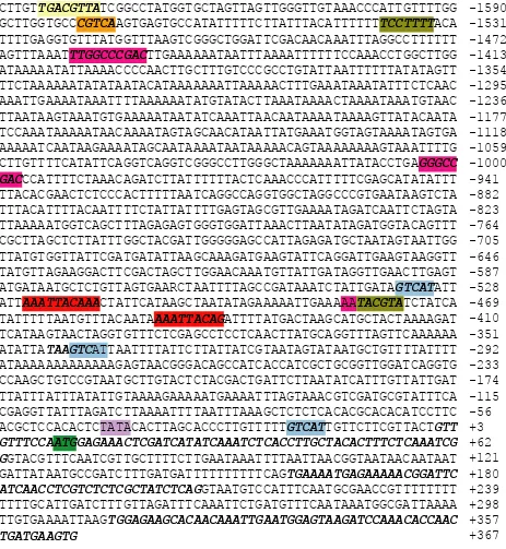
Isolation of the putative UDPGp promoter. The 5’end of a UDPGp gene was isolated and cloned from cotton as described in Materials and Methods. The genomic exon sequences that have identity to the cDNA are shown in Figure 4 in bold italics. Over all, the coding region was identical to the UDPGp EST from fiber suggesting this is the UDPGp gene expressed in fiber. Two introns were identified in
Figure 2. Alignment of the 3′ cDNA ends representing UDPGp mRNAs isolated from the indicated tissues. The alignments
start at the stop codon (in bold) that ends the open reading frame. Differences among the sequences are highlighted.
fiber TGAGAGAGGCCTGTCTACCAGCTTAAGTTTCCCCGATTTTGGTGTGTTGCAGTAGATAACG
embryo TGAGAGAGGCCTGTTTACCAGCTTAAGTTTCCCCGATTTTGGTGTGTTGCAGTAGATAACG
leaf TGAGAGAGGCCTGTCTACCAGCTTAAGTTTCCCCGATTTTGGTGTGTTGCAGTAGATAACG
fiber AACGCATCTTTTATATAAATAGGAAGTAAAATAAAATAAAAAAACCTGGAACAGAAGTAGT
embryo AACGCATCTTTTATATAAATAGGAAGTAAAATAAAATAAAAAAACCTGGAACAGAAGTAGT
leaf AACGCATCTTTTATATAAATAGGAAGTAAAATAAAATAAAAAAACCTGGAACAGAAGTAGT
fiber ATTTGCGTTTTATATCACATATATATGTTGTATGTCTTGCGGGAGTTTCCCTTGAATTACT
embryo ATTTGCGTTTTATATCACATATATATGTTGTATGTCTTGCGGGAGTTTCCCTTGAATTACT
leaf ATTTGCGTTTTATATCACATATATATGTTGTATGTCTTGCGGGAGTTTCCCTTGAATTACT
fiber ATTTTTCGAGGTATGATGAAAAACAGTGTTCTGAATGTTGTATCTACTTTTTCCCCCAAA
embryo ATTTTTCGAGGTATGATGAAAA
Figure 3. Alignment of the deduced sequence of the UDPGp protein from G. hirsutum with the UDPGp protein from D. discoideum (AF150929). Asterisks mark the location of sequence identity between the two deduced proteins.
G.hirsutum ---MEKLEHLKSAVAALSEISE
D.discoideum MMKPDLNSPLPQSPQLQSFGSRSSDLATEDLFLKKLEAISQTAPNETVKNEFLN * *
G.hirsutum NEKNGFINLVSRYLSGEAQHIEWSKIQTPTDEVVVPYDTLSPSPDDPAETKKLL
D.discoideum KEIPSINKLFTRFLKNRKKVIDWDKINPPPADMVLNYKDLP--AITEQRTSELA * * * * ** * * *
G.hirsutum DKLVVLKLNGGLGTTMGCTGPKSVIEVRNGLTFLDLIVIQIENLNSKYGCNVPL
D.discoideum SKLAVLKLNGGLGTTMGCTGPKSVIEVRSEKTFLDLSVQQIKEMNERYNIKVPL * ************************ ***** * ** * * ***
G.hirsutum VLMNSFNTHDDTLKIVDKYSNSNIEIHTFNQSQYPRLVVEDFAPLPSKGQHGKD
D.discoideum VLMNSFNTHQETGKIIQKYKYSDVKIHSFNQSRFPRILKDNLMPVPDKLFGSDS ********* ** ** * ** **** **
G.hirsutum GWYPPGHGDVFPSLMNSGKLDAFLSQGKEYVFVANSDNLGAIVDMKILNHLVQN
D.discoideum EWYPPGHGDVFFALQNSGLLETLINEGKEYLFISNVDNLGAVVDFNILEAMDKN ********** * *** * **** * ***** ** * *
G.hirsutum KNEYCMEVTPKTLADVKGGTLISYEGKVQLLEIAQVPDEHVNEFKSIEKFKIFN
D.discoideum KVEYIMEVTNKTRADVKGGTLIQYEGKAKLLEIAQVPSSKVEEFKSIKKFKIFN * ** **** ** ********* **** ******** * * *** ******
G.hirsutum TNNLWVNLNATKRLVEADALK-MEIIPNPKEVNGIKVLQLETAAGAAIRFFDHA
D.discoideum TNNIWVNLKAMDRILKQNLLDDMDIIINPKVADGKNIIQLEIAAGAAIEFFNNA *** **** * * * ** *** * *** ****** ** *
G.hirsutum IGINVPRSRFLPVKATSDLLLVQSDLYTLVDGFVIRNKDRANPTNPSIELGPEF
D.discoideum RGVNVPRSRFLPVKSTSDLFIVQSNLYSLEKGVLVMNKNRPFTTVPLVKLGDNF * *********** *** **** ** * * **** * * *
G.hirsutum KKVGNFLSRFKSIPSIIELDSLKVTGDVWFGAGIVLKGKVSIAAKPGVKLEIPD
D.discoideum KKVSDYQARIKGIPDILELDQLTVSGDITFGPNMVLKGTVIIVANHGSRIDIPE ** * * ** * *** * * ** ** **** **
G.hirsutum
GAVIEKKEINVPEDI---D.discoideum GSEFENKVVSGNLHCGAL * * *
the genomic clone that have the expected conserved GT splice donor sites and AG splice acceptor sites (Simpson and Filipowicz, 1996). The longer EST sequences were used to identify the region where transcription initiates, which is shown by convention as nucleotide +1, because the 5’RACE product was shorter than the EST in the database.
Sequences 5’of the transcription start site were evaluated for conserved motifs that bind to transcription factors by two programs that identify plant cis-regulatory elements. The programs identi-fied numerous conserved motifs. Motifs identiidenti-fied in other plants as conferring root-specific expres-sion (Yamaguchi-Shinozaki and Shinozaki, 1993) and endosperm-specific expression (Takaiwa et al., 1991; Kim and Wu, 1990) were identified, which are consistent with the root and seed expression of cotton UDPGp shown previously (Kim and Wu, 1990) (Figure 4). Motifs consistent with
expres-sion associated with wounding were also identified (Pastuglia et al., 1997); however, wounded leaves have about half the steady state levels of UDPGp mRNA of intact leaves (Figure 1). Interestingly, two elements associated with sugar repression of gene expression are present in the cotton UDPGp puta-tive promoter (Toyofuku et al., 1998; Morita et al., 1998). Responsive elements were found for methyl jasmonate, abscisic acid (Baker et al., 1994; Hattori et al., 1997), auxin (Hagan et al., 1991), and other hormones (data not shown).
DISCUSSION
Figure 4. The 5’ end of a gene homologous to an UDPGp EST (AI727382). The exons shown in bold italics were identical to the EST. Numbering is relative to the first base of the putative transcription start site. Conserved transcription factor binding motifs are highlighted in different colors in the putative promoter region. Yellow: root specific motif and auxin responsive element (Yamaguchi-Shinozakin and Shinozakin, 1993); orange: methyl jasmonate responsiveness (Rouster et al. 1997); blue: endosperm specific motif (Kim and Wu, 1990; Takaiwa et al., 1991); red: wound induced (Pastuglia et al., 1997); olive green: sugar repression (Morita et al., 1998; Toyofuku et al., 1998); pink: ABA responsiveness; green: transla-tion initiatransla-tion (Hattori et al., 1997; Baker et al., 1994) and purple: TATA box.
CTTGTTGACGTTATCGGCCTATGGTGCTAGTTAGTTGGGTTGTAAACCCATTGTTTTGG -1590 GCTTGGTGCCCGTCAAGTGAGTGCCATATTTTTCTTATTTACATTTTTTTCCTTTTACA -1531 TTTTGAGGTGTTTATGGTTTAAGTCGGGCTGGATTCGACAACAAATTTAGGCCTTTTTT -1472 AGTTTAAATTTGGCCCGACTTGAAAAAATAATTTAAAATTTTTTCCAAACCTGGCTTGG -1413 ATAAAAATATTAAAACCCCAACTTGCTTTGTCCCGCCTGTATTAATTTTTTATATAGTT -1354 TTCTAAAAAATATATAATACATAAAAAAATTAAAAACTTTGAAATAAATATTTCTCAAC -1295 AAATTGAAAATAAATTTTAAAAAATATGTATACTTAAATAAAACTAAAATAAATGTAAC -1236 TTAATAAGTAAATGTGAAAAATAATATCAAATTAACAATAAAATAAAAGTTATACAATA -1177 TCCAAATAAAAATAACAAAATAGTAGCAACATAATTATGAAATGGTAGTAAAATAGTGA -1118 AAAAATCAATAAGAAAATAGCAATAAAATAATAAAAACAGTAAAAAAAAGTAAATTTTG -1059 CTTGTTTTCATATTCAGGTCAGGTCGGGCCTTGGGCTAAAAAAATTATACCTGAGGGCC -1000
GACCCATTTTCTAAACAGATCTTATTTTTTACTCAAACCCATTTTTCGAGCATATATTT -941 TTACACGAACTCTCCCACTTTTTAATCAGGCCAGGTGGCTAGGCCCGTGAATAAGTCTA -882 TTTACATTTTACAATTTTCTATTATTTTGAGTAGCGTTGAAAATAGATCAATTCTAGTA -823 TTAAAAATGGTCAGCTTTAGAGAGTGGGTGGATTAAACTTAATATAGATGGTACAGTTT -764 CGCTTAGCTCTTATTTGGCTACGATTGGGGGAGCCATTAGAGATGCTAATAGTAATTGG -705 TTATGTGGTTATTCGATGATATTAAGCAAAGATGAAGTATTCAGGATTGAAGTAAGGTT -646 TATGTTAGAAGGACTTCGACTAGCTTGGAACAAATGTTATTGATAGGTTGAACTTGAGT -587 ATGATAATGCTCTGTTAGTGAARCTAATTTTAGCCGATAAATCTATTGATAGTCATATT -528
ATTAAATTACAAACTATTCATAAGCTAATATAGAAAAATTGAAAAATACGTATCTATCA -469
TATTTTTAATGTTTACAATAAAATTACAGATTTTATGACTAAGCATGCTACTAAAAGAT -410 TCATAAGTAACTAGGTGTTTCTCGAGCCTCCTCAACTTATGCAGGTTTAGTTCAAAAAA -351 ATATTATAAGTCATTAATTTTATTCTTATTATCGTAATAGTATAATGCTGTTTTATTTT -292 ATAAAAAAAAAAAAAGAGTAACGGGACAGCCATCACCATCGCTGCGGTTGGATCAGGTG -233 CCAAGCTGTCCGTAATGCTTGTACTCTACGACTGATTCTTAATATCATTTGTTATTGAT -174 TTATTTATTTATATTGTAAAAGAAAAATGAAAATTTAGTAAACGTCGATGCGTATTTCA -115 CGAGGTTATTTAGATCTTAAAATTTTAATTTAAAGCTCTCTCACACGCACACATCCTTC -56 ACGCTCCACACTCTATACACTTAGCACCCTTGTTTTTGTCATTGTTCTTCGTTACTGTT +3
GTTTCCAATGGAGAAACTCGATCATATCAAATCTCACCTTGCTACACTTTCTCAAATCG +62
GGTACGTTTCAATCGTTGCTTTTCTTGAATAAATTTTAATTAACGGTAATAACAATAAT +121 GATTATAATGCCGATCTTTGATGATTTTTTTTTTCAGTGAAAATGAGAAAAACGGATTC +180
ATCAACCTCGTCTCTCGCTATCTCAGGTAATGTCCATTTCAATGCGAACCGTTTTTTTT +239
TTTTGCATTGATCTTTGTTAGATTTCAAATTCTGATGTTTCAATAAATGGCGATTAAAA +298 TTGTGAAAATTAAGTGGAGAAGCACAACAAATTGAATGGAGTAAGATCCAAACACCAAC +357
TGATGAAGTG +367
activity reported previously in fiber (Wäffler and Meier, 1994). The increase in UDPGp mRNA is greater than the increase in SuSy mRNA during the same developmental time period. It is possible that the difference in the mRNA levels of UDPGp
phosphorylated to alter activity. It remains possible that UDPGp participates in providing substrate for cellulose biosynthesis in fiber. UDP-glucose also acts as a donor for glycosylation of a variety of substrates, including carbohydrates and proteins (Bocca et al., 1999).
Analysis of the mRNA encoding UDPGp in fiber, seedlings, and seed indicated the same gene was expressed in these tissues. This mRNA encoded a deduced protein with sequence similarities to UDPGps from other plant species. Alignment of the cDNA with the 5’ end of a genomic clone con-firmed that the genomic clone encoded the UDPGp expressed in fiber. Two introns with conserved splic-ing motifs and 10 bp of untranslated 5’region be-fore the translational start site were in the region of the overlap. The promoter of this gene located 5’to the putative transcriptional start site must contain sequences that account for expression of UDPGp in a variety of cotton tissues. Consistent with the observed expression of UDPGp were motifs that potentially direct expression in roots (Yamaguchi-Shinozaki and (Yamaguchi-Shinozaki, 1993) and endosperm (Kim and Wu, 1990). There were also motifs that could direct expression of the UDPGp mRNA in wounded tissue (Pastuglia et al., 1997), but elevated levels of UDPGp mRNA upon wounding were not observed in our experiments. The reduction of UD-PGp mRNA upon wounding suggests that this gene does not play an important role in the biosynthesis of callose, an important glucan that is likely to be synthesized at wound sites. It is possible that there might be a local elevation of UDPGp at the site of wounding and callose deposition that would be undetected by our assay.
In addition to motifs highlighted in Figure 4, other motifs directing expression during heat shock, and in response to light were also found (data not shown). Further analysis of the UDPGp promoter will define sequence motifs that direct expression of genes in cotton fiber, as well as in other tissues, where it has been demonstrated that this gene is expressed.
ACKNOWLEDGEMENT
The authors thank Pameka Johnson and Sheron Simpson for excellent technical assistance. This work was supported by the USDA-ARS CRIS # 6402-21000-026-00D.
DISCLAIMER
Mention of trade names or commercial products in this article is solely for the purpose of providing specific information and does not imply recommen-dation or endorsement by the USDA.
REFERENCES
Amor, Y., C. H. Haigler, M. Wainscott, M. Johnson, and D. P. Delmer. 1995. A membrane-associated from of sucrose synthase and its potential role in synthesis of cellulose and callose in plants. Proc. Natl. Acad. Sci. USA 92: 9353-9357.
Baker, S. S., K. S. Wilhelm, and M. F. Thomashow. 1994. The 5’-region of Arabidopsis thaliana cor15a has cis-acting elements that confer cold-, drought-, and ABA-regulated gene expression. Plant Mol. Biol. 24: 701-713.
Bishop, J. D., B. C. Moon, F. Harrow, D. Ratner, R. H. Gomer, R. P. Dottin, and D. T. Brazill. 2002. A Second UDP-glucose Pyrophosphorylase Is Required for Differ-entiation and Development in Dictyostelium discoideum. J. Biol. Chem. 277: 32430-32437.
Bocca, S., R. Kissen, J. Rojas-Beltran, F. Noel, C. Geb-hardt, S. Moreno, P. du Jardin, and J. Trandecarz. 1999. Molecular cloning and characterization of the enzyme UDP-glucose: protein transglycosylase from potato. Plant Physiol. Biochem. 37: 809-819.
Graves, D. and J. M. Stewart. 1988. Chronology of the differ-entiation of cotton (Gossypium hirsutum L.) fiber cells. Planta 175: 254-258.
Hagan, G., G. Martin, and J. T. Guilfoyle. 1991. Auxin-in-duced expression of the soybean GH3 promoter in trans-genic tobacco plants. Plant Mol. Biol. 17: 567-579. Haigler, C. H., M. I. Datcheva, S. P. Gogan, V. V. Salnikov, S.
Hwang, K. Martin, and D. P. Delmer. 2001. Carbon parti-tioning to cellulose synthesis. Plant Mol. Biol. 47: 29-51. Hannah, L. C. and O. E. Nelson. 1976. Characterization of
ADP-glucose pyrophosphorylase from shrunken-2 and brittle-2 mutants of maize. Biochem. Genet. 14: 547-560. Hattori, T., T. Terada, and S. Hamasuna. 1997. Regulation of
the Osem gene by abscisic acid and the transcriptional activator VP1: analysis of cis-acting promoter elements required for regulation by abscisic acid and VP1. Plant J. 7: 913-925.
Kawagoe, Y. and D. P. Delmer. 1997. Pathways and genes involved in cellulose biosynthesis. Genet. Engineer 19: 63-87.
Kim, S. Y. and R. Wu. 1990. Multiple protein factors bind to the promoter region of a rice glutelin promoter region. Nucleic Acids Res. 18: 6845-6852.
Kleczkowski, L. 1994. Glucose activation and metabolism through UDP-glucose pyrophosphorylation in plants. Phytochem. 37: 1507-1515.
Lescot, M., P. Déhais, Y. Moreau, P. Rouzé , and S. Rombauts. 2002. PlantCARE: a database of plant cis-acting regula-tory elements and a portal to tools for in silico analysis of promoter sequences. Nucleic Acids Res. 30: 325-327. (Available on-line at http://intra.psb.ugent.be:8080/Plant-CARE) (Verified 14 Apr. 2004).
Morita, A., T. Umemura, M. Kuroyanagi, Y. Futsuhara, P. Perata, and J. Yamaguchi. 1998. Functional dissection of a sugar-repressed α-amylase gene (Ramy1A) promoter in rice embryos. FEBS Lett. 423: 81-85.
Muller-Rober, B., U. Sonnewald, and L. Willmitzer. 1992. Inhibition of the ADP-glucose pyrophosphorylase in transgenic potatoes leads to sugar-storing tubers and in-fluences tuber formation and expression of tuber storage protein genes. EMBO J. 11: 1299-1238.
Pastuglia, M., D. Roby, C. Dumas, and J. M. Cock. 1997. Rapid induction by wounding and bacterial infection of an S gene family receptor-like kinase gene in Brassica oleracea. Plant Cell 9: 49-60.
Ragheb, J. A. and R. P. Dottin. 1987. Structure and sequence of a UDP glucose pyrophosphorylase gene from Dictyo-stelium discoideum. Nucleic Acids Res. 15: 3891-3906. Rouster, J., R. Leah, J. Mundy, V. Cameron-Mills. 1997.
Iden-tification of a methyl jasmonate-responsive region in the promoter of a lipoxygenase 1 gene expressed in barley grain. Plant J. 11: 513-523.
Simpson, G. and W. Filipowicz. 1996. Splicing of precursors to mRNA in higher plants: mechanism, regulation and sub-nuclear organization of the spliceosomal machinery. Plant Mol. Biol. 32: 1-41.
Takaiwa, F., K. Oono, D. Wing, and A. Kato. 1991. Sequence of three members and expression of a new major sub-family of glutelin genes from rice. Plant Mol. Biol. 17: 875-885.
Taliercio, E. and J. D. Ray. 2002. Identification of transcripts translated on free or membrane-bound polyribosomes by differential display. Plant Mol. Biol. Rep. 269a-296f. Toyofuku, K., T. Umemura, and J. Yamaguchi. 1998. Pro-moter elements required for sugar-repression of the RAmy3D gene for alpha amylase in rice. FEBS Lett. 428: 275-280.
Wäffler, U. and H. Meier. 1994. Enzyme activities in develop-ing cotton fibers. Plant Physiol. Biochem. 32: 697-702. Yamaguchi-Shinozaki, K. and K. Shinozaki. 1993.
Arabi-dopsis DNA encoding two desiccation-responsive rd29 genes. Plant Physiol. 101: 1119-1120.


