IJPAR |Vol.7 | Issue 3 | July - Sep -2018
Journal Home page: www.ijpar.com
Research article Open Access
In-vitro diffusion studies
and anticancer activity of cytarabine loaded
nanocochleates against MCF-7 cell lines
Niranjana Vaidhya Anantharaman
1*, Ubaidulla Udhumansha
2, Grace Rathnam
3Department of Pharmaceutics, C. L. Baid Metha College of Pharmacy, Thoraipakkam, Chennai-97.
*Corresponding Author: Niranjana Vaidhya AnantharamanEmail: ubaidnkl@gmail.com
ABSTRACT
Cochleates are a relatively new type of crystalline particles having a large and continuous lipid bilayer sheets rolled up in a spiral structure of less internal aqueous phases. Cytarabine is used in the treatment of the leukemia. It can bind with the cross-linking agent like calcium chloride to form the coiled structure. The In-vitro diffusion studies was performed by altering the various concentrations of cross-linking agent, drug, cholesterol: the formulation has its effect by change in the release pattern and the entrapment efficiency. In this study the concentration of the crosslinking agent is related to the release pattern.
The Nanocochleates is used to increase bioavailability, to achieve the maximum therapeutic efficacy, to reduce side effects and adverse effects. Nanocochleates was prepared by ionic crosslinking method. The in-vitro release was studied at different pH conditions 7.4 and acidic pH 5.3. The for mulation shows maximum entrapment efficiency of 86.22 ± 3.41% and good stability. Prolonged drug release was observed in pH 5.3 when compared to pH 7.4. Particle size was found in the range of 0.94 to 1.8 µm and the zeta potential was -7.3 ±3.3mV. Moreover, the stability studies of the prepared formulation show excellent stability. The in-vitro MTT assay was performed using the MCF-7 breast cancer cell lines. CYT- NCs were more cytotoxic than CY solution at 24hrs. These results suggest that CY-NCs can be explored for treatment of leukemia owing to their ability of providing sustained drug release, stability and improved cytotoxicity in MCF -7 cell line.
Keywords:
Cytarabine, Nanocochleates, In-vitro cell line- MCF-7, Cross-linkers.INTRODUCTION
Cochleates exhibit efficient incorporation of hydrophobic or even hydrophilic drugs into the lipid layers or internal phases, and they were reported to have a potential for slow release for drugs. Better clinical efficacy activity is attributed mainly to the sustained release properties of cochleates. This enhanced activity may also be
attributed to the membrane fusion capability of cochleates and an increased contact by increasing the number of particles due to their size and the high tension at the bilayer edges of cochleates. This effect can also create a kind of driving force for the molecule to penetrate efficiently [1, 2, 3]. The interaction with the tissue or cell membrane can be also important; the contact time between cochleates
and cell membrane can be also increased because of structure or composition similarities. Penetration into the tumor or enhancing activity at the site of action may also be high because of Enhanced Permeability and Retention (EPR) effect. The effect was found to be high with cochleates than liposomes, and this was attributed to the shape difference [4, 5, 6].
Many factors were found in nanocochleates formulation to represent a better activity including slow drug release, longer release time, penetration enhancing effect of cochleates, similarities of cochleate composition with cell membranes, alteration of distributions, eliminations and half-life. To further advancement of the therapeutic utility of cytarabine, the present study investigates the potential of nanocochleates as vehicle for systemic delivery of cytarabine. Such formulation has not been reported previously [7, 8].
When calcium ions bind with
phosphatidylcholine head groups, nanocochleate internal water space is replaced, resulting in a restructuring of the lipid bilayer and formation of membrane discs that fuse into lipid-based supramolecular assemblies composed of rolled sheets of lipid bilayers and calcium. When drugs are incorporated into nanocochleates, the unique multilayered cylindrical structures provide protection from drug degradation in hazardous environmental conditions [9, 10].
It is of interest to consider what properties of calcium chloride with a positive charge are important to give some of these systems the unique property of promoting nanocochleate structures. One feature to consider is the charge on the lipid. Because the negative charge on the lipid appears to play a key role in the formation of nanocochleates, but it has also been shown to influence dehydration of the lipid membrane interface and facilitate coiling of the lipid bilayer when bridged by cationic molecules [11, 12, 13].
Nanocochleate formulation technology is particularly applicable to macromolecules as well
as small molecule drugs that are
hydrophobic/hydrophilic, positively charged, negatively charged, and that possess poor oral bioavailability. Cytarabine is colourless and odourless crystalline powder substance. It has the molecular weight of 243.2g/mol. It is freely soluble in water and DMSO, insoluble in ethanol. It has the LogP value of -2.8. which indicates poor
permeability in membrane, which helps in the preparation of the nanocochleate formulation an ideal one [14]. The release study of the nanocochleate was done by using the buffer solution with neutral pH and the acidic pH, to mimic the biological environment. The drug release is monitored for the 24 hrs. The release rate is proportional directly to the entrapment efficiency. Cytarabine administered orally will turn into biologically inactive metabolite, so mode of administration is better for parenteral. For parenteral administration: Sterile isotonic aqueous or non-aqueous solutions, dispersions, suspensions or emulsions, or sterile powders which may be reconstituted into sterile injectable solutions or dispersions just prior to use [15].
MATERIALS AND METHODS
Cytarabine, cholesterol and dialysis bag (molecular weight cut off 12,000 D) were purchased from Sigma Aldrich Chemical Private Ltd, Bangalore, India. Soya Phosphatidyl Choline (SPC) was gifted from Lipoid GmbH Ludwigshafen, Germany. Ethanol, Ethylene Diamine Tetra Acetic Acid (EDTA) and calcium chloride were of analytical grade. Water were of HPLC grade and procured from Merck, Mumbai, India. All other solvents used for study were of analytical grade.
Preparation
Cytarabine-loaded small unilamellar vesicles were prepared by Ethanol Injection method. Briefly, specific amount of cholesterol and Soya Phosphatidyl Choline (SPC) were dissolved in 2 ml of ethanol and heated up to the phase transition temperature (40°C) of SPC. This ethanolic solution was rapidly injected into 10 ml of the phosphate buffer (pH 7.4) containing cytarabine, which was kept under stirring at 500 rpm using Teflon coated beads and it was continued until complete evaporation of ethanol under vacuum. Further, phosphate buffer was added to adjust the volume of final lipid vesicles suspension to 10 ml. Now the vesicles were passed through a membrane filter of 0.22 µm to obtain a cytarabine-loaded small unilamellar vesicle suspension with uniform size.
Particle size analysis
The particle size was determined by laser diffraction technique by particle size analyser (Malvern 2000 SM; Malvern Instruments, Malvern, UK). To determine the particle size, the samples must be prepared by taking CYT-NCs formulation and it was diluted with the distilled water. 10µl of the sample is taken in a 2ml of the standard volumetric flask and make up the volume. The sample is then placed inside the plastic cuvette for analysis.
Zeta potential
The Zeta Potential determination was based on particle electrophoretic mobility in aqueous medium. The Zeta Potential characterizes the surface charge and gives the information about long term stability. At higher Zeta Potential, the particle aggregation is less likely due to electric repulsion. Similarly, the zeta potential of the NCs was also found to know its surface potential charge. Zeta potential was determined using Zetasizer HSA 3000 (Malvern Instrument Ltd., UK). Samples are placed in clear disposable zeta cells and results are recorded.
Entrapment efficiency for CYT entrapped
NC
SEntrapment efficiency is determined by using the centrifuge apparatus of Hermle (Inkarp), Germany. 1ml of cytarabine NCs were introduced into polypropylene centrifuge tube. The tube was centrifuged at 15000 rpm for 30 min at 4°C and the supernatant and pellets were separated; 50µl of pH 9.5 EDTA was added to the cochleate pellet to allow the opening of the cochleates into vesicles and the release of CYT. The resulting solution was suitably diluted with phosphate buffer (pH 7.4) and vortexed completely. Then the absorbance was determined at 272 nm. The free CYT concentration in the supernatant was also measured and percentage of encapsulation efficiency was calculated using following Equation (1).
Equation: 1= Encapsulation efficiency (%) = Amount of drug entrapped in the cochleates / Total amount of the drug × 100
In-vitro
diffusion study
The In-vitro release of CYT was studied at physiological temperature of 37℃ and CYT-NCs at
pH of 7.4 (the physiological pH) and pH of 5.3 (the endosomal pH of cancer cells) using dialysis bag diffusion technique. Briefly, formulation equivalent to 10 mg of CYT-NCs was introduced into a dialysis bag (cellulose membrane, molecular weight cut off 120,000Da), hermetically sealed and immersed into 50ml of Na2HPO4-KH2PO4 buffer
solution with pH of 7.4 and 5.3. The entire system was kept at 37±0.5℃ with continuous magnetic stirring at 100 rpm/min. At selected time intervals, sample was removed and replaced with the buffer solution. The amount of CYT in the buffer solution was quantified using UV–VIS absorption spectrophotometer at 272nm.
Stability testing
The formulated CYT entrapped nanocochelates were studied for the stability using the stability chamber of Thermolab scientific by following the ICH guidelines. The CYT-NCs was separated into three portions and kept well closed in glass container. One portion was kept at room temperature, the second at 37°C, and the third at 4°C for 6 months in a stability chamber. At monthly intervals for the maximum period of six months, these portions were then evaluated for their physical stability, drug content and release characteristics to know the storage stability of the formulation.
In-vitro
anticancer activity
and dissolved in DMSO solution. The colour developed is then determined in an ELISA reader at 570 nm.
Procedure
The MCF-7 cells were plated separately in 96 well plates at a concentration of 1 × 10⁵ cells/well. After 24 h, cells were washed twice with 100 µl of serum-free medium and starved for an hour at 37oC. After starvation, cells were treated with different concentrations of test compound (25-500µg/ml) for 24 h. At the end of the treatment period the medium was aspirated and serum free medium containing MTT (0.5 mg/ml) was added and incubated for 4 h at 37ºC in a CO2 incubator.
The 50% inhibitory concentration value (IC50) of
the NCs was identified for normal fibroblast cell line. The MTT containing medium was then discarded and the cells were washed with PBS (200 µl). The crystals were then dissolved by adding 100 µl of DMSO and this was mixed properly by pipetting up and down. Spectrophotometrically absorbance of the purple blue formazan dye was measured in a microplate reader at 570 nm (Biorad 680). Cytotoxicity was determined using Graph pad prism5 software. The cell growth was measured using the Equation (2).
Equation: 2 = Cell growth (%) = Average absorbance of the test well / Average absorbance of the control wells × 100
Using the six absorbance measurements (time zero (Tz), control growth (C) and test growth in the presence of drug at various concentration levels (Ti), the percentage growth was calculated at each of the drug concentration levels. Percentage growth inhibition was calculated as: [(Ti - Tz) / (C - Tz)] × 100 for concentrations where Ti -Tz or (Ti - Tz) is positive or zero; as [(Ti - Tz) / Tz] × 100 for concentrations where Ti -Tz or (Ti - Tz) is negative. Growth inhibition of 50 % (GI50) was calculated from equation [(Ti - Tz) / (C - Tz)] × 100 = 50 as the drug concentration resulting in a 50% reduction in the net protein increase (as measured by MTT staining) in control cells during the drug incubation.
RESULTS AND DISCUSSION
Particle size
The average particle size of Cytarabine entrapped NCs was found between 0.94 to 1.8 µm. Average diameter (Z-Average) of cochleates was found out to be 1009 nm with peak obtained at 522.6 nm, this difference in the average diameter and peak can be attributed to the elongated shaped structure of cochleate particle. Size distribution report of the formulation also shows maximum particle intensity in the range of 450 -620 nm. The graph is shown below in Figure No.1.
Fig. No.1 The particle size of the cytarabine entrapped NCs.
Zeta potential
Zeta potential for NCs was determined using Zeta sizer HSA 3000 (Malvern Instrument Ltd.,
found to be negative charge. The measurement was carried out at 633 nm using a 173° scattering angle at a temperature of 25°C. Poly dispersity index (PDI) values ranging from 0 to 1 reflects the polydispersity of the suspension with the lower value indicating a more monodispersed
suspension. Before putting the fresh sample, cuvettes are washed with the distilled water and rinsed using the sample to be measured before each experiment. The CYT-NC showed negative zeta potential of-7.3 ±3.3mv due to the anionic nature of the employed lipids as seen in Figure No.2(b).
Fig. No.2 The zeta potential of the cytarabine NCs (a) zeta potential graph, (b) Histogram of the NCs.
Entrapment efficiency (E.E.)
CYT-NCs formulations showed good entrapment efficiency (E.E) of 86.22 ± 3.41%. The Cytarabine encapsulation efficiency of calcium induced nanocochleates was evaluated by adding various amounts of CYT at 5, 10 and 15(%w/w). A lipid to drug ratio of 10% gave the highest encapsulation efficiency. The vesicles size increased when CYT concentration increased from 5 to 15mg. However, further increase up to 10mg resulted in low encapsulation efficiency. CYT, a hydrophilic bioactive gets easily solubilised into the aqueous phase of vesicles. The cohesive forces between the vesicles reduced due to insertion of CYT, thus increasing the vesicle size. At still higher concentration of CYT, however, the vesicles size increased but with lower encapsulation efficiency due to exclusion of components from the lipid vesicles as shown in Table No.1. The formulation CYT-NC presented good encapsulation efficiency; hence the maximum amount that can be loaded into the phospholipid vesicles was fixed at 10 mg. Drug concentration is inversely proportional
to the E.E. CYT-loaded vesicles (CYT-SUV) obtained by ethanol injection were converted into nanocochleates using calcium chloride. The addition of calcium ions to SUV induces fusion of lipid membrane and the formation of planar sheets, which eventually coil around an initial point of folding to form rod shape nanocochleates.
Divalent cations Ca2+, Mg2+, Ba2+ andZn2+ can be used for preparing nanocochleates. It has been reported that Ca2+ forms a more tightly packed, highly ordered and less hydrated structure than does Mg2+ with phospholipids. Also, it is required in much lower concentration than Mg2+. Additionally, it is well documented that Ca2+ plays a vital role in natural membrane fusion phenomena while other cations listed above are ineffective in most such systems. Hence, it is most compatible with the body. Thus, calcium is the most suitable divalent cation for preparing Nanocochleates and hence used in the present work. The increase in particle size due to free drug encapsulation and demonstrated better encapsulation efficiency as compared to SUVs.
Table No. 1 Entrapment Efficiency and Size of the Nanocochleates
Type of Formulations Drug Entrapment (%) Size (µm)
F1 (5mg) 68.2 0.90
F2 (10mg) 86.6 0.98
In-vitro
diffusion study
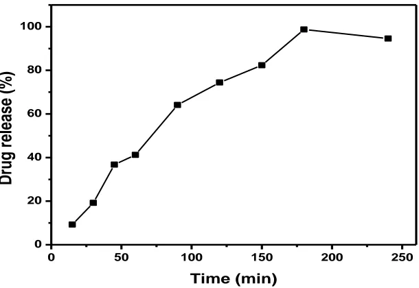
The results of release profile revealed that cytarabine solution could freely diffuse in the dispersion form causing 100% drug release within 5hr as shown in the Figure No 3. However, CYT release from nanocochleates showed a biphasic pattern with initial burst release (10%) within the first 1hr followed by controlled release up to 24hr. In this in-vitro release study, the initial burst release may be due to the dissolution of excess drug absorbed on the surface of the nanocochleates, while the further controlled release could be caused by diffusion of the drug. The drug encapsulated into the inner core compartment stayed firmly inside the nanocochleates showing a very slow release even at sink conditions with 10% of the initially incorporated drug still being associated with the nanocochleates even after 24 hr.
We found that at pH 7.4, CYT released in a slower and controlled manner from CYT-NC. It is described to the hydrogen-bonding interaction between CYT and nanocochleates, which is more prominent in the neutral condition, resulting drug release in a controlled manner upto 24 hr as shown in Figure No.4(a). On the other hand, the release pattern under acidic media pH 5.3 indicates that the amount of CYT released was much higher than at neutral conditions. It was found that around 88% of the total bound CYT was released from the CYT-NC after 24 hr as shown in Figure No.4(b). Under acidic conditions, the amine (–NH2) group of CYT get protonated resulting in the partial dissociation of hydrogen-bonding interaction, hence the amount of released CYT from nanocochleates is much higher. It is shown in the graph below. For the anti-cancer drug like cytarabine the release should be more for the better activity.
Fig. No. 3 In-vitro release of the plain drug cytarabine
0 50 100 150 200 250
0 20 40 60 80 100
Dr
u
g
r
elea
se
(
%)
Fig.No.4(a) In-vitro release of CYT-NCs from the pH 7.4
Fig. No. 4(b) In-vitro release of CYT-NCs from the pH 5.3
Stability studies
Short term stability studies were conducted for the optimized formulations as per ICH guidelines. The storage conditions used for stability studies
were at 37°C±2°C/75%±5% RH and
25°C±2°C/60%±5%RH and 4℃±2°C. Sample of CYT-NCs were analyzed after 0 and 6 months for
its physical characters, drug content and release properties were performed followed by in-vitro release. The NCs were analyzed and report was shown in Table No. 2. The results revealed that no considerable different in all the parameters were observed.
0 20 40 60 80 100 120
0 5 10 15 20 25 30
%
D
rug
R
el
ea
se
d
Time (Hrs)
0.5 % CaCl2
1% CaCl2
1.5% CaCl2
0 20 40 60 80 100 120
0 5 10 15 20 25
%
D
rug
R
e
le
as
e
d
Time (Hrs)
pH 5.3
0.5% CaCl2
1% CaCl2 1.5% CaCl2 pH
Table No. 2 Stability study profile of the NCs
In-vitro
anticancer activity
The In-vitro anticancer activity of CYT-loaded nanocochleates was investigated and compared with free drug in dispersion and blank-nanocochleates against human breast cancer MCF-7 cells using in-vitro MTT assay. The results illustrated in the below Figure no. 5 indicated that CYT-loaded nanocochleates demonstrated superior anticancer activity than the free drug dispersion and blank nanocochleates. The GI50 of CYT-loaded nanocochleates was lower (8.42 ± 0.43 µg/ml) than that of free CYT dispersion (19.32 ± 1.2 µg/ml) and blank nanocochleates (> 95 µg/ml). The CYT -NCs were shown significant cancer cell killing efficiency when compared to cytarabine solution (P value < 0.0001).
The in-vitro anticancer study demonstrated that the GI50 concentration of CYT-NC is less than free CYT and blank NC. This can be attributed to higher accumulation of the drug via direct interaction or phagocytosis i.e. fusion between nanocochleates and the cancer cell membrane followed by controlled release of the drug. Many
Fig. No. 5 MTT cell lines assay for pure drug, blank and CYT-NCs.
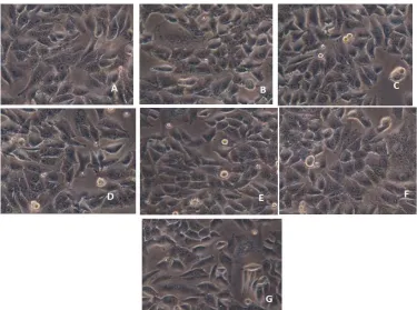
Fig. No. 6 The MCF-7 cell lines of the breast cancer cell lines by using MTT assay, (a) Control cells, (b) 12.5 ug/ml treated cells, (c) 25 ug/ml treated cells, (d) 50 ug/ml treated cells, (e) 75 ug/ml treated cells, (f) 100 ug/ml
treated cells, (g) 200 ug/ml treated cells.
CONCLUSION
The cytarabine nanocochleate was prepared by altering the concentration of the calcium chloride. The particle is uniform in size and shape. The
surface charge of the particles from the zeta potential was found to be -7.3 ±3.3Mv. The in-vitro diffusion study showed the better release of drug takes place in the pH of 5.3 of nearly 86% of the
0
20
40
60
80
100
120
12.5
25
50
75
100
200
%
Cell
Viabi
lity
Concentration (µg/ml)
drug is released in to the site. The entrapment efficiency of the NCs was found 80%. In vitro drug release pattern showed initial fast release (15.99 ± 1.2 % in 1 h) followed by sustained release up to 24 h. stability report of the NCs revealed that no
change in the assay, physical appearance and drug release. From the above results it can be concluded that the developed NCs might have potential to target cancer cells.
REFERENCES
[1]. Ramasamy, T., Khandasamy, U., Hinabindhu, R., Kona, K., Nanocochleate – A new drug delivery system. Fabad J. Pharm. Sci. 2009, 34, 91-101.
[2]. A comprehensive cancer control program for BC, BC Cancer Agency Cancer Drug Manual© Cytarabine
Developed:http://www.bccancer.bc.ca/drug-database-site/Drug%20Index/Cytarabine_monograph_1May2014.pdf. 2014.
[3]. Panwar V., Mahajan V., Panwar A. S., Darwhekar G. N, Jain D. K. Nanocochleate: As Drug Delivery Vehicle. International Journal of Pharmacy and Biological Sciences. 1(1), 2011, 31-38.
[4]. Bothiraja C, Rajput N, Poudel I, Rajalakshmi S, Panda B, Pawar A. Development of novel biofunctionalized chitosan decorated nanocochleates as a cancer targeted drug delivery platform. Artif Cells Nanomed Biotechnol. 25, 2018, 1-15.
[5]. Poudel I, Ahiwale R, Pawar A, Mahadik K, Bothiraja C. Development of novel biotinylated chitosan-decorated docetaxel-loaded nanocochleates for breast cancer targeting. Artif Cells Nanomed Biotechnol. 26, 2018, 1-12.
[6]. Tamargo B, Monzote L, Piñón A, Machín L, García M, Scull R, Setzer WN. In Vitro and In Vivo Evaluation of Essential Oil from Artemisia absinthium L. Formulated in Nanocochleates against Cutaneous Leishmaniasis. Medicines (Basel). 9, 4(2), 2017.
[7]. Liu M, Zhong X, Yang Z. Chitosan functionalized nanocochleates for enhanced oral absorption of cyclosporine A. Sci Rep. 23(7), 2017, 41322.
[8]. Biodelivery sciences International Retrieved 2006 from http://www.biodeliverysciences.com/A bout Nanocochleate Technology. html 2005.
[9]. Nagarsekar K, Ashtikar M, Steiniger F, Thamm J, Schacher FH, Fahr A. Micro-spherical cochleate composites: method development for monodispersed cochleate system. J Liposome Res. 27(1), 2017, 32-40.
[10]. Jin, T., Zarif, L., & Mannino, R. Nanocochleate formulations, process of preparation and method of delivery of pharmaceutical agents, U.S. Patent 6, 2000, 153, 217.
[11]. Gould-Fogerite, S., & Mannino, R. J. Cochleate delivery vehicles, U.S. Patent 5(994), 1997, 318.
[12]. Shivli Nomani, Jeyabalan Govinda Samy. Simultaneous loading of two antidiabetic agents in a nanoliposomal system: formulation development and characterization. Int J Adv Pharmacy Med Bio allied Sci. 3(3), 2015, 122-131.
[13]. Zarif, L., & Perlin, D. Amphotericin B Nanocochleates: From Formulation to Oral Efficacy. Drug Delivery Technology, 2(4), 2002, 34-37.
[14]. Delmarre, D., Lu, R., Tatton, N., Krause-Elsmore, S., Gould-Fogerite, S., & Mannino, R. J. Formulation of Hydrophobic Drugs Into Cochleate Delivery Vehicles: A Simplified Protocol & Formulation Kit. Drug Delivery Technology, 4(1), 2004, 64-69.
[15]. O'Donnell, Francis E. JR., Gould-Fogerite, S., & Mannino, R. J., Apoprotein cochleate compositions U.S. Patent Application 2006/0019870 A1. 2006.
[16]. Mannino, R. J., Gould-Fogerite, S., Krause-Elsmore, S. L., Delmarre, D., & Lu, R. Novel encochleation methods, cochleates and methods of use, U.S. Patent Application 2005/0013854 A1. 2005.
[17]. Zarif, L., Jin, T., Segarra, I., Mannino, R. J. Hydrogel-isolated cochleate formulations, process of preparation and their use for the delivery of biologically relevant molecules, U.S. Patent 6592894. (2003).