Dental Caries Indices used for Detection, Diagnosis, and Assessment IJOCR
Dental Caries Indices used for Detection, Diagnosis, and
Assessment of Dental Caries
1Deoyani Doifode, 2Alkesh Godhane, 3Kirti S Pattanshetti, 4Shreya Sanklecha, 5Rituraj Kesri, 6Chayan Jain
IJOCR
REVIEW ARTICLE 10.5005/jp-journals-10051-0153
Currently, dental caries is described to arise from a variety of disease states starting from initial subclinical lesion and continue to subsurface changes to clinically detectable lesions which can be seen as small cavities with considerable dentinal involvement at later stages.4,5 Dental caries is a disease that occurs after tooth eruption, as it usually starts just after the teeth eruption into the oral cavity.
It affects both males and females of all ages and all socioeconomic groups. A method of detection and esti-mation of dental caries lesions as a result of dental caries disease which is reliable, reproducible, and practical has been a big problem for a long period of time.3,6 Carious lesions can be present on all surfaces of primary, perma-nent, and mixed dentition.
The lesions on the surface of tooth can then be calcu-lated based on:
• Type of the teeth (incisors, canines, premolars, and molars) or
• Surfaces (occlusal, proximal, and free smooth surfaces). Mixed dentition stage: This starts from 6 to 12 years of age, when the permanent teeth erupt and the primary teeth are exfoliating. The mixed dentition period is the initial stage in which association between the number of caries lesions and the primary and permanent teeth can be determined.
Several studies in the past have shown direct association between caries experience between the primary and perma-nent teeth.7,8 In a prospective clinical trial caries studies, the exfoliated tooth presents a special problem, as the tooth and tooth surfaces need to be included in both of the examina-tions.9 Various indices are used for detecting dental caries in both primary and permanent dentition, depending on the involvement of coronal tissue or root caries.
Klein and Palmer10 and Knutson introduced Decayed-Missing-Filled (DMF) Index for dental caries in permanent teeth. The World Health Organization11 has included the index in the oral health assessment form which is used for conducting national oral health surveys. The main advan-tage of this index is that it is simple to use, valid, and reli-able, and is still being used very commonly for evaluating and comparing caries status among population groups.
DENTAL CARIES INDEX
In this index, the absence or presence of dental caries is presented as the DMF index in an individual or group,
1Professor and Head, 2,3Associate Professor, 4,5Senior Lecturer 6Postgraduate Student
1-6Department of Pedodontics and Preventive Dentistry, Maitri
College of Dentistry & Research Centre, Durg, Chhattisgarh, India Corresponding Author: Deoyani Doifode, Professor and Head Department of Pedodontics and Preventive Dentistry, Maitri College of Dentistry & Research Centre, Durg, Chhattisgarh, India Phone: +919923433189, e-mail: drdeoyani@yahoo.co.in ABSTRACT
Background: Due to increasing problem of oral health diseases worldwide, there is urgent need of comprehensive data collec-tion system. Tradicollec-tionally, carious lesions were detected and evaluated using physical criteria that consist of size, depth, and existence or absence of cavitations. Untreated caries in children were increasing like epidemic and there is an urgent need to determine a system that can assess initiation of dental caries to advanced stages so as to determine the disease diagnosis and decide on appropriate clinical treatment. Paucity of consistent and standardized criteria to detect caries which are applicable universally is a major issue in caries measurement. This may result in confusion among clinicians, educators, and researchers in interpretation of data from various research studies, leading to apparently conflicting results.
Aim: This article is presented with the aim of studying in detail about different dental caries indices used for detection, diag-nosis, and estimation of dental caries.
Conclusion: Reliable, reproducible, and realistic detection and estimation of dental caries lesions as a result of dental caries have been a challenge for a long time. There are many promising newer dental caries indices, which will help in identifying caries at early or precavitated stage and accurate diagnosis of dental caries.
Keywords: Dental caries, Detection, Diagnosis.
How to cite this article: Doifode D, Godhane A, Pattanshetti KS, Sanklecha S, Kesri R, Jain C. Dental Caries Indices used for Detection, Diagnosis, and Assessment of Dental Caries. Int J Oral Care Res 2018;6(1):77-81.
Source of support: Nil
Conflict of interest: None
BACKGROUND
at the tooth level (i.e., DMFT), and in case of tooth surface level, it ranges from 0 to 128 (i.e., DMFS).
Since its discovery in 1938, the DMF index has been used very usually as a measure to detect dental caries. Burt and Eklund12 have described details of DMF index and its limitations.
There are two major issues with the DMF index in the current time as caries estimation moves toward detection of precavitated or early lesions:
• This index is unable to determine restorative or pre-ventive treatment need.
• In various studies, it has been reported that DMF indices may not be comparable unless detection crite-ria and examination methods are clearly described.13 The DMF index score shows a total caries experience of an individual and it does not differentiate between untreated decay and well-restored tooth, as equal weights were given to decayed, missing because of caries, filled tooth, or tooth surface.12 Despite various problems, the DMFT index is universally used until the criteria for dental caries status is defined more evidently.
Due to these limitations, there is an urgent need for a more uniform description of caries measurement criteria and examination methods.13-15 The DMFT/S index is used for long period of time to measure dental caries in cases where decay extends into the dentin.11,16 Over the years, for various reasons, DMFT index has also been criticized17: • Lack of reliability in diagnosis of caries.
• During examination, it is very difficult for any exam-iner to confirm the reason for extraction.
• Secondary carious lesions with restorations are not counted.
• It is difficult to estimate caries activity by using DMFT. • Carious lesions in enamel are not counted.
• DMF values do not reveal teeth/surfaces at risk. • Missing teeth, untreated caries, or restored teeth are
given equal weight.
• DMF index has a limitation of overestimation of caries experience in teeth restored with preventive resin restorations or cosmetic restorations.
• DMFT index is of a very little use in estimating treat-ment needs.
• This index does not include sealants.
Significant caries index (SiC) measurement shows that DMF index produces skewed distribution of dental caries experience.18 The SiC calculates mean DMFT of one-third of the population having highest DMFT values, and online Microsoft Excel application was used for estimation. Bratthall19 introduced this index in order to measure people with the highest dental caries experience in the population under investigation.
dental caries experience in a population, mainly in developed countries leading to wrong conclusion that the caries situation for the whole population is good, while in reality, several individuals still have caries.
In a research study conducted in Nevada, the problem of skewed distribution of dental caries experience was analyzed in detail; in this study, it was confirmed that dental caries was a frequent chronic disease among youths of Nevada, and the mean SiC score calculated was considerably higher than DMFT scores among all surveys conducted in the year across and among comparison groups (p < 0.001).
The conclusion of the study was that both caries indices (DMF and SiC) should be used in combination which may help to determine inequalities in oral health more precisely in various groups in the entire population and within the whole community in order to recognize the need for special preventive oral health interventions.20 The calcula-tion of SiC is done by selecting individuals based on their DMFT values; among these, the mean DMFT among one-third population having highest caries scores is calculated. This value obtained is the SiC index. Therefore, by using the SiC index, preventive or control measures can be done by concerned authority among this subgroup.
As SiC index is an expansion of DMF index and follows the same criteria as that of DMF index, it may have limitation in determining dental caries among population subgroup in the same manner as that of DMF index. The SiC index is more appropriate to be used in population groups having low caries level and skewed distribution.
To determine the serious outcomes of untreated caries, another caries index can be used known as pulp-ulcer-fistula-abscess (PUFA) index. To find out pulpal involve-ment (p), ulceration (u), fistula (f) formation, and abscess formation (a) in the primary dentition, PUFA index is used, while PUFA scores are similar in the permanent dentition. These two indices are becoming appropriate tools for measuring oral health and planning strategy.21 As compared with DMF index, PUFA index records the advanced stages of untreated caries lesions, which result in greater impact on concerned decision-makers.
The scoring method of PUFA index (PUFA index scoring system) is as follows:
P/p—this refers when opening of pulp chamber is evident or complete decay of crown part of tooth structure and tooth is left with only root fragments; in this situation, pulpal involvement is recorded. In this index, no probing is done to diagnose pulpal involvement.
Dental Caries Indices used for Detection, Diagnosis, and Assessment IJOCR
fragments have caused traumatic ulceration of the sur-rounding soft tissues ex tongue or buccal mucosa.
F/f—fistula is scored when pus-releasing sinus tract which is related to a tooth having pulpal involvement is present.
A/a—abscess is present in a situation when a pus containing swelling is present which is related to a tooth having pulpal involvement.
International Caries Detection and Assessment System
It includes early enamel caries lesions categorized accord-ing to the stage of their progression.22,23 Various in vitro and clinical studies were conducted to test the reproduc-ibility and validity of the International Caries Detection and Assessment System (ICDAS).24-26 The ICDAS is now globally recommended for dental health surveys.12 In 2004, the International Consensus Workshop on Caries Clinical Trials described the exact meaning of caries diagnosis (which means a summation of all available data by human professional), and later divided it for carious lesion detection (which describes some objective method of finding whether or not disease is there) and lesion assessment (which aims to classify or monitor a lesion after it has been detected).9
There is major decline in caries prevalence and severity among developed countries due to preventive measures, such as water fluoridation.17,18 Due to low prevalence of caries and its slow progression, there has been an increase in the prevalence of precavitated caries (reversible condition) lesions as compared with cavitated lesions (irreversible condition).21 Thus, it is necessary to develop criteria to detect caries at initial stages (precavi-tated), as compared with frank cavitated and irreversible lesion only.20 Current agreement is to find dental caries at the precavitated lesion stages.9
The epidemiologic assessment of disease prevalence and treatment need is affected by the stage at which it is detected in a population and decision of a dental clini-cian and practitioners.8,23 An important problem in caries estimation is the absence of rational and uniform criteria applicable across the world, which may lead to confusion and difficulty in interpretation of data among clinicians, educators, and researchers from different studies which may lead to conflicting results.
A new index called the ICDAS came into existence in 2002 by a group of cardiologists and epidemiologists for caries diagnosis which was proposed as a way to put forward a globally accepted caries detection system. The index was based on visual examination with the help of WHO probe. The short form of this index is ICDAS.24 This index is a variation of a previous visually ranked
dental caries lesion scoring system, which detects occlusal lesions in permanent teeth and evaluates their depth with adequate accuracy and reproducibility.25-27
The ICDAS classifies the caries process into five stages, ranging from first visible signs of dental caries in enamel to excessive cavity with visible dentin that replicate dif-ferent phases in the occurrence of dental caries on tooth surfaces and the different levels of treatment, ranging from preventive to operative care.
The aim of ICDAS is to present a flexible, yet reli-able, system in caries estimation so that clinicians and researchers have the choice based on the phase of caries process and other features according to the requirements of their research or practice. Validity and reproducibility of ICDAS have been field tested.27 It consists of a 2-digit recognition system (X–Y). In first step, the situation of the surfaces is recorded or described as unrestored, sealed, restored, or crowned.
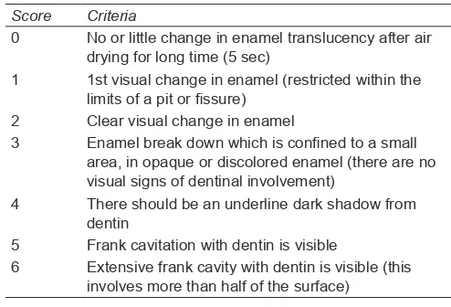
Then, a 2nd code is given (Y), which ranges from estimation of the first visual changes, that is, cavity in the enamel to extensive cavitation. The description and examples of each code are presented in Table 1.28 Before examination, thorough cleaning of teeth should be done and then with the help of light illumination, an air syringe, plane buccal mirror, and a WHO periodontal probe, if necessary, examinations must be performed.
As enamel in primary teeth is very thin, it is highly difficult to differentiate precisely between lesions associ-ated with the outer or inner half of the enamel by using the ICDAS index.29
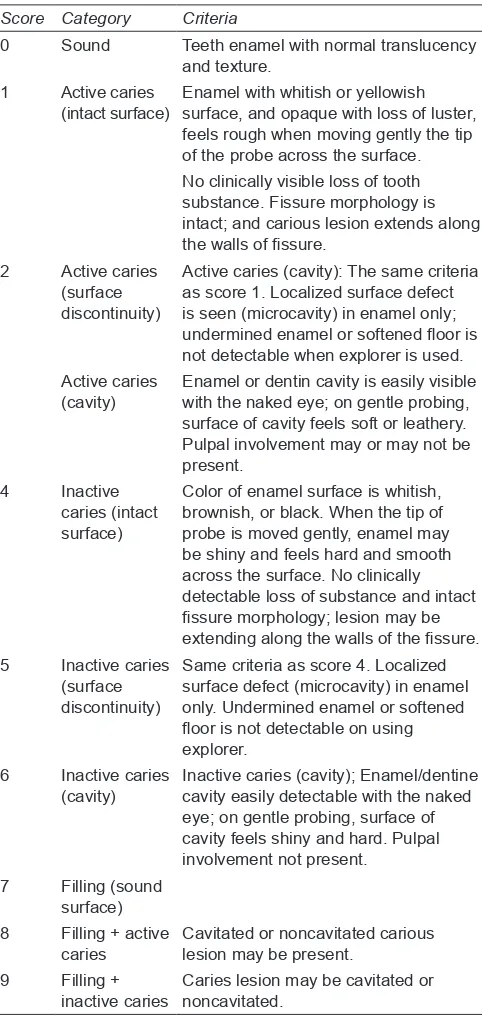
Nyvad’s system (Table 2)30,31 is another system to estimate caries activity of noncavitated and cavitated caries. This system has predictive and construct validity (the different conditions of caries lesions project different outcomes) related to caries lesion activity status.30 In this system, a separate score can be given to all observed char-acteristics of the lesion, ultimately classifying the lesion as inactive or active. A lesion is classified as active if at least
Table 1: Description and score of ICDAS
Score Criteria
0 No or little change in enamel translucency after air drying for long time (5 sec)
1 1st visual change in enamel (restricted within the
limits of a pit or fissure)
2 Clear visual change in enamel
3 Enamel break down which is confined to a small
area, in opaque or discolored enamel (there are no visual signs of dentinal involvement)
4 There should be an underline dark shadow from dentin
5 Frank cavitation with dentin is visible
one characteristic of the lesion is similar to active lesion. Various original studies used plaque, as it indicates caries activity. A standard probe is used in this system to deter-mine roughness. In various recent studies, examination was performed by investigators using Nyvad’s scoring criteria precisely as published.
But, in order to homogenize the methodology used in the examinations, various modifications of Nyvad system were done and compared with the original version, such as inspection was performed after prophylaxis and WHO probe was used.31 The depth of lesions on primary teeth was accurately measured using the Nyvad index.31 As seen in ICDAS, microcavities are those cavitations
demineralization among primary teeth.
CONCLUSION
Reliable, valid, and practical determination and assess-ment of dental caries lesions as an outcome of dental caries disease have been a challenge for a long time.
There are many promising newer dental caries indices which will help in identifying caries at early or precavi-tated stage and accurate diagnosis of dental caries.
REFERENCES
1. Last, JM. A dictionary of epidemiology. 4th ed. New York: Oxford University Press; 2001.
2. Ismail AI, Hasson H, Sohn W. Dental caries in the second millennium. J Dent Educ 2001 Oct;65(10):953-959.
3. Whelton H. Overview of the impact of changing global pat-terns of dental caries experience on caries clinical trials. J Dent Res 2004 Jul;83(Spec No C):C29-C34.
4. ten Bosch JJ, Angmar-Mansson B. Characterization and valida-tion of diagnostic methods. Monogr Oral Sci 2000;17:174-189. 5. Fejerskov, O.; Kidd, EA. Dental caries: the disease and its
clinical management. Copenhagen: Munksgaard; 2003. 6. Thylstrup A. When is caries caries, and what should we do
about it? Quintessence Int 1998 Sep;29(9):594-598.
7. Kidd EA, Fejerskov O. What constitutes dental caries? Histo-pathology of carious enamel and dentin related to the action of cariogenic biofilms. J Den Res 2004 Jul;83(Spec No C):C35-C38. 8. Pitts NB. Modern concepts of caries measurement. J Dent Res
2004;83(Spec No C):C43-C47.
9. Pitts NB, Stamm JW. International consensus workshop on caries clinical trials (ICW-CCT)—final consensus statements: agreeing where the evidence leads. J Dent Res 2004 Jul;83(Spec No C):C125-C128.
10. Klein H, Palmer CE, Knutson JW. Studies on dental caries. Pub Health Rep 1938 Aug;53(31):751-765.
11. World Health Organization. Oral health surveys—basic methods. 4th ed. Geneva: World Health Organization; 1997.
12. Burt, BA.; Eklund, SE. Dentistry, dental practice, and the com-munity. 6th ed. Philadelphia (PA): Elsevier Saunders; 2005. 13. Burt BA. How useful are cross-sectional data from surveys
of dental caries? Community Dent Oral Epidemiol 1997 Feb;25(1):36-41.
14. Lawrence HP, Beck JD, Hunt RJ, Koch GG. Adjustment of the M-component of the DMFS index for prevalence studies of older adults. Community Dent Oral Epidemiol 1996 Oct;24(5):322-331.
15. Boradbent JM, Thomson WM. For debate: problems with the DMF index pertinent to dental caries analysis. Community Dent Oral Epidemiol 2005 Dec;33(6):400-409.
16. National Institute for Dental and Craniofacial Research (NIDCR). Oral health of United States adults. Bethesda (MD): NIDCR; 1987. pp. 161-165.
17. Marthaler TM. Changes in dental caries 1953-2003. Caries Res 2004 May-Jun;38(3):173-181.
18. Petersen PE. The World Oral Health Report 2003: continuous improvement of oral health in the 21st century—the approach
0 Sound Teeth enamel with normal translucency and texture.
1 Active caries
(intact surface) Enamel with whitish or yellowish surface, and opaque with loss of luster, feels rough when moving gently the tip of the probe across the surface. No clinically visible loss of tooth substance. Fissure morphology is intact; and carious lesion extends along
the walls of fissure.
2 Active caries (surface discontinuity)
Active caries (cavity): The same criteria as score 1. Localized surface defect is seen (microcavity) in enamel only;
undermined enamel or softened floor is
not detectable when explorer is used. Active caries
(cavity) Enamel or dentin cavity is easily visible with the naked eye; on gentle probing, surface of cavity feels soft or leathery. Pulpal involvement may or may not be present.
4 Inactive caries (intact surface)
Color of enamel surface is whitish, brownish, or black. When the tip of probe is moved gently, enamel may be shiny and feels hard and smooth across the surface. No clinically detectable loss of substance and intact
fissure morphology; lesion may be extending along the walls of the fissure.
5 Inactive caries (surface discontinuity)
Same criteria as score 4. Localized surface defect (microcavity) in enamel only. Undermined enamel or softened
floor is not detectable on using
explorer. 6 Inactive caries
(cavity) Inactive caries (cavity); Enamel/dentine cavity easily detectable with the naked eye; on gentle probing, surface of cavity feels shiny and hard. Pulpal involvement not present.
7 Filling (sound surface) 8 Filling + active
caries Cavitated or noncavitated carious lesion may be present. 9 Filling +
Dental Caries Indices used for Detection, Diagnosis, and Assessment IJOCR
of the WHO Global Oral Health Programme. Community Dent Oral Epidemiol 2003 Dec;31(Suppl 1):3-23.
19. Bratthall D. Introducing the Significant Caries Index together with a proposal for a new oral health goal for 12-year-olds. Int Dent J 2000 Dec;50(6):378-384.
20. Ditmyer M, Dounis G, Mobley C, Schwarz E. Inequalities of caries experience in Nevada youth expressed by DMFT index vs. Significant Caries Index (SiC) over time. BMC Oral Health 2011 Apr;11:12-21.
21. Monse B, Heinrich-Weltzien R, Benzian H, Holmgren C, van Palenstein Helderman W. PUFA—an index of clinical conse-quences of untreated dental caries. Community Dent Oral Epidemiol 2010 Feb;38(1):77-82.
22. Ismail AI. Clinical diagnosis of precavitated carious lesions. Community Dent Oral Epidemiol 1997 Feb;25(1):13-23. 23. Bader JD, Brown JP. Dilemmas in caries diagnosis. J Am Dent
Assoc 1993 Jun;124(6):48-50.
24. Rimmer PA, Pitts NB. Effects of diagnostic threshold and overlapped approximal surfaces on reported caries status. Community Dent Oral Epidemiol 1991 Aug;16(4):166-170. 25. Pitts N. “ICDAS”—an international system for caries
detec-tion and assessment being developed to facilitate caries epi-demiology, research and appropriate clinical management. Community Dent Health 2004 Sep;21(3):193-198.
26. Bader JD, Shugars DA, Bonito AJ. Systematic reviews of selected dental caries diagnostic and management methods. J Dent Educ 2001 Oct;65(10):960-968.
27. Ekstrand KR, Ricketts DN, Kidd EA, Qvist V, Schou S. Detec-tion, diagnosing, monitoring and logical treatment of occlusal caries in relation to lesion activity and severity: an in vivo
examination with histological validation. Caries Res 1998 Feb;32(4):247-254.
28. Ismail AI, Sohn W, Tellez M, Amaya A, Sen A, Hasson H, Pitts NB. The International Caries Detection and Assessment System (ICDAS): an integrated system for measuring dental caries. Community Dent Oral Epidemiol 2007 Jun;35(3): 170-178.
29. Mortimer KV. The relationship of deciduous enamel structure to dental disease. Caries Res 1970;4(3):206-223.
30. Nyvad B, Machiulskiene V, Baelum V. Reliability of a new caries diagnostic system differentiating between active and inactive caries lesions. Caries Res 1999 Jul-Aug;33(4): 252-260.
31. Braga MM, Mendes FM, Martignon S, Ricketts DN, Ekstrand KR.