Current Research in Microbiology and Biotechnology
Vol. 5, No. 5 (2017): 1258-1265Research Article Open Access
I
ISSSSNN:: 22332200--22224466
Study the effect of levan produced from
Pseudomonas
putida
on phagocytic activity
Alaa Raheem Kazim, Alyaa Razoki Hussein Allami
⃰
and Hala Mouayed Radif
Department of Biology, College of Science, Baghdad University, Baghdad, Iraq.
* Corresponding author:Alyaa Razoki Hussein Allami; e-mail: [email protected]
ABSTRACT
Levan is one of few natural polysaccharides and the basic unit of it is fructose, it can be produced from: plants, fungi, yeast and some bacterial genera. Levan had many beneficial effects on human and animal health therefore levan is best known to have anti-tumour, antioxidant and anti inflammatory effects. It has been reported that levan strongly induces IL-12 and TNF-α in macrophages. This article aimed to study the optimal conditions to increase the production of levan that extracted from local isolates of Pseudomonas putida, and determination the role of levan as phagocytic enhancer against Candida albicans. The result showed that Pseudomonas putida had the ability to produce levan when cultured in mineral salt medium and the levan was extracted from the production medium as off white gummy precipitate. The highest levan dry weight was (4.65 mg/100ml), when the production medium was contained 20% sucrose, and the optimum pH was 7, temperature 370C and
incubated for 48 hrs. FT-IR analysis of levan dry weight exhibited the essentials groups found in the chemical structure of carbohydrates which it were (C-H, C-O, C=O and O-H), also the result showed the effect of levan on phagocytosis processes, it showed that the phagocytic index was 70% when used levan in concentration of 200µg ∕ ml while the phagocytic index was 45% in control (without levan).
Keywords:
Levan, Mineral salt medium, FT-IR, Candida albicans, Phagocytic index.1. INTRODUCTION
Levan is a homopolymer of fructose commonly reffered as polyfructose [1], it is made from repeating fructose sub-units which form a main chain with β-(2→6) fructofuranosidic bonds and occasionally with β-(2→1) branching this backbone make levan a unique biopolymer, being at the same time one of the few natural polymers in which carbohydrate is found in the furanose form [2]. It can be found in plants and many microbial strains, microbial levans are much larger than those produced by plants with multiple branches with molecular weights ranging from 2 to 100 million Da. and more beneficial in: economical, industrially, food, medicine, pharmaceutical, cosmetic and commercial industrial sectors, while plant levans generally have molecular weights ranging from 2000 to 33,000 Da. [3, 4].
Levan is also manufactured as an exopolysaccharide, usually from sucrose-based substrates by a variety of microorganisms. The main reaction involved in its biosynthesis is the transfructosylation and is carried out by an extracellular enzyme namely levan sucrose
(sucrose 6-fructosyltransferase) [5,6] therefore
levansucrase (LSC) is considered to be a key point of microbial levancreation [7]. Many microorganisms, including Gram negative and Gram positive bacteria, yeasts and molds are capable to produce levan. The most well-known microbial levan producers belong to
the genera: Zymomonas, Bacillus, Acetobacter,
Aerobacter, Pseudomonas, Erwinia, Gluconobacter,
Streptococcus, Corynebacterium [7].
Many studies reported levan’s multiple beneficial effects on human and animal health therefore levan is best known to have anti-tumour, antioxidant and anti Received: 18 September 2017 Accepted: 05 October 2017 Online: 09 October 2017
inflammatory effects. It has been reported that levan strongly induces IL-12 and TNF-α in macrophages [8].
Phagocytosis is the primary function of macrophages, which leads to increase a various range of antimicrobial and cytotoxic responses including generation of respiratory bust, secretion of inflammatory mediators
and antigen presentation [9]. Park et al. (2008)
reported that the phagocytic activity of macrophages was significantly increased when treated the call with different concentration of levan [10].
This paper was aimed to study the optimal conditions to increase the production of levan that extracted from
local isolates of Pseudomonas putida, and determination
the role of levan as phagocytic enhancer against
Candida albicans.
2. MATERIALS AND METHODS
2.1 Collection of samples
Twenty-five different samples from foods and soils were taken and collected in sterile container, transported to the laboratory until use.
2.2 Isolation and identification of bacteria
Half-gram of each sample was added to 4.5 ml of sterilized peptone water, mixed thoroughly and serial dilutions were done, after that 100 µl from each dilution were taken and cultured on MacConkey agar, incubated at 37°C for 24h. Nutrient agar plates were prepared and streaked with bacterial colonies to obtained pure culture. For completed identification of bacterial isolates, several morphological, cultural characteristic and biochemical tests were done.
2.3 Screening for levan - producing Pseudomonas spp. isolates on solid medium
Sucrose mineral salt agar was streaked with 100 µl of activated bacterial culture suspension, incubated at
370C for 48h. Colonies with viscosity appearance
indicate for levan production. The highest viscosity colonies were selected and underwent further steps of screening [11].
2.4 Quantitative screening in liquid medium
The highest viscosity colonies on sucrose mineral salt agar was inoculated in 10 ml of Brain Heart Infusion
broth (BHI), incubated at 370C for 24h. The absorbency
was measured at 600nm after incubation periods. Mineral salt broth was prepared and inoculated with100 µl of activated bacterial culture [12].
2.5 Extraction of levan
Levan was extracted from production medium
according to Szwengiel et al. (2004) [13], mineral salt
broth with 20% of sucrose was prepared and inoculated with activated bacterial culture, incubated at
370C for 48h. After incubation periods, cells were
separated from the broth medium by centrifugation at 6000 rpm for 30 min. Cell free supernatant was mixed with 95% ethanol at a ratio (1:4 v/v) and allowed to
stand overnight at 40C. after that the precipitated
sediment (levan) was collected and dissolved in 10 ml of distilled water (D.W.) and heated in boiling water bath for 10 min. to inhibited the activity of exo- enzymes, cooled by tab water, ethanol was added again to precipitated levan at ratio (1:4 v/v).The off white gummy precipitate was collected in glass Petri dish and left till dryness, dried levan was collected in clean screwed vial after scraping it from Petri dish to determine levan dry weight.
2.6 Analysis of Levan by Fourier Transform Infrared Spectroscopy (FT-IR)
To complete the diagnosis of levan, Fourier Transform Infrared (FTIR) spectroscopy was carried out to identify the functional groups, Each 1 mg dried sample
was mixed with 200 mg of KBr (Spectranal) and
pressed under vacuum to form thin tablet. The tablet was immediately analyzed with a spectrophotometer [14].
2.7 Determination the optimal conditions for levan production
Different conditions were used to determine the optimum one for increasing the production of levan, these conditions include;
Effect of different:
1. Carbon sources
Mineral salt broth containing 20% of different carbon sources (lactose, glucose) were prepared and inoculated with 1% of activated bacterial culture broth
(optical density, 0.1), incubated at 37°C for 48h. levan
was extracted and dry weight was weighted after
incubation periods, andcompared with the dry weight
of levan that extracted from mineral salt broth containing 20% of sucrose previously prepared and inoculated with 1% of activated bacterial culture.
2. Nitrogen sources
Mineral salt broth media with optimum carbon source were prepared with additional of two nitrogen sources (yeast extract and peptone) at concentration (1%), and inoculated with 1% of activated bacterial culture broth,
incubated at 370C for 48h. after incubation periods,
levan was extracted and dry weight was weighted.
3. pHs value
Mineral salt broth media with 20 % of optimum carbon
source were prepared with different pHsvalue (5, 6, 7,
8, 9 and 10), and inoculated with 1% of activated
bacterial culture broth, incubated at 370C for 48h. levan
was extracted after incubation periods and dry weight was weighted.
4. Temperature
Mineral salts broth media with optimum source of (carbon, nitrogen) and optimum pH value were prepared and inoculated with 1% of bacterial culture
broth, incubated at different temperatures (370C, 450C
and 500C) for 48h, after incubation, levan was
2.8 Study the effect of levan in phagocytic activity on Candida albicans in vitro
A. Preparation of Candida albicans suspension
Sabouraud Dextrose Agar was prepared and inoculated
with Candida albicans, incubated at 370C for 24 hrs,
after incubation period 50 ml of Brain Heart Infusion broth (BHI) was inoculated with a single colony of
Candida albicans, incubated at 370C for (3-4) days.
Candida albicans cellswere collected by centrifugation
at a 3000 rpm for 15 min., washed twice with Hanks solution, cell were harvested and suspended in Hanks solution [15].
B. Preparation of levan suspension
Levan was dissolved in normal saline to obtain
concentration (200µg / ml).
C. Blood sample collection
Blood was collected from vein by using syringe, and then put in container contained anticoagulant. This blood sample was used to calculate the percentage of phagocytic cells.
D. Determination the phagocytosis index
Phagocytosis index was determined against Candida
albicans before and after the addition of levan
suspension to blood sample according to Furth et al.
(1985)[16] as following:
1. A volume of 250 µl of blood was mixed with 250 µl
(1× 10 6 cell/ ml) of Candida albicans suspension
before and after the additional of levan suspension to blood sample.
2. The mixture was incubated for 30 minutes in 370C,
and mixed every 5 minutes.
3. Smear of mixture was done by putting one drop of
mixture on a glass slide by Pasture pipette, left the slide to dry.
4. One drop of absolute methanol was added to fix the
mixture.
5. One drop of Leishman stain was added, left for 15
minutes, after that, washed the slide with D.W, left it to dry, and examined under the microscope.
6. Phagocytosis index was calculated by counting 200
phagocytic and non phagocytic cells.
Phagocytosis index = number of phagocytic cells × 100 200 phagocytic and non phagocytic cells
3. RESULTS AND DISCUSSION
3.1 Isolation and identification of Pseudomonas putida
Twenty-five samples were collected from different foods and soil; forty-six bacterial isolates from 96
bacterial isolates were belonged to Pseudomonas spp.
depending on morphological examination of
Pseudomonas spp. on MacConkey agar. Pseudomonas
isolates exhibited fluorescing colonies, fluorescing colonies subculturing to fresh MacConkey agar plates, the purified isolates were used to inoculate on several media for biochemical testing and pigment production
on nutrient and MacConkey agar; semi solid media were used for investigation for the motility of bacterial isolates. Bacterial isolates exhibited positive results for (oxidase, catalase, motility, and nitrate reduction) testes, and negative results for (indole and Voges Proskauer) testes. On nutrient agar; bacterial colonies appeared as large oval, convex and rough colonies with
metallic iridescent and enclitic by serrated growth[17].
Microscopic examination of bacterial isolates evidenced it was Gram negative, rod, non spore former, and motile with one or more flagella [18].
3.2 Screening for levan producing Pseudomonas spp.
Levan production medium was inoculated with 100 µl of activated bacterial culture suspension, incubated at
370C for 48h. for screening their ability to grow with
viscosity appearance in the presences of sucrose in production media, the viscous appearance of bacterial isolates indicate for levan production. Bacterial colonies distinguished by gummy appearance and adhering on the agar surface due to levan production [19].
Thirty-two from forty-six of Pseudomonas isolates
exhibited viscous appearance and adhesive to the production medium and the viscosity differed among
the Pseudomonas isolates.
3.3 Quantitative screening in liquid medium
The colonies with the highest viscosity appearance on production medium were harvested for further screening of levan production in mineral salts broth
medium. It was found that seven Pseudomonas isolates
possessed the highest viscosity appearance on solid
medium, there were selected and underwent further screening in mineral salt broth medium. The highest levan dry weight was (4.65 mg/100ml), it was extracted from the more viscous isolate.
3.4 Identification of the highest levan production isolate
The more viscous isolate which gave the maximum production of levan dry weight (4.65 mg/100ml) was preferred and identification by VITEK 2 compact. The results approved that the bacterial isolate was
Pseudomonasputida as showed in figure1.
3.5 Extraction of levan from Pseudomonas putida
For production and extraction of levan from
Pseudomonas putida, mineral salts broth medium was
inoculated with activated Pseudomonas putida and
levan was extracted from the mineral salts broth after the incubation period by centrifugation the mineral salts broth at 6000 rpm for 30 min. and addition four volume of ethanol to the culture supernatant at several steps during extraction process. The off white viscous precipitate was collected in glass Petri dish and weight after dryness. Levan dry weight was (4.77gm/100ml).
which lead to separation of levan from the production
medium and increased the purification of product[20].
Figure 1: VITEK2 compact identification for Pseudomonas putida
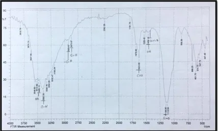
3.6 Analysis of Levan by FT- IR Spectroscopy
The functional groups present in levan structure that
extracted from Pseudomonasputida was determined by
FTIR Spectroscopy, FTIR analyzed exhibited the presence of (C-O, C-H, O-H and C=O) in the chemical structure of levan, C=O and C-O stretching group in
(1623.95 cm-1, 1139.85 cm-1) respectively, C-H group
stretching in (2929.67cm-1 and 2875.67 cm-1), and OH
group stretching in (3485.13 cm-1 and 3392.55 cm-1),
(C-H, O-H) bending group in (1456.16 cm-1 and 1334.68
cm-1) respectively as showed in figure (2).
The functional groups in the chemical structure of carbohydrates are (C-H, C-O, C=O and O-H) proved that levan lack the functional groups present in lipid and proteins and consist only from carbohydrate without lipid or protein in there structures [21].
3.7 Determination the optimal conditions for levan production
A. Effect of carbon sources
Levan production may influences by the nature and concentration of carbon substrate. For determination the effect of kinds of sugar on levan production; media containing glucose, lactose and sucrose as carbon sources were used for production of levan from
Pseudomonas putida.
The results showed the highest levan production was in medium containing sucrose then medium containing lactose, its dry weight was 3.2 gm ∕100 ml and the lowest levan production was in mineral salts medium containing glucose, its dry weight was 0.1gm ∕ 100 ml as showed in figure (3).
Figure 3: Production of levan from Pseudomonas putida in mineral salts media containing 20% of different carbon sources incubated at 370C for 48hrs, pH7 and the inoculums size 1%.
Levansucrase is an extracellular enzyme which catalyze the formation of the fructose polymer levan after induction by sucrose and it was found that the most important feature for the production of levansucrase is that at least 5% sucrose must be added to the growth medium to enhanced the synthesis of enzyme and the production of enzyme is increase in the present of sucrose [22]. The production of exopolysaccharide from bacteria is an energy reserve and defense against high concentration of sucrose [23].
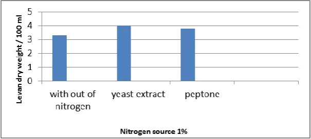
B. Effect of nitrogen source on levan production
The results revealed that the presence of yeast extract and peptone in the production medium inoculated with
Pseudomonas putida increases levan dry weight to 4.0
gm ∕ 100ml and 3.8 gm ∕ 100ml respectively, while in the absence of these nitrogen sources the levan production was 3.3gm ∕ 100ml as dry weight as showed in figure (4).
Figure 4: Production of levan from Pseudomonas putida in mineral salts media containing 20% of sucrose and 1% of different nitrogen sources incubated at 370C for 48hrs, pH7 and the inoculums size 1%.
Nitrogen source is one of the important factors that enhances and increases bacterial growth. The increasing in bacterial growth may be caused
increasing in levan production. Melo et al. (2007)
reported that the influence of initial concentration of yeast extract in the production of levan. Yeast extract
improve cellular metabolism, subsequently leading to enhancement of the substrate consumption [1].
The results exhibited that the amount of levan dry weights was varied depending on the pHs of culture medium as showed in figure (5).
It was indicated that the production of levan was influenced by the pHs of inoculation culture, at low pH less than 7 and high pH more than 7 the levan dried
weight produced from Pseudomonas putida was very
low, and the highest production was gained in pH 7.
The optimum pH for bacterial growth and levansucrase synthesis was between (6-7) [24]. When the production medium inoculated with the bacterial culture, the pH of the medium was decreased due to the formation of acids and exopolysaccharide (levan) may be hydrolyzed in acidic pH, thus its necessary to maintaining the pH above 5.5 [25].
During the first 24 hrs of the fermentation process, the pH of the inoculated medium decreased from 7.0 to about 6.8. In the second day of fermentation, pH reduction was faster and was probably caused by acid production by bacteria which were in their log phase of growth therefore the reduction in pH of the medium caused a decrease in levan production [20].
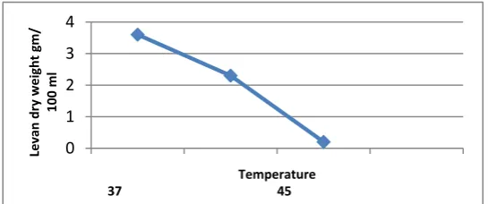
D. Effect of Temperature on levan production
It was observed that the highest dry weight of levan
was 3.6 gm ∕ 100ml at 370C, while the dry weight of
levan was 2.3gm ∕ 100ml at 450C, and the lowest dry
weight of levan was 0.2gm ∕ 100ml at 500C as showed in
figure (6).
These finding indicated that the optimum temperature
for levan production was 370C and this temperature
may be the optimum temperature for the synthesis and activity of levansucrase (enzyme that responsible for levan production). The amount of levan produced from
Bacillus subtilis at 370C was higher than at 320C, this
temperature may performed a significant effect on the secretion of the levansucrase enzyme into the medium, and it may cause a change in the kinetics of the enzyme itself [13].
3.8 Effect of levan on phagocytic activity
Human blood cells were treated with levan in order to increase their phagocytic ability. Phagocytic index increased in the presence of levan in compared with control (without levan). The result showed that the phagocytic index was 70% when used levan in concentration of 200µg ∕ ml while the phagocytic index was 45% in control (without levan), this results indicated that levan increases the phagocytosis of phagocytic cells and this may be due to its structure or the nature of its bond β-(2→6) fructose, it may binds to specific receptors on phagocytic cells and enhances their phagocytic ability as shown in figure (7).
Low concentration of levan (1µg ∕ ml) showed no effect on phagocytic activity of macrophages While
macrophages treated with levan from Zymomonas
mobilis at concentration (2-100µg ∕ ml) have higher
phagocytic activity than untreated with levan[10].
0 1 2 3 4
Le
va
n
d
ry
w
e
ig
h
t
gm
/
100
ml
pH
5 6 7 8 9 10
Figure 5: Effect of pH on levan production from Pseudomonas putida in mineral salts media containing 20% of sucrose and without nitrogen source, incubated at 370C for 48hrs, the inoculums size 1%
0 1 2 3 4
Levan
d
ry
weight
gm/
100
ml
Temperature
37 45 50
Figure 7: The phagocytosis of Candida albicans from phagocytic cells after treatment with levan under oil immersion, A- phagocytic cells with non phagocytosis; B- Phagocytic cells with phagocytosis.
5. REFERENCES
1. Melo, I. R.; Pimentel, M. F.; Lopes, C. E. and Calazans, G. M.T.
(2007). Application of fractional factorial design to levan
production by Zymomonas mobilis. Brazilian Journal of
Microbiology. 38: 45-51.
2. Shih Ing-Lung; Chen Li-Dar; Wu Jane-Yii. (2010). Levan
production using Bacillus subtilis natto cells immobilized on
alginate. Carbohydrate Polymers. 82: 111-117.
3. Silbir, S.; Dagbagli, S.; Yegin, S.; Baysal, T. and Goksungur, Y.
(2014). Levan production by Zymomonas mobilis in batch and
continuous fermentation systems. Carbohydrate Polymers. 99: 454-461.
4. Sarilmiser, K.; Ates, O.; Ozdemir, G.; Arga, K. Y. and Őner, E.T.
(2015). Effective stimulating factors for microbial levan
production by Halomonas smyrnensis AAD6T. Journal of
Bioscience and Bioengineering. 119(4): 455-463.
5. Han, Y. W. and Clarke, M. A. (1996). Production of fructan
(levan) polyfructose polymers using Bacillus polymyxa. US
Patent no. 5547863.
6. Srikanth, R.; Reddy, C. H. S. S.; Siddartha, G.; Ramaiah, M. J. and Uppuluri, K. B. (2015). Review on production, characterization and applications of levan. Carbohydrate Polymers. 120: 102-114.
7. Donot, F.; Fontana, A.; Baccou, J. C. and Galindo, S. S. (2012).
Microbial exopolysaccharides: main examples of synthesis, excretion, genetics and extraction. Carbohydrate Polymers. 87: 951-962.
8. Xu, Q.; Yajima, T.; Li, W.; Saito. K.; Ohshima, Y. and Yoshikai,Y.
(2006). Levan (beta-2, 6-fructan), a major fraction offermented soybean mucilage, displays immunostimulating properties via Toll-like receptor 4 signalling: induction of interleukin-12 production and suppression of T-helper type2
response and immunoglobulin E production. Clin ExpAllergy.
36: 94-101.
9. Adams, D. O. and Hamilton, T. A. (1984). The cell biology of
macrophage activation. Annu Rev Immunol.2: 283-318.
10.Park, S.; Jang, K. H.; Kim, M. H.; Lim, J. D.; Han, E. T.; Jang, S. A.; Kim, K.; Poy, S. and Sohn, E. H. (2008). The differential immunodulatind effects of levan and DFA-IV on macrophage function. J. Food Sci. Nutr. 13: 1-6.
11.Rhee, S. K.; Song, K. B.; Kim, C. H.; Park, B. S. and Jang, E. K .
(2005). Levan. In: Polysaccharides and Polyamides in the food industry. (ed. Steinbuchel, A. and Rhee, S. K.). Wiley VCH Verlag. Gmbh& Co. KGaA. Weinheim. pp. 325-349.
12.Jang, K. H.; Eun-Kyung, J.; Seung- Hwan, K.; In- Hwan, K.; Soon-Ah, K.; Issac, K.; Young-Il, P.; Young-Jun, K.; Sang-Do, H. and Chul, H. K. (2006). High-Level Production of
Low-Branched Levan from Pseudomonas aurantiaca S-4380 for
the Production of di-β-D-Fructofuranose Dianhydride IV. J. Microbiol. Biotechnol. 16(1): 102–108.
13.Szwengiel, A.; Czarnecka, M.; Roszyk, H. and Czarnecki, Z.
2004. Levan production by Bacillus subtilis DSM347 strain.
EJPAU. 7(2): 1-9.
14.Naja, G. M.; Mustin, C. and Volesky,B. (2005). A high
resolution; a new approach to studying binding site of
microbial biosorbent. Water Research 39: 579-588.
15.Cech, P. and Lehrer, R. I. (1984). Heterogeneity of Human
neutrophil phagolysosomes: functional consequences for candidadial activity. Blood. 64(1): 147-151.
16.Furth, R; Tneda, L. and Leijilt, P. (1985). In vitro
determination phagocytosis and intra-cellular killing by poly morphonuclear and mononuclear phagocytosis, In: Hand
book of experimental Imunology. (3rd ed.), Black well
© 2017; AIZEON Publishers; All Rights Reserved
This is an Open Access article distributed under the terms of the Creative Commons Attribution License which permits unrestricted use, distribution, and reproduction in any medium, provided the original work is properly cited.
17.Meyer, J. M.; Geoffroy, V. A.; Baida, N.; Gardan, L.; Izard, D. and
Lemanceau, P. (2002). Siderophore typing, a powerful tool for the identification of fluorescent and non fluorescent Pseudomonads. Appl. Environ. Microbiol. 68: 2745-53.
18.Laura, F. and Mauro, S. (2007). Characterization of
Pseudomonas spp. isolated from foods. Ann Microbiol 57:
39-47.
19.Ghaly, A. E.; Arab, F.; Mohmoud, N. S. and Higgins, J. (2007).
Production of levan by Bacillus licheniformis for used as soil
sealant in earthen manure storage structures. American Jornal of Biochemistry. 3(2): 47-52.
20.Marzieh, M. N.; Behnaz, L.; Ladan, A. and Mohammad, B. H.
(2010). Microbial Production of Levan using Date Syrup and Investigation of Its Properties. International Scholarly and Scientific Research & Innovation. 4(8): 603-609.
21.Grube, M.; Bekers, M.; Upite, D. and Kaminska, E. (2002).
IR-Spectroscopic studies of Zymomonas mobilis and levan
precipitate. Vibrational Spectroscopy. 28 (2): 277-285. 22.Charles, R. L. and Shetty, J. K. (1995). Bacillus licheniformis
NRRL B- 18962 capabale for producing levan sucrose in the absence of sucrose. United State Patent, No. 5380661.
23.Borsari, R. R. J.; Celligoi, M. A. P. C.; Buzato, J. B. and Silva, R. S. F. (2006). Influence of carbone source and the fermentation process on levan production by Zymomonas mobilis analyzed by the surface response method. Cienc. Tecnol. Aliment. Campinas. 26(3): 604-609.
24.Shih, I.; Shieh, Y.; Yu, C. and Hsieh, C. (2005). Selective production and characterization of levan by Bacillus subtilis
(Natto) Takahashi. Journal of Agricultural and Food Chemistry. 53:8211-8215.
25.Han, Y. W. and Watson, M. A. (1992.) Production of microbial
levan from sucrose, sugarcane juice and beet molasses. J. Ind. Microbiol. 9:257–260.