1271
Journal of Thoracic Oncology • Volume 7, Number 8, August 2012
ORIGINAL ARTICLE
Introduction: Commonly reported complications after concurrent chemoradiotherapy (CCRT) in patients with stage III non–small-cell lung cancer (NSCLC) include febrile neutropenia, radiation esopha-gitis, and pneumonitis. We studied the incidence of tumor cavitation and/or “tumor abscess” after CCRT in a single-institutional cohort. Methods: Between 2003 and 2010, 87 patients with stage III NSCLC underwent cisplatin-based CCRT and all subsequent follow-up at the VU University Medical Center. Diagnostic and radiother-apy planning computed tomography scans were reviewed for tumor cavitation, which was defined as a nonbronchial air-containing cav-ity located within the primary tumor. Pulmonary toxicities scored as Common Toxicity Criteria v3.0 of grade III or more, occurring within 90 days after end of radiotherapy, were analyzed.
Results: In the entire cohort, tumor cavitation was observed on computed tomography scans of 16 patients (18%). The histology in cavitated tumors was squamous cell (n = 14), large cell (n = 1), or adenocarcinoma (n = 1). Twenty patients (23%) experienced pulmo-nary toxicity of grade III or more, other than radiation pneumonitis. Eight patients with a tumor cavitation (seven squamous cell carci-noma) developed severe pulmonary complications; tumor abscess (n = 5), fatal hemorrhage (n = 2), and fatal embolism (n = 1). Two patients with a tumor abscess required open-window thoracostomy post-CCRT. The median overall survival for patients with or without tumor cavitation were 9.9 and 16.3 months, respectively (p = 0.09). Conclusions: With CCRT, acute pulmonary toxicity of grade III or more developed in 50% of patients with stage III NSCLC, who also had radio-logical features of tumor cavitation. The optimal treatment of patients with this presentation is unclear given the high risk of a tumor abscess. Key Words: NSCLC, Stage III, Concurrent chemoradiotherapy, Tumor cavitation, Toxicity.
(J Thorac Oncol. 2012;7: 1271–1275)
C
oncurrent chemoradiotherapy (CCRT) is considered to be the standard of care for fit patients who present with a stage III non–small-cell lung cancer (NSCLC).1 Thesurvival gains observed with CCRT relative to the sequen-tial administration of both modalities have been attributed to improved loco-regional tumor control.2 Increased toxicity
observed with CCRT includes acute esophagitis and neutro-penia, with grade III/IV neutropenia reported in range of 32% to 43% of patients treated using cisplatin-etoposide and concurrent radiotherapy.3–5 A retrospective analysis of late
complications after CCRT to 66 Gy or higher, reported pul-monary complications including bronchial stenosis and fatal hemoptysis but neither tumor cavitation nor fatal infections were mentioned.6
About 10% to 20% of all lung carcinomas present with radiological cavitation, which is believed to be due to tumor necrosis as a consequence of ischaemia and/or bron-chial obstruction.7 Earlier reports had observed that
cavi-tated tumors undergoing treatment with chemotherapy and/or radiotherapy could result in potentially “serious, difficult-to-treat infectious complications.”8,9 Awareness of tumor
cavita-tion has increased since this finding was linked to the toxicity and efficacy of antiangiogenic agents in NSCLC.10,11
CCRT has been our routine treatment strategy for stage III NSCLC since 2003.12 After encountering isolated cases
of cavitation developing in such, we evaluated the incidence, treatment, and outcome of tumor cavitation in consecutive patients with stage III NSCLC who underwent CCRT with cisplatin-based chemotherapy and concurrent thoracic radio-therapy, between 2003 and 2010.
PATIENTS AND METHODS
Patients treated at our institution were eligible for the present retrospective analysis if all baseline and follow-up imaging, and the complete details on pulmonary toxicity and follow-up imaging were available. Between 2003 and 2010, a total of 243 patients with stage III NSCLC underwent treatment with cisplatin-based CCRT, including 22 patients who were treated in a phase II trial conducted between 2005 and 2006.13
Of these 243 patients, we excluded 71 who underwent surgery after induction CCRT. Also excluded were 85 patients who underwent radiotherapy at our center, but whose systemic
Copyright © 2012 by the International Association for the Study of Lung Cancer ISSN: 1556-0864/12/0708-1271
Tumor Cavitation in Patients With Stage III Non–Small-Cell
Lung Cancer Undergoing Concurrent Chemoradiotherapy
Incidence and Outcomes
Erik C. J. Phernambucq, MD,* Koen J. Hartemink, MD, PhD,† Egbert F. Smit, MD, PhD,‡
Marinus A. Paul, MD, PhD,† Pieter E. Postmus, MD, PhD,‡ Emile F. I. Comans, MD, PhD,§ and
Suresh Senan, MRCP, FRCR, PhD*
Departments of *Radiation Oncology, †Thoracic Surgery, ‡Pulmonary Diseases, and §Nuclear Medicine & PET research, VU University Medical Center Amsterdam, The Netherlands.
Disclosure: The authors declare no conflict of interest.
Address for correspondence: Erik C. J. Phernambucq, MD, VU University Medical Center, Department of Radiation Oncology, P.O. Box 7057, 1007 MB Amsterdam, The Netherlands. E-mail: E.Phernambucq@vumc.nl
Journal of Thoracic Oncology
7
8
Copyright © 2012 by the International Association for the Study of Lung Cancer
1556-0864
JTO
JTO202127
Tumor Cavitation in
NSCLC
Phernambucq et al.
August
1271
1275
10.1097/JTO.0b013e3182582912
chemotherapy and radiological follow-up was performed at other referral hospitals. The remaining 87 patients with a stage III NSCLC are the subject of this report because all imaging, chemotherapy, and follow-up were conducted at our center after definitive CCRT.
Details of the treatment scheme for patients treated out-side trials have been described previously, and eligibility was mainly based on fitness to receive full-dose cisplatin-based chemotherapy, as well as the percentage volume of lung tissue receiving a dose of 20 Gy or more (V20).12 The chemotherapy
schedule consisted of a cycle of induction cisplatin 80 mg/m2 on
day 1, and gemcitabine 1250 mg/m2 on days 1 and 8, followed by
cisplatin 80 mg/m2 (days 21 and 42) and etoposide 100 mg/m2
(days 21–23 and 42–44) during concurrent thoracic radio-therapy. Involved-field radiotherapy commenced on day 22, with a minimum dose of 46 Gy upto 66 Gy in 2 Gy-fractions (5 days/week). The remaining 22 patients with stage III NSCLC were treated with CCRT in a phase II trial,13 receiving
weekly cisplatin and docetaxel on days 1, 8, 15, 22, 29, and 36 at a dose of 20 mg/m2. Radiotherapy commenced on day 1 in
once-daily fractions of 1.8 Gy to a maximum dose of 59.4 Gy. All diagnostic and radiotherapy planning computed tomography (CT) scans before the start of CCRT were reviewed by two authors (EP and SS) for tumor cavitation, which was defined as an air-containing cavity within the pri-mary tumor and which was not identifiable as an airway. At the time these patients were treated, the institutional policy was not influenced by the presence of cavities in the tumor.
Subsequently, patient/treatment characteristics, details of pulmonary toxicity of grade III or more (Common Toxicity Criteria v3.0) within 90 days after the end of radiotherapy, and follow-up data were derived from an institutional database. Progression-free and overall survival was calculated using the Kaplan-Meier method and calculated from start of chemother-apy to September 2, 2011. Results are presented as median and range or number and percentage. A p value less than 0.05 was considered statistically significant.
RESULTS
Incidence of Tumor Cavitation and Survival Characteristics of the 87 eligible patients are summa- rized in Table 1. Sixteen patients (18%) had a cavity in the primary tumor before the start of CCRT, with maximal cav-ity diameters ranging between 0.5 and 4.5 cm. In this patient group, the histology was squamous cell carcinoma (SCC) (n = 14), adenocarcinoma (n = 1), or large-cell undifferenti-ated carcinoma (n = 1). The CT images showing individual tumor cavitations from six patients are presented in Appendix 1. All but three patients with tumor cavitation, completed their planned chemotherapy, and received a minimal radiotherapy dose of 46 Gy. Median overall survival for all 87 patients was 15.2 months, and for patients with or without tumor cavita-tion, the corresponding values were 9.9 and 16.3 months, respectively (log-rank p = 0.09) (Fig. 1). Survival for patients with SCC and other histologies did not differ significantly (log-rank p = 0.273).
Among patients who did not have pretreatment tumor cavitation, two subsequently developed a cavitation in the tumor within 3 months of completing CCRT. In addition, three more patients developed pulmonary cavitations in the setting of a postobstructive pneumonia.
Pulmonary Toxicity of Grade III or More, and Follow-Up of Patients
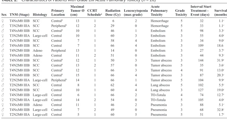
Eight of 16 patients (50%) with pretreatment tumor cavitation developed acute pulmonary toxicity of grade III or more, other than radiation pneumonitis, as opposed to only 12 of 71 (17%) in those without tumor cavitation. Table 2 sum-marizes details of all 20 patients developing acute pulmonary toxicity of grade III or more. Two patients with large cavi-tated tumors died of massive pulmonary hemorrhage 1 month after start of the treatment. Another patient died 2 weeks after the end of radiotherapy (46 Gy) from pulmonary embolism. Infection, presenting as an air-fluid level in the tumor cavita-tion (tumor abscess) on radiological imaging, was observed in the five remaining patients. Of these, one patient was hospital-ized and treated with intravenous (IV) administration of antibi-otics during CCRT, but died shortly after treatment. The other four were hospitalized 10, 13, 21, and 87 days, respectively, after the end of radiation treatment. Of the latter, two patients recovered after IV administration of antibiotics, and subse-quently developed intrathoracic disease progression after 18.5 and 27 months. Two remaining patients underwent (multiple) surgical intervention(s), which consisted of open-window tho-racostomy and muscle transposition into the thoracic cavity.
Aspergillus fuminatus was reported in pathological specimens derived from cavities of both patients. Despite surgical inter-vention, one patient died of postoperative complications and the other died of massive bleeding from the ipsilateral pulmo-nary artery 8.9 months after surgery.
The eight patients with baseline tumor cavitation, who did not develop any acute pulmonary toxicity of grade III or more, had intrathoracic disease progression within 7.3 months (n = 3), brain metastasis manifesting between 4.6 and 15.5 months (n = 3), or bone metastasis after 3.4 months (n = 1) as first site of recurrence. Only one patient was alive after 48.8 months without progression of disease.
DISCUSSION
The use of bi- and trimodality treatment strategies in patients with stage III NSCLC has increased awareness of the potential for severe treatment-related toxicity. We observed an 18% incidence of pretreatment tumor cavitation in 87 consecutive patients with stage III NSCLC at our center, with half developing acute pulmonary toxicity of grade III or more, including five patients with grade V complications. Despite a lack of consistent published data on the prognostic role of pretreatment tumor cavitation,7–9 patients in our study cohort
with cavitated tumor developed serious complications during or after CCRT.
The etiology of the observed toxicity is unclear. CCRT increases the likelihood of high-grade neutropenia; in our cohort, six of 10 patients who developed pneumonia of grade
III or more and/or abscess, had experienced leucocytopenia of grade III or more. Both the tumor cavitation (in SCC) and leu-cocytopenia may have increased the risk of developing infec-tious complications. However, 48 other patients with grade III/IV leucocytopenia did not experience these problems, a group that included 20 with SCC. Of the total cohort, five patients developed an infection and/or abscess in the tumor cavity, representing 6% of all patients.
To the best of our knowledge, no previous reports have highlighted such a high incidence of complications related to tumor cavitation during or after CCRT in phase III trials or
recent meta-analyses.2–5,14 Publications of randomized
clini-cal trials may have failed to identify some cliniclini-cal toxicities, as a result of patient selection and insufficient follow-up15. In
addition, regional differences in the incidence of squamous-cell tumors may also have contributed to under-reporting. For example, only 33% of patients in a large phase III CCRT trial from the United States had SCC,4 whereas 43% of patients in
the present study had SCC. In the Netherlands, the proportion of non-SCCs increased between the periods 1989 to 1993 and 2004 to 2009, from 42% to 67% for male patients and from 67% to 81% for female patients.16
Yet another explanation for the observed tumor cavitations and complications could be the relatively large primary tumor size in our patients, as 30% had a tumor measuring 8 cm or larger. A recent report suggests that many European centers consider tumors measuring more than 8 to 10 cm to be suitable for only palliative treatment.17
Similarly, a recent European Organisation for Research and Treatment of Cancer (EORTC) study excluded patients if the tumor volume required a radiation field encompassing 12 cm or more of the esophagus in the high-dose region.18
High-dose radiotherapy may contribute to tumor cavita-tion because of radiacavita-tion-induced decreases in regional lung perfusion,19 and central tumor necrosis has also been observed
after two fractions of 18 Gy in patients undergoing stereotactic radiotherapy.20 Although fluoro-2-deoxy-D-glucose positron
emission tomography may also identify central tumor necrosis, baseline tumor cavitation identified on fluoro-2-deoxy-D-glu-cose positron emission tomography has not been shown to be a poor prognostic feature.21 However, it should be pointed out
that patients in the last mentioned report did not undergo CCRT. A study of transthoracic aspirates from cavitating tumors showed that six of seven febrile patients had positive aspiration cultures at diagnosis.22 Since 2009, cone-beam CT scans are
rou-tinely performed during the course of radiotherapy at our center, and this can potentially facilitate the earlier detection of tumor cavitation and/or abscess during CCRT. Early identification of tumor cavitation may indicate a need for treatment using appro-priate antibiotics. Surgical management of lung abscess has been previously described in 247 patients, excluding abscesses sec-ondary to carcinoma.23 Despite conservative treatment in all, 119
and 58 patients underwent subsequent surgical drainage or pul-monary resection, respectively. Surgical drainage cured 67.2% of patients, 11.8% improved but needed additional interven-tions, and 21% died. Two of the patients in our report underwent open-window thoracostomy for acute complications of a tumor abscess within 5 months post-CCRT, after failure to respond to the IV administration of antibiotics. Nevertheless, one died of postoperative complications. Currently, we consider early surgi-cal drainage when conservative treatment strategies fail.
Some limitations of the present study must be recog-nized, one of which is the fact that it is restricted to patients in whom all follow-up was performed within the VU University Medical Center. Although overall survival between patients with or without tumor cavitation was not significantly differ-ent in this relatively small study, our findings suggest caution when using CCRT in this setting.
We postulate that primary surgical resection with post-adjuvant chemoradiotherapy in patients who are potentially
TABLE 1. Patient and Treatment Characteristics (n = 87) Characteristic Patients Sex Male 61 (70%) Female 26 (30%) Age (yrs) Median (range) 60 (42–80) Clinical staging Stage IIIA 36 (41%) Stage IIIB 51 (59%) Histology Squamous-cell carcinoma 37 (43%) Adenocarcinoma 28 (32%) Large-cell carcinoma 22 (25%)
Maximum size of primary tumor (n=83)
All tumors (median) 6 cm
Squamous-cell carcinoma (median) 7 cm Percentage of tumors ≥8 cm 26 (30%) Concurrent chemotherapy
Cisplatin-etoposide (3-weekly) 75 (86%) Cisplatin-docetaxel (weekly) 12 (14%) Total radiation dose (Gy)
<60 38 (44%)
60 21 (24%)
>60 28 (32%)
Leucocytopenia grade ≥III (n)
Squamous-cell carcinoma 25
Nonsquamous-cell carcinoma 29
Radiation pneumonitisa
Grade II 13
Grade III 4
aScored according to Common Toxicity Criteria v3.0.
FIGURE 1. Overall survival for patients with or without tumor cavitation. ©+, patients with cavitated tumors; ©−, patients with noncavitated tumors.
resectable could be a strategy to prevent complications of tumor cavitation. In addition, administration of weekly low-dose che-motherapy is potentially a less toxic alternative. Finally, evi-dence of infection in a developing cavity requires aggressive antibiotic treatment and probably early surgical intervention.
APPENDIX 1
Pretreatment tumor cavitation in six study patients.
REFERENCES
1. Alberts WM. Diagnosis and management of lung cancer executive summary: ACCP evidence-based clinical practice guidelines (2nd Edition). Chest 2007;132:1S–19S.
2. Aupérin A, Le Péchoux C, Rolland E, et al. Meta-analysis of concomitant versus sequential radiochemotherapy in locally advanced non-small-cell lung cancer. J Clin Oncol 2010;28:2181–2190.
3. Hanna N, Neubauer M, Yiannoutsos C, et al. Phase III study of cispla-tin, etoposide, and concurrent chest radiation with or without consolida-tion docetaxel in patients with inoperable stage III non-small-cell lung cancer: the Hoosier Oncology Group and U.S. Oncology. J Clin Oncol 2008;26:5755–5760.
4. Kelly K, Chansky K, Gaspar LE, et al. Phase III trial of maintenance gefitinib or placebo after concurrent chemoradiotherapy and docetaxel consolidation in inoperable stage III non-small-cell lung cancer: SWOG S0023. J Clin Oncol 2008;26:2450–2456.
5. Albain KS, Swann RS, Rusch VW, et al. Radiotherapy plus chemotherapy with or without surgical resection for stage III non-small-cell lung cancer: a phase III randomised controlled trial. Lancet 2009;374:379–386. 6. Lee CB, Stinchcombe TE, Moore DT, et al. Late complications of
high-dose (>/=66 Gy) thoracic conformal radiation therapy in combined
modality trials in unresectable stage III non-small cell lung cancer. J Thorac Oncol 2009;4:74–79.
7. Chaudhuri MR. Primary pulmonary cavitating carcinomas. Thorax 1973;28:354–366.
8. Pentheroudakis G, Kostadima L, Fountzilas G, et al. Cavitating squamous cell lung carcinoma-distinct entity or not? Analysis of radiologic, histo-logic, and clinical features. Lung Cancer 2004;45:349–355.
9. Mouroux J, Padovani B, Elkaïm D, Richelme H. Should cavitated bron-chopulmonary cancers be considered a separate entity? Ann Thorac Surg 1996;61:530–532.
10. Crabb SJ, Patsios D, Sauerbrei E, et al. Tumor cavitation: impact on objec-tive response evaluation in trials of angiogenesis inhibitors in non-small-cell lung cancer. J Clin Oncol 2009;27:404–410.
11. Sandler AB, Schiller JH, Gray R, et al. Retrospective evaluation of the clinical and radiographic risk factors associated with severe pulmonary hemorrhage in first-line advanced, unresectable non-small-cell lung can-cer treated with Carboplatin and Paclitaxel plus bevacizumab. J Clin Oncol 2009;27:1405–1412.
12. Phernambucq EC, Spoelstra FO, Verbakel WF, et al. Outcomes of con-current chemoradiotherapy in patients with stage III non-small-cell lung cancer and significant comorbidity. Ann Oncol 2011;22:132–138. 13. Maas KW, El Sharouni SY, Phernambucq EC, et al. Weekly
chemora-diation (docetaxel/cisplatin) followed by surgery in stage III NSCLC; a multicentre phase II study. Anticancer Res 2010;30:4237–4243. 14. O’Rourke N, Roque IFM, Farre Bernado N, et al. Concurrent
chemora-diotherapy in non-small cell lung cancer. Cochrane Database Syst Rev 2010:CD002140.
15. Ocana A, Amir E, Seruga B. Clinical research: show us the data. J Clin Oncol 2011;29:1099–1100.
16. Van der Drift MA, Karim-Kos HE, Siesling S, et al. Progress in standard of care therapy and modest survival benefits in the treatment of non-small cell lung cancer patients in the Netherlands in the last 20 years. J Thorac Oncol 2012;7:291–298.
TABLE 2. Characteristics of Patients with Grade ≥III Acute Pulmonary Toxicity (n = 20)
Sex TNM (Stage) Histology Primary Location
Maximal Tumor Ø
(cm) CCRT Schedulea
Radiation
Dose (Gy) Leucocytopenia (max grade) Acute Pulmonary
Toxicity Grade Toxicity
Interval Start Treatment -
Event (days) Survival (months)
♀ T4NxM0 IIIB SCC Centralb 13 1 6 2 Hemorrhage 5 32 1.1c
♂ T3N2M0 IIIA SCC Peripheralb 12 2 29 0 Hemorrhage 5 33 1.1c
♂ T4NxM0 IIIB SCC Centralb 10 1 46 1 Embolism 5 98 3.3c
♀ T3N2M0 IIIA Large-cell Central 10 1 60 3 Embolism 3 55 4.0c
♂ TxN3M0 IIIB SCC Central 7 1 58 4 Embolism 3 34 9.0c
♂ T4NxM0 IIIB SCC Central 7 1 66 4 Embolism 3 109 18.6
♂ T4N2M0 IIIB Adeno Peripheral 13 1 14 0 Embolism 3 27 3.7c
♀ T4NxM0 IIIB Adeno Central 11 2 51 0 Embolism 3 44 9.3c
♂ T4N3M0 IIIB SCC Centralb 12 1 50 3 Tumor abscess 3 144 31.9c
♂ T4N2M0 IIIB SCC Centralb 13 2 57 0 Tumor abscess 5 35 3.6c
♂ T4N2M0 IIIB SCC Centralb 12 1 66 3 Tumor abscess 4 91 13.0c
♀ T4NxM0 IIIB SCC Centralb 15 1 66 4 Tumor abscess 3 87 20.3c
♂ T2N2M0 IIIA Large-cell Peripheralb 14 1 66 1 Tumor abscess 5 104 9.9c
♂ T2N2M0 IIIA SCC Central 8 1 62 4 Lung abscess 5 102 5.5c
♂ T4N2M0 IIIB SCC Central 10 1 60 4 Lung abscess 3 127 19.0c
♂ T4N3M0 IIIB Large-cell Central 6 1 66 2 TO-fistula 4 74 12.7c
♀ T2N2M0 IIIA Large-cell Central 14 2 54 0 TO-fistula 5 105 4.0c
♀ T4NxM0 IIIB Adeno Central 11 1 46 2 Pneumonia 3 88 5.1c
♂ T4NxM0 IIIB Large-cell Central 7 2 49 0 Pneumonia 3 68 32.8c
♀ T2N2M0 IIIA Large-cell Central 5 1 60 5 Pneumonia 5 51 1.7c
TNM, tumor-node-metastasis; SCC, squamous-cell carcinoma; Large-cell, large-cell undifferentiated carcinoma; Adeno, adenocarcinoma; CCRT, concurrent chemoradiotherapy; TO-fistula, trachea-esophageal fistula.
aCisplatin-etoposide (1) or cisplatin-docetaxel (2). bPatients with tumor cavitation.
17. Sundstrøm S. Palliative external beam thoracic radiation therapy of non-small cell lung cancer. In B Jeremić (Ed.), Advances in Radiation Oncology in Lung Cancer. Berlin Heidelberg, PA: Springer-Verlag, 2011. 18. Belderbos J, Uitterhoeve L, van Zandwijk N, et al. Randomised trial of
sequential versus concurrent chemo-radiotherapy in patients with inoper-able non-small cell lung cancer (EORTC 08972-22973). Eur J Cancer 2007;43:114–121.
19. Zhang J, Ma J, Zhou S, et al. Radiation-induced reductions in regional lung perfusion: 0.1-12 year data from a prospective clinical study. Int J Radiat Oncol Biol Phys 2010;76:425–432.
20. Devisetty K, Salama JK. Tumor necrosis and cavitation after stereotactic body radiation therapy. J Thorac Oncol 2010;5:1100–1102.
21. Coffey JP, Hill JC. 18F-fluoro-2-deoxy-D-glucose standardized uptake value in cavitating non-small-cell lung carcinoma. Nucl Med Commun 2008;29:1040–1045.
22. Liao WY, Liaw YS, Wang HC, Chen KY, Luh KT, Yang PC. Bacteriology of infected cavitating lung tumor. Am J Respir Crit Care Med 2000;161:1750–1753.
23. Klepser RG, Davis EW. The surgical management of lung abscess. Dis Chest 1950;17:172–180.