An EEG-based brain-computer interface for gait training
Dong Liu
1,2∗, Weihai Chen
1, Kyuhwa Lee
2, Zhongcai Pei
1, and Jos´e del R. Mill´an
2 1. School of Automation Science and Electrical Engineering, Beihang University (BUAA), 100191, Beijing, ChinaE-mail: [email protected]
2. Defitech Chair in Brain-Machine Interface (CNBI), ´Ecole Polytechnique F´ed´erale de Lausanne (EPFL), Campus Biotech H4, 1202, Geneva, Switzerland
E-mail: [email protected]
Abstract: This work presents an electroencephalography (EEG)-based Brain-computer Interface (BCI) that decodes cerebral activities to control a lower-limb gait training exoskeleton. Motor imagery (MI) offlexion and extension of both legs was distinguished from the EEG correlates. We executed experiments with 5 able-bodied individuals under a realistic rehabilitation scenario. The Power Spectral Density (PSD) of the signals was extracted with sliding windows to train a linear discriminate analysis (LDA) classifier. An average classification accuracy of 0.67±0.07 and AUC of 0.77±0.06 were obtained in online recordings, which confirmed the feasibility of decoding these signals to control the gait trainer. In addition, discriminative feature analysis was conducted to show the modulations during the mental tasks. This study can be further implemented with different feedback modalities to enhance the user performance.
Key Words:Brain-computer Interface (BCI), electroencephalography (EEG), motor imagery (MI), gait training
1 INTRODUCTION
Brain-computer Interfaces (BCIs) are direct communica-tion and control systems between the brain and an external device, bypassing the physiological output pathways of pe-ripheral nerves and muscles [1]. For severely disabled in-dividuals suffering from illnesses such as amyotrophic lat-eral sclerosis (ALS), spinal cord injury (SCI) or strokes, BCI provides a promising approach for them to interact with the surroundings. In the past decades, non-invasive EEG-based BCI has been developed in both experimental contexts and clinical trials, e.g., brain-actuated intelligen-t wheelchair [2], powered lower-limb exoskeleintelligen-ton [3] and robotic arm [4].
EEG-based BCI can be categorized into spontaneous and evoked paradigms, according to the types of control signal-s. Evoked BCI exploits brain patterns in response to visual, auditory or tactile stimulus. Typical examples are steady state visually evoked potential (SSVEP) and P300. SSVEP is brain activity modulated in the visual cortex evoked by repetitive visual stimuli which has distinctive frequencies. It has been used to control a mobile robot [5]. Further-more, P300, elicited about 300 ms after the desired target stimulus presented within a random sequence of nontarget stimuli, can be implemented for the user to navigate un-known and evolving scenarios [6]. The advantages of these systems include high information transfer rate (ITR), min-imum training sessions and high accuracy. However, the subjects are easy to be fatigue with the consecutive stimu-lus and they have to keep following the cues all the time in order to transfer the information, which limits the
applica-This work was supported by the National Natural Science Founda-tion of China under Grant No. 61573047, No. 51675018, and the China Scholarship Council under Contract 201406020025.
bility of evoked BCIs.
Spontaneous BCI, on the other hand, is fully operated by the user without any external stimulus. The subjec-t can send subjec-their commands volunsubjec-tarily subjec-to insubjec-teracsubjec-t wisubjec-th the devices, e.g., neuroprosthesis and wheelchair. One of the most commonly-used neurophysiological signatures in such BCI systems is the modulation of sensorimotor rhythms (SMRs) during motor imagery (MI). SMRs can display related desynchronisation (ERD) and event-related synchronization (ERS) [7], which reflects the pow-er changes in cpow-ertain frequency bands, e.g., mu (7−13 Hz) and beta bands (15−30 Hz). Imagination of kinaesthet-ic movement of left hand, right hand and/or both feet can produce distinctive lateralized patterns on the primary sen-sory and motor cortex. Therefore, MI of left and right hand has been widely exploited as EEG correlates in BCI frame-works for motor training. Much attention has been focused on the development of brain-controlled robots for assistive strategy and rehabilitation tools using upper-limb MI [8]. More recently, interest has been devoted to studies about the gait rehabilitation. Brain-actuated robotic devices have been shown as an alternative to conventional physical ther-apy. In these paradigms, either healthy subjects or patients were required to imagine movement of the arms to mod-ulate EEG activity to generate motor commands to con-trol the lower-limb movement. For instance, MI of hand movement was used to control the Lokomat (Hocoma AG, Volketswil, Switzerland), a commercially available robot-ic walker [9]. Another work by Lee et al used MI of both hands to build a cascaded ERD classifier, to control the Rex (Rex Bionics LTD, Auckland, New Zealand), a hand-free, self-supporting robotic mobility device [10].
Signal acquisition
Feature engineering
Action generation
Subject External device
Classification/regression
Feedback interface
Figure 1: Architecture of a brain-controlled system. In the current study, the external device is a lower-limb gait train-ing exoskeleton.
tasks would be an intuitive yet challenging method for gait rehabilitation oriented BCI. Cortical homunculus, a physi-cal representation of functional region within the brain, has shown a relatively small brain region mapping to leg move-ments [11]. The cortical areas of legs, feet and toes are located closely at primary motor cortex from the frontal lobe. Besides, Brodmann cortical areas show that left and right legs are controlled by overlapping regions from the primary somatosensory cortex. As a result, the implemen-tation of discriminative features from MI of left and right legs are difficult to be applied in non-invasive BCI. Howev-er, a recent study has shown that the direction of imagined visual motion can be decoded using functional magnetic resonance imaging (fMRI) [12]. Similar patterns were per-formed in human gait therapy, e.g., repetitive and cyclic knee and ankle movement. Other lower-limb BCIs include sitting and standing classification from pre-movement s-tates [13] and ankle dorsiflexion MI detection from readi-ness potential, a type of slow cortical potentials (SCPs) [14].
The main goal of this work is to build an EEG-based BCI with lower-limb MI as control signals. In contrast to hand MI, we used MI of directional movement of both legs to differentiateflexion and extension. There are three char-acteristics of the proposed framework. First, directly us-ing MI of legflexion and extension is natural and intuitive, which is in consistent with the physical gait training proce-dures. Second, this paradigm is a spontaneous BCI, with-out time-locked to any external stimulus. The subject can initialize the task in a self-paced manner and get involved for a long period with full manipulation. Third, visual feed-back was provided to form a closed loop. The subjects can modulate their mental strategies accordingly in order to en-hance the performance. Furthermore, we performed fea-ture analysis to show the discriminate power in the classifi -cation of legflexion and extension. Another classification was conducted between mental task and rest period (base-line) in order to show the brain pattern modulations. In this work, we willfirst describe our BCI framework as a pattern recognition system. Then we will continue by materials and methods used for classification of legflexion and extension, as well as feature analysis. Results will be shown followed by the discussion and conclusion.
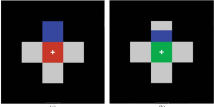
(a) (b)
Figure 2: Visual cues presented to the subject during the experiment: (a) Baseline period and (b) MI period.
2 MATERIALS AND METHODS
2.1 A brain-controlled system
A BCI is an artificial intelligence system which collects and analyzes biological neural signals. To date, specif-ic patterns in brain signals can be identified with a pat-tern recognition architecture, composed offive consecutive stages: signal acquisition, feature engineering, classifi ca-tion/regression, action generation and feedback interface, as shown in Figure 1.
Signal acquisition captures the brain signal using neu-roimaging techniques, e.g., EEG in the current work. Other processes, e.g., notchfiltering, band passfiltering and arti-fact rejection, might also be integrated in this module. Fea-ture engineering include feaFea-ture extraction and feaFea-ture se-lection. Typical features include signal envelope or ampli-tude in the time domain, power or phase lag in the frequen-cy domain, and Mahalanobis distance. Feature selection is performed to overcome the curse of dimensionality, as well as to find the subject-specific patterns corresponding to the mental tasks. Classification/regression are the main part as a decoder, which are typical supervised machine learning techniques for discrete and continuous output to-wards various applications. Actions are generated based on the decision made from the decoder, and then transferred to devices such as wheelchair, speller and even drone. A cer-tain set of feedback, e.g., visual, tactile and proprioceptive, is provided to inspect the real-time performance of the user. In this work, thefive modules were merged to translate the brain signals to control the robot, and visual feedback was presented for online testing.
2.2 Experimental protocol
Five subjects (three females, mean age 24.68±1.04) partic-ipated in the experiments. They were all naive BCI users with normal or corrected-to-normal vision. None of them reported any known neurological or psychiatric disease. The experimental protocol was approved by the local re-search ethical committee and all participants gave their in-formed consent.
Each subject did two sessions of recordings: offline for training and online for testing. Each session consists of five successive runs with a rest period in between. A cus-tomized gait training exoskeleton called the legoPress (by LSRO, EPFL, Lausanne, Switzerland) was implemented in
Figure 3: BioSemi-32 layout, and other configurations can be found at http://www.biosemi.com/headcap.htm.
this study. This robot is a simple and cost-effective device for gait retraining. More importantly, it can simulate the real movement of leg extension andflexion as a natural ap-proach for inducing sensory feedback. In each run, trials of extension andflexion were randomized and balanced and a total of 60 trials were performed (around 10 min). This yielded 300 trials for one session and each recording was around 1.5 h including experiment preparation and robot familiarization.
During the experiment, the subject was seated in the lego-Press with a monitor in front of the robot. The visu-al cues were presented with a customized Python scrip-t (hscrip-tscrip-tps://c4science.ch/diffusion/1299/) and synchronized with the lower-level routines. As shown in Figure 2, vi-sual bars pointing upwards indicated leg extension MI and downwards for legflexion MI. The preparation period in each trial was 2 s followed by MI period lasting 4 s for training sessions. In testing sessions, the MI period de-pended on the user performance and the time out was 5 s. The interval between these two sessions for each subject was around 2 weeks in order to simulate the realistic reha-bilitation scenario.
2.3 Signal acquisition and preprocessing
EEG signals were recorded from 32 channels according to the international 10/20 system using a Biosemi Active T-wo system. The sampling frequency was set to 2048 Hz. Ground was replaced by the Driven Right Leg (DRL) pas-sive electrode and all signals were referenced to the Com-mon Mode Sense (CMS) active electrode placed 1 cm to the left of POz. The layout of bioSemi32 cap can be seen in Figure 3. Before recording, the signals were inspect-ed with a GUI (eegviewer), as shown in Figure 4. Between runs, the waveforms were also checked by the experimenter to ensure the quality of signal.
For signal preprocessing, peripheral channels werefirst re-moved, since they were prone to body movement and phys-iological electrical artifact, e.g., electromyogram (EMG) and electrooculogram (EOG). We kept 17 channels: F3, FC1, FC5, C3, CP1, CP5, P3, Pz, P4, CP6, CP2, C4, FC6,
Figure 4: Customized GUI to show the EEG signals in real time.
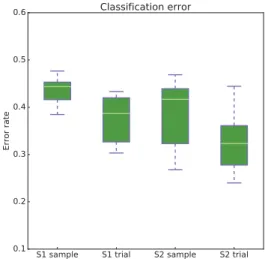
Figure 5: Sample-based and trial-based classification error from offline and online sessions, respectively.
FC2, F4, Fz, and Cz to reduce the signal contamination. In order to increase the Signal-to-Noise Ratio (SNR), a s-mall Laplacian spatialfilter was used by removing the local activity of neighboring electrodes as formula (1).
ei(t) =ei(t)−1/Nk Nk
j=1
ej(t) (1)
whereei(t)was the value ofith channel andNk was the
number of considered nearest neighbor channels. The DC component of the spatiallyfiltered signal was further re-moved as follows,
ei(t) =ei(t)−ei(t−T, t) (2)
whereei(t−T, t)was the mean value from timet−T to tin theith channel, andTis the length of time window. 2.4 Feature selection
As there were power changes in the EEG signals during MI tasks, we extracted the features in the frequency domain. The Power Spectral Density (PSD) was calculated over 1 s windows (T = 1)overlapped for 0.75 s using Hamming windows of 500 ms for all the channels, with 62.5 ms in-crements. The PSD was estimated in the range of 4 to 48
Figure 6: Classification result of one subject across the val-idation sets for session 1.
Hz with a resolution of 1 Hz. Therefore, the number of fea-tures was 765, given the remaining EEG channels (17) and the frequency components (45). We did a log transforma-tion on the features in order tofit the normality assumption for the following analysis.
After feature extraction, we performed feature selection us-ing Canonical Variate Analysis (CVA), also known as mul-tivariate discriminant analysis [15]. CVA was used to ex-tract Canonical Discriminant Spatial Patterns (CDSPs) on the original feature space, e.g., powers after log transfor-mation. Given the two mental tasks, i.e., legflexion and extension, CVA maximized the separability between the t-wo classes. We kept 10 features for further classification. 2.5 Classification
We used linear discriminant analysis (LDA) to classify the two mental tasks. LDA is a simple and well-known classi-fication approach based on normal distribution assumption and Bayes rule. To calculate the sample-based accuracy, we relied on a 5-fold cross validation. It is worth noting that the feature selection aforementioned was only performed on training data. Furthermore, chronological order of the data was maintained in order to yield a better,and less opti-mistic, estimation of accuracy.
In addition to sample-based classification, trial-based per-formance, also known as BCI command accuracy, was e-valuated based on the posterior probabilities from the slid-ing windows. An evidence accumulation shown in formu-la (3) was implemented as an exponential smoothingfilter, in order to eliminate effects from the outliers.
pt=α∗pt−1+ (1−α)∗pt (3)
whereptwas the integrated probability at timetandαwas
the smoothing factor. Asαdefined a trade-off between the command speed and confidence, we empirically set it to 0.95 in the current study. When the integrated probability reached the user-specific threshold, an action was generat-ed and the command would send to the robot to execute eitherflexion or extension movement accordingly. Final-ly, we reported the results in terms of both accuracy, and
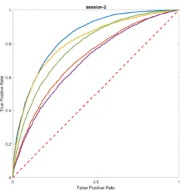
Figure 7: Classification result of one subject across the val-idation sets for session 2.
Table 1: Average AUC for each subjects in the two sessions
subject ID s1 s2 s3 s4 s5 session-1 0.76 0.73 0.73 0.74 0.71 session-2 0.84 0.71 0.81 0.70 0.78
sensitivity-specificity in the receiver operating characteris-tics (ROC) space.
2.6 Feature analysis
A post-processing of feature analysis was conducted to estimate the discriminant power (DP) for the two mental tasks. Given the number of features as cand number of classes ask, the feature matrixTwas defined as:
T =
k i=1
Ti (4)
The normalized eigenvalues were defined as γu= kλ−u1
u=1λu
(5) whereλuwas the eigenvalues of the CDSP matrix. DP of
single feature can be computed as formula (6).
DPe= 100∗ N i=1 k−1 u=1γuitieu2 N i=1 c e=1 k−1 u=1γuitieu2 (6) whereNwas the number of runs, andti
euwas the element
from feature matrix withe = 1, ..., c. The calculation of DP took the stability of features among data chunks into consideration, since it penalized the features not consisten-t during runs. We reporconsisten-ted consisten-the DP wiconsisten-th boconsisten-th consisten-topographic and heat maps between MI of leg extension and flexion. Furthermore, DP between focus and rest was shown to es-timate the brain patterns during the mental tasks.
3 RESULTS
3.1 classification
Figure 5 depicts the classification error from both session-s. As expected, the BCI command accuracy was signifi -cantly better than sample-based performance (two-sample
Figure 8: Heat maps to shown the DP of a typical subject. Peripheral channels were removed for the analysis.
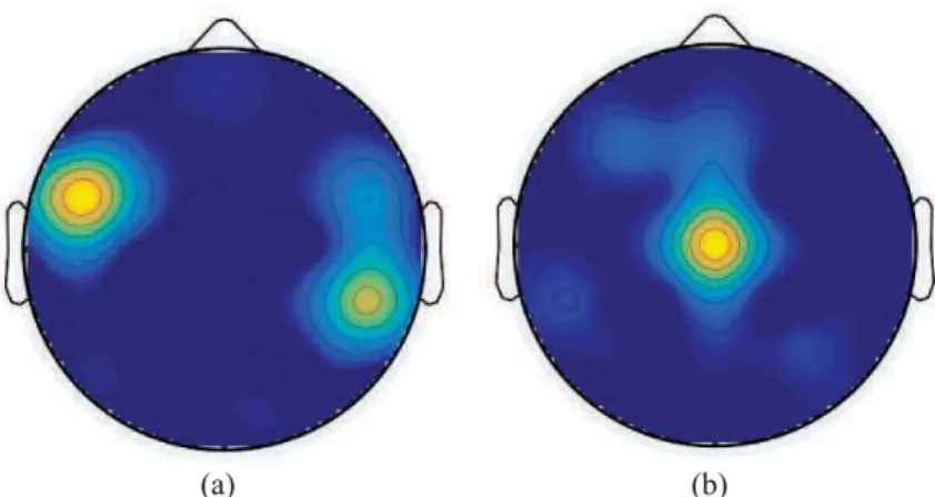
Figure 9: Topographic maps to shown the DP of a typical subject. The DP of peripheral channels was set to 0.
t-test, p<0.01 and p = 0.017, respectively). No signif-icant difference was found (two-sample t-test, p> 0.05) between these two sessions. The average BCI command accuracy of offline and online sessions were 0.63±0.05 and 0.67±0.07, respectively. Furthermore, chance level was calculated based on binomial distribution [16], which yielded an accuracy of 0.56. The performance of all sub-jects was significantly better than the chance level, with an accuracy of 0.76 in the best case.
Results of binary classification with ROC curves are shown in Figure 6 and 7. Each curve represents the performance of one subject averaged across 5-fold cross validation. The chance level was calculated by shuffling the labels for the training data and performing 1000 times 5-fold cross vali-dation, as shown in dotted red lines. The mean Area Under the Curve (AUC) values across the cross validation are il-lustrated in Table 1. The mean AUC of the two sessions were 0.73±0.02 and 0.77±0.06, respectively.
3.2 Feature analysis
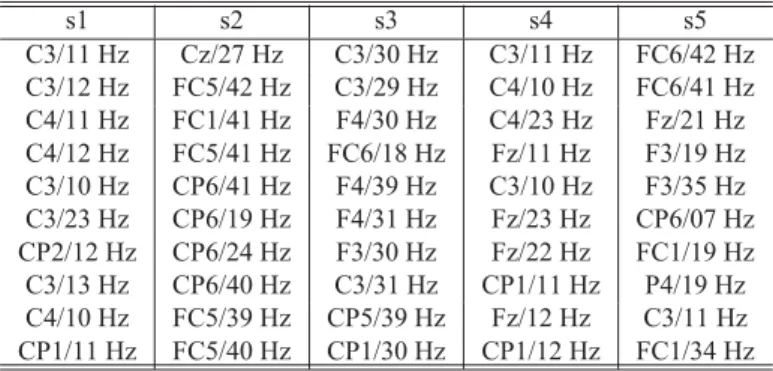
Based on CVA, DP for the classification was estimated for each subject. Discriminative features of a typical subject are shown in both heat maps and topoplots, as displayed in Figure 8 and 9, respectively. The DP is distributed at prima-ry motor cortex, mainly in the mu band. C3 and C4 are the discriminant channels for the classification. Features, i.e., channel and frequency component pairs, selected by CVA from offline and online sessions are shown in Table 2 and Table 3, respectively. Although the DP is highly subject-specific, mu and beta bands located at frontal and central brain areas are the most frequently selected features.
Table 2: Features selected by CVA for all subjects in the offline session. s1 s2 s3 s4 s5 C3/11 Hz Cz/27 Hz C3/30 Hz C3/11 Hz FC6/42 Hz C3/12 Hz FC5/42 Hz C3/29 Hz C4/10 Hz FC6/41 Hz C4/11 Hz FC1/41 Hz F4/30 Hz C4/23 Hz Fz/21 Hz C4/12 Hz FC5/41 Hz FC6/18 Hz Fz/11 Hz F3/19 Hz C3/10 Hz CP6/41 Hz F4/39 Hz C3/10 Hz F3/35 Hz C3/23 Hz CP6/19 Hz F4/31 Hz Fz/23 Hz CP6/07 Hz CP2/12 Hz CP6/24 Hz F3/30 Hz Fz/22 Hz FC1/19 Hz C3/13 Hz CP6/40 Hz C3/31 Hz CP1/11 Hz P4/19 Hz C4/10 Hz FC5/39 Hz CP5/39 Hz Fz/12 Hz C3/11 Hz CP1/11 Hz FC5/40 Hz CP1/30 Hz CP1/12 Hz FC1/34 Hz
Table 3: Features selected by CVA for all subjects in the online session. s1 s2 s3 s4 s5 CP6/29 Hz Cz/23 Hz FC6/28 Hz P4/07 Hz F3/29 Hz FC5/30 Hz Cz/20 Hz FC6/29 Hz C4/10 Hz FC5/30 Hz CP6/30 Hz Cz/21 Hz FC6/30 Hz FC5/15 Hz F3/30 Hz FC5/28 Hz Fz/04 Hz FC6/27 Hz CP5/10 Hz F3/26 Hz FC5/29 Hz Cz/24 Hz FC6/26 Hz C4/11 Hz F3/22 Hz CP6/28 Hz Cz/22 Hz FC6/25 Hz CP6/07 Hz F3/25 Hz FC5/27 Hz F3/17 Hz CP6/30 Hz CP6/10 Hz FC6/28 Hz FC5/26 Hz Cz/25 Hz FC6/24 Hz C3/07 Hz FC6/27 Hz FC6/29 Hz Fz/05 Hz CP6/28 Hz Fz7/07 Hz FC5/22 Hz FC5/24 Hz CP5/14 Hz CP6/29 Hz P4/22 Hz F3/21 Hz
Furthermore, the DP of focus vs. rest was estimated, to compare with the leg extension vs. flexion, as shown in Figure 10. The channels selected in the leg extension vs. flexion are FC5 and CP6, located in Brodmann Area 40 and 44, with the function of motor planning and somatosensory integration. On the other hand, the features of MI vs. rest are located in the the primary motor cortex, especially at Cz, which is in consistent with previous works of lower-limb MI [7].
4 Discussion
Brain-actuated robotics usually exploit spontaneous EEG correlates, e.g., MI, as control signals. To design a BCI for gait rehabilitation, directly performing lower-limb MI, e.g., leg extension andflexion is a natural and intuitive strategy. In this study, we built classifiers and executed experiments in a closed loop to tackle this issue. The goal is to evaluate whether we canfind significant difference between MI of leg extension andflexion, as well as discriminative features employed in the mental tasks. Although the DP is highly subject-specific and session-specific, we showed promising results with both sample-based and BCI command accura-cy. The results reveal that it is possible to distinguish MI of leg extension andflexion, based on the proposed pattern recognition framework.
In the current work, CVA was used for feature selection and LDA classifier was built to differentiate the mental tasks. We assume that the log transformed PSD follows a Gaus-sian distribution and the two classes are linearly separable. However, the brain signal might be highly nonlinearity and more sophisticated machine learning techniques, e.g., di-mensionality reduction and classification, could be
intro-(a) (b)
Figure 10: Topographic maps to compare the DP between leg extension vs.flexion (a) and focus vs. rest (b). duced to enhance the capability of the model. Besides,
heathy subjects participated in the experiments as a proof of concept. Our ultimate users would be motor disabled subjects, e.g., paraplegic patients, to help them with the proposed brain-controlled gait trainer for therapy.
The main limitation of a MI-based BCI is the long period required for training. In this study, each subject was record-ed with two sessions with visual ferecord-edback providrecord-ed during online testings. The users learned fast on the modulation of their brain patterns for the mental tasks, although none of them had any experience with MI. Further improvement might be achieved with more experimental sessions. Still, a large number of subjects, including end-users, should be incorporated in further work to reach a stronger conclusion [17]. In addition to visual feedback, multiple modalities of feedback, e.g., proprioceptive or functional electrical stim-ulation (FES) would be introduced and compared in further work.
ACKNOWLEDGEMENT
We would like to thank Dr. Mohamed Bouri from LSRO, EPFL, Switzerland, for modification on the gait training ex-oskeleton. We also appreciate all subjects who participated in the recordings.
REFERENCES
[1] J. R. Wolpaw, N. Birbaumer, D. J. McFarland, G. P-furtscheller, and T. M. Vaughan, Brain-computer interfaces for communication and control, Clinical neurophysiology 113, no. 6: 767-791, 2002.
[2] J. d. R. Mill´an, F. Gal´an, D. Vanhooydonck, E. Lew, J. Philip-s, and M. Nuttin, Asynchronous non-invasive brain-actuated control of an intelligent wheelchair, Proc. 29th Annual Intl. Conf, 2009.
[3] K. Lee, D. Liu, L. Perroud, R. Chavarriaga, and J. D. R. Mil-l´an, Endogenous control of powered lower-limb exoskeleton, Wearable Robotics: Challenges and Trends, pp. 115-119, 2017.
[4] J. Meng, S. Zhang, A. Bekyo, J. Olsoe, B. Baxter, and B. He, Noninvasive Electroencephalogram Based Control of a Robotic Arm for Reach and Grasp Tasks, Scientific Reports 6: 38565, 2016.
[5] X. Gao, D. Xu, M. Cheng, and S. Gao, A BCI-based environ-mental controller for the motion disabled, IEEE Transactions on Neural Systems and Rehabilitation Engineering, 11: 137-140, 2003.
[6] I. Iturrate, J. M. Antelis, A. Kubler, and J. Minguez, A nonin-vasive brain-actuated wheelchair based on a P300 neurophys-iological protocol and automated navigation, IEEE Transac-tions on Robotics 25, no. 3: 614-627, 2009.
[7] G. Pfurtscheller, F. L. Da Silva, Event-related EEG/MEG synchronization and desynchronization: basic principles, Clinical Neurophysiology, 110: 1842-1857, 1999.
[8] G. Dornhege, J. d. R. Mill´an, T. Hinterberger, D. McFar-land, and K. Muller, Toward brain-computer interfacing, MIT press, 2007.
[9] A. R. Donati, S. Shokur, E. Morya, D. S. Campos, F. L. Brasil, Long-term training with a brain-machine interface-based gait protocol induces partial neurological recovery in paraplegic patients, Scientific Reports 6, 2016.
[10] K. Lee, D. Liu, L. Perroud, R. Chavarriaga, and J. D. R. Mill´an, A brain-controlled exoskeleton with cascaded event-related desynchronization classifiers, Robotics and Au-tonomous Systems, 2016.
[11] W. Penfield and E. Boldrey, Somatic motor and sensory rep-resentation in the cerebral cortex of man as studied by elec-trical stimulation, Brain: A journal of neurology, 1937. [12] T. C. Emmerling, J. Zimmermann, B. Sorger, M. A. Frost,
and R. Goebel, Decoding the direction of imagined visual motion using 7 T ultra-highfield fMRI, Neuroimage, 125, 61-73, 2016.
[13] T. C. Bulea, S. Prasad, A. Kilicarslan, and J. L. Contrerasvi-dal, Sitting and standing intention can be decoded from s-calp EEG recorded prior to movement execution, Frontiers in Neuroscience, 8(6), 2014.
[14] I. K. Niazi, N. Jiang, O. Tiberghien, J. F. Nielsen, K. Drem-strup, and D. Farina, Detection of movement intention from single-trial movement-related cortical potentials, Journal of neural engineering, 8(6): 066009, 2011.
[15] F. Gal´an, P. W. Ferrez, F. Oliva, J. Guardia, and J. d. R. Mil-l´an, Feature extraction for multi-class BCI using canonical variates analysis, IEEE International Symposiumon on Intel-ligent Signal Processing, pp. 1-6, 2007.
[16] M. Gernot, R. Scherer, C. Brunner, R. Leeb, and G. P-furtscheller, Better than random: A closer look on BCI re-sults, International Journal of Bioelectromagnetism 10, pp. 52-55, 2008.
[17] R. Leeb, S. Perdikis, L. Tonin, A. Biasiucci, M. Tavella, M. Creatura, and J. d. R. Mill´an, Transferring brainCcom-puter interfaces beyond the laboratory: successful applica-tion control for motor-disabled users, Artificial Intelligence in Medicine 59: 121-132, 2013.