0066-4804/07/$08.00
⫹
0
doi:10.1128/AAC.00377-07
Copyright © 2007, American Society for Microbiology. All Rights Reserved.
Randomized, Double-Blind, Placebo-Controlled Trial of Cephalexin
for Treatment of Uncomplicated Skin Abscesses in a Population
at Risk for Community-Acquired Methicillin-Resistant
Staphylococcus aureus
Infection
䌤
Priya M. Rajendran,
1David Young,
2* Toby Maurer,
1Henry Chambers,
3Francoise Perdreau-Remington,
3Peter Ro,
4and Hobart Harris
2Department of Dermatology,
1Department of Surgery,
2and Department of Medicine,
3University of
California San Francisco School of Medicine,
4San Francisco, California
Received 20 March 2007/Returned for modification 10 July 2007/Accepted 27 August 2007
Empirical use of beta-lactam antibiotics, the preferred agents for treating uncomplicated skin and soft tissue
infections, may no longer be appropriate for these infections because of the increasing prevalence of community
strains of methicillin-resistant
Staphylococcus aureus
(MRSA). Retrospective studies, however, suggest that
outcomes are good even when beta-lactams are used. We conducted a randomized, double-blind trial of 166
outpatient subjects comparing placebo to cephalexin at 500 mg orally four times for 7 days after incision and
drainage of skin and soft tissue abscesses. The primary outcome was clinical cure or failure 7 days after
incision and drainage.
S. aureus
was isolated from 70.4% of abscess cultures. Of the isolates tested 87.8% were
MRSA, 93% of which were positive for Panton-Valentine leucocidin genes. Clinical cure rates were 90.5% (95%
confidence interval, 0.82 to 0.96) in the 84 placebo recipients and 84.1% (95% confidence interval, 0.74 to 0.91)
in the 82 cephalexin recipients (difference in the two proportions, 0.0006; 95% confidence interval,
ⴚ
0.0461 to
0.0472;
P
ⴝ
0.25). The 90.5% cure rate observed in the placebo arm and 84.1% cure rate observed in the
cephalexin arm provide strong evidence that antibiotics may be unnecessary after surgical drainage of
uncomplicated skin and soft tissue abscesses caused by community strains of MRSA.
There is evidence that antibiotics may not be necessary for
treatment of uncomplicated skin and soft tissue infections
(SSTIs) such as abscesses, including those caused by
methicillin-resistant
Staphylococcus aureus
(MRSA) (10, 14). In recent
studies, treatment outcomes for SSTIs were the same whether
or not the antibiotic prescribed was actually active against the
cultured organism (10, 11, 14, 20). When MRSA isolates were
cultured from SSTI patients, infections resolved even when
beta-lactam antibiotics, which are presumably inactive against
MRSA, were used (11, 20, 30). It is standard practice to treat
patients with SSTIs with an antibiotic (30). A 2005 survey
found that 87% of health care providers continue to prescribe
antibiotics after incision and drainage of abscesses, and a
beta-lactam antibiotic is typically used (24). Overuse of antibiotics
has adverse consequences, including untoward side effects and
financial costs, and may contribute to the spread of
antibiotic-resistant organisms (9, 15, 18, 25).
The alarming increase in the prevalence of
community-as-sociated MRSA may be a consequence of years of antibiotic
misuse. The virulence of community MRSA strains, which
typically carry genes for Panton-Valentine leucocidin (PVL),
has been of particular concern (3, 5, 23). PVL has been
asso-ciated with furunculosis; severe SSTIs, including necrotizing
fasciitis (19); and necrotizing pneumonia (10, 16). These
de-velopments have necessitated a reassessment of the
manage-ment and antimicrobial therapy of staphylococcal infections.
The purposes of this study were (i) to compare a current
“standard-of-care” antibiotic, cephalexin, to placebo after
sur-gical incision and drainage of uncomplicated skin abscesses;
(ii) to establish the prevalence of MRSA in the population
under study; and (iii) to prospectively determine whether
dis-cordance between therapy and isolate susceptibility affected
outcome.
MATERIALS AND METHODS
Study setting.This study was conducted at the ISIS Clinic at the San Francisco General Hospital from November 2004 to March 2005. The patient population at this clinic has high rates of illicit drug injection use; homelessness; and infection with hepatitis C, hepatitis B, or human immunodeficiency virus (HIV) (30). The clinic provides uninterrupted, 7-days-per-week treatment from 9 a.m. to 5 p.m. on weekdays and 9 a.m. to 12 p.m. on weekends and holidays for all patients on a walk-in, outpatient basis (12).
Study population.All patients presenting to the ISIS Clinic are assessed by a surgeon to determine if an abscess is present and if surgical drainage is necessary. All study participants were recruited from the group of patients considered to have a surgically drainable abscess (Fig. 1). We included patients with medical comorbidities such as intravenous drug use, hepatitis B and C, HIV infection, and diabetes. Subjects were eligible for enrollment if they were over the age of 18 years and had an abscess that the attending surgeon believed required surgical intervention and was severe enough that a duration of 5 or more days of anti-biotic therapy was anticipated. Diagnostic criteria for an abscess were as follows: (i) acute onset within 7 days prior to enrollment; (ii) purulent drainage or purulent aspirate; (iii) erythema, induration (ⱖ2 cm in diameter), or tenderness; and (iv) evidence of loculated fluid at time of enrollment. Our clinical standard is to prescribe antibiotics when two or more of these criteria are met. Subjects were ineligible for enrollment if they were unlikely to survive through the treat-ment period and evaluations or had toxic shock syndrome or toxic shock-like syndrome, shock or hypotension, oliguria (urine output of⬍20 ml/h) not
re-* Corresponding author. Mailing address: Box 0807, SFGH 3A,
Uni-versity of California, San Francisco, San Francisco, CA 94143-0807.
Phone: (415) 206-4643. Fax: (415) 206-5484. E-mail: dyoung@sfghsurg
.ucsf.edu.
䌤
Published ahead of print on 10 September 2007.
sponsive to fluid challenge, an incisional wound extending into visceral compart-ments, suspected or proven contiguous bone or joint involvement, ischemic ulcers or wounds associated with severe arterial insufficiency or gangrene, infec-tion of prosthetic materials or venous catheters that could not be removed as part of the treatment of the current infection, infection of a full-thickness burn wound or burn wound that was⬎20% of total body area, allergy to penicillin or cephalexin, or renal compromise requiring adjusted dosing of cephalexin.
Febrile patients were included. The current standard of care at ISIS does not include routine blood drawings, and thus, white blood cell counts were not a component of the study criteria.
The Committee on Human Research at the University of California approved the study, and all patients gave written informed consent.
The drug trial is registered under identifier NCT00187759 at http://www .clinicaltrials.gov.
Study protocol.At the initial visit, participants underwent a directed history and a physical examination, including complete examination of the skin. Ab-scesses were measured, and local anesthetic was injected along the line of the anticipated incision. Using a no. 11 blade, the attending surgeon used a sawing motion to create a wide opening and completely drain the pus. The cavity was probed for loculations and further drained if necessary. The cavity was packed from deep to superficial levels with plain gauze for healing by secondary inten-tion. Participants were then randomized to treatment assignments by the phar-macist, using a block randomization scheme to generate a 1:1 ratio of subjects in each group. Assignments were placed in sequentially numbered, sealed enve-lopes, which were opened and recorded after all other data had been entered.
The antibiotic treatment regimen consisted of the current standard of care in the community, oral cephalexin at 500 mg four times daily for 7 consecutive days. Patients in the placebo group received oral placebo capsules identical in appear-ance to the cephalexin capsules for the same length of time. All subjects were seen daily in the clinic by a nurse who assessed wound healing and changed dressings until the wound showed the following signs of healing: absence of purulent wound drainage, erythema, fluctuance, localized warmth, pain/tender-ness, and edema/induration. Patients were also asked to return 7 days after initial study enrollment for their follow-up visit. All patients, investigators, and clinic staff were blinded to study group assignment.
Microbiologic cultures and susceptibility testing.Wound cultures were ob-tained from patients during the incision and drainage procedure. The intact skin over the abscess was cleansed with 10% povidone-iodine solution (Smith and Nephew, Largo, FL) prior to incision. A sterile Dacron swab (Becton, Dickinson
& Co., Cockeysville, MD) was rotated within the cavity immediately after sur-gical incision. Wet swabs were sent to the clinical microbiology laboratory at San Francisco General Hospital for routine aerobic and anaerobic culture and anti-microbial susceptibility testing. Since staphylococci and streptococci are the two most prevalent organisms in SSTIs, the laboratory tested only for these. MICs were determined by Microscan (Microscan Walkaway instrument; Dade Inter-national, West Sacramento, CA). Guidelines from the Clinical and Laboratory Standards Institute (formerly NCCLS) were used throughout to assess suscep-tibility (22). Samples identified as MRSA were tested for the presence of PVL genes using the method of Lina et al. (16).
Clinical response. The primary study outcome was either clinical cure or clinical failure, which was determined according to a predetermined set of cri-teria and based on the clinical judgment of trained nurse practitioners. There is currently no validated instrument available to assess cure or failure of an abscess; the goal of this study was not to create such an instrument but rather to reflect clinical practice. Determination of clinical cure was made by one of the study investigators (P.R.) and five trained study nurses at the 1-week follow-up visit if there was resolution of the following signs and symptoms—purulent wound drainage, erythema, fluctuance, localized warmth, pain/tenderness, and edema/ induration—such that no further antibiotics or surgical procedure was needed. Treatment failure, defined as the presence of any of those symptoms, was con-firmed by a nurse practitioner in the ISIS Clinic if the infection did not resolve as expected and thereby required antibiotics or a surgical procedure anytime after study entry. All those who assessed study outcome were trained by a study investigator. Patients who came to the clinic after the designated 1-week fol-low-up visit were also included. Treatment adherence was based on patient self-report.
Patients were instructed not to take any other antibiotics except the study drug during the study period. If they reported taking additional antibiotics for their abscess or for any unrelated condition, treatment was considered a failure and patients were followed in the clinic until their infection resolved.
Statistical analysis.This was an intent-to-treat analysis of all patients who enrolled in the study. The primary clinical end point was the proportion of patients whose wounds were considered to be clinically cured. Early stopping rules were set in the event that the overall cure rate was high, irrespective of treatment group. Patients who missed their follow-up visit and whose treatment was not already considered a failure were contacted via telephone by one of the study investigators, and their medical records were reviewed. If sufficient infor-mation was available to determine clinical outcome, these patients were included in the analysis. Patients who did not return for follow-up, who could not be contacted by phone, and whose outcome could not be determined from chart review were deemed failures.
The sample size was designed to provide 80% power to detect a difference of 10% or more between the two groups (6). A one-tailed Fisher exact test with a 5% level of significance was used to compare the primary outcomes, as placebo was not expected to be better than cephalexin.
Statistical analyses were performed using Microsoft Access 2000 and SAS software, versions 8.2 and 9.1. Cure rates are presented as proportions with corresponding 95% confidence intervals. Dichotomous variables were analyzed using Fisher’s exact test. Ordinal variables were analyzed using the Kruskal-Wallis test. Continuous variables were analyzed with Mann-Whitney tests. Two-sidedPvalues of⬍0.05 were set to indicate statistical significance for all other comparisons.
RESULTS
Characteristics of the study subjects.
A total of 166 eligible
subjects were enrolled and randomized to receive either
cepha-lexin or placebo (Fig. 1). Subjects randomized to the two study
groups were similar with respect to baseline characteristics
(Table 1). A total of four subjects were lost to follow-up.
Clinical response was determined in the clinic for 89% of the
166 subjects eligible for analysis. Of the 14 subjects who did not
return for their 1-week follow-up visit, outcome was
deter-mined by chart review of hospital records alone (
n
⫽
8) and by
chart review coupled with a telephone conversation with one of
the study investigators (
n
⫽
2). Treatment adherence data
were available for 113 patients: 72.2% (
n
⫽
39) reported taking
cephalexin as instructed and 78.0% (
n
⫽
46) reported taking
placebo as instructed. No major adverse events (events
result-ing in death, life-threatenresult-ing situations, or permanent
disabil-ities) were reported for either study group. No disseminated
disease such as pneumonia or meningitis was reported.
Clinical outcome.
There was no difference in the clinical
cure rate between subjects receiving placebo and those
receiv-ing cephalexin (90.5% [
n
⫽
76/84] versus 84.1% [
n
⫽
69/82];
95% confidence interval, 0.82 to 0.96 versus 0.74 to 0.91,
re-spectively; difference in the two proportions, 0.0006; 95%
con-fidence interval for this difference,
⫺0.0461 to 0.0472;
P
⫽
0.25). Of the 21 subjects in the treatment failure group, four
were lost to follow-up (Table 2). Of the 17 subjects who were
followed up, 11 from the cephalexin group and six from the
placebo group were subsequently prescribed antibiotics. Four
from the cephalexin group underwent another surgical
proce-dure, and two, also from the cephalexin group, were
hospital-ized.
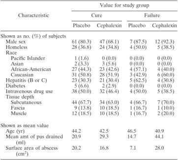
Patients who were cured and those who were not did not
differ with respect to demographics, medical comorbidities, or
abscess characteristics (Table 3). Among those with HIV
in-fection, all six of those on placebo and 8/13 on cephalexin were
cured (
P
⫽
0.128).
Pathogen distribution and susceptibility.
There was no
dif-ference in pathogen distribution between study groups (Table
4). Of the 162 patients for whom cultures were obtained,
Staphylococcus aureus
was isolated from 114 (114/162
⫽
70.4%). Of these 114
S. aureus
isolates, 99 were tested for
antibiotic susceptibilities; 87 (87/99
⫽
87.8%) were MRSA. Of
these 87 MRSA isolates, 86 were tested for the presence of the
PVL gene and 80 were positive for PVL (80/86
⫽
93.0%).
Susceptibility data for the isolates tested are shown in Table 5.
There was no difference in the distribution as well as clinical
cure rates in patients with PVL-positive MRSA between the
placebo group and the cephalexin group (Table 6). Of the
seven
Streptococcus
species-only isolates, the three/five
pa-tients on placebo were cured and two/two papa-tients on
cepha-lexin were cured.
TABLE 1. Baseline characteristics of the 166 study subjects
included in the analyses, by study group
Characteristic Value for study group: Pvalue Cephalexin Placebo
Age, median (range), yr 43 (22–66) 45 (22–88) 0.3191 Male sex (%) 72.0 81.0 0.2313 Race (%) 0.2431 American Indian 6.2 0 Pacific Islander 1.5 1.5 Asian 4.6 1.5 African-American 38.5 46.2 Caucasian 49.2 50.8 Homeless (%) 35.4 38.1 0.7493 Diabetes (%) 2.4 6.0 0.4432 Hepatitis B or C (%) 30.5 33.3 0.7408 HIV infection (%) 15.9 7.1 0.0915 Abscess size of⬎5 cm in dimension (%) Length 17.7 18.1 1.0000 Width 19.2 24.1 0.5668 Depth 1.4 5.3 0.3678
Surface area, mean (range), cm2 18.7 (1–144) 19.0 (1–150) 0.3676 Tissue depth (%) Subcutaneous 64.1 67.7 0.9048 Fascia 17.2 14.1 Muscle 18.8 18.3 Underlying skin disease (%) Folliculitis 38.8 40.2 1.0000 Atopic dermatitis 0.05 0.01 1.0000
TABLE 2. Reasons for clinical failures by study group
Reason No. of subjects in study group: Cephalexin (n⫽13) Placebo (n⫽8)
Nurse practitioner thought abscess
was not healing as expected
8
3
Patient was prescribed antibiotics
for new, nonadjacent abscess
0
2
Patient returned to emergency
department complaining of
worsening abscess
2
0
Patient started on antibiotics for
unrelated medical condition
during study period
0
1
Primary care provider urged
patient to withdraw from study
due to HIV infection
1
0
Loss to follow-up
2
2
TABLE 3. Characteristics of the 166 subjects, by clinical outcome
Characteristic
Value for study group Cure Failure Placebo Cephalexin Placebo Cephalexin Shown as no. (%) of subjects
Male sex 61 (80.3) 47 (68.1) 7 (87.5) 12 (92.3) Homeless 28 (36.8) 24 (34.8) 4 (50.0) 5 (38.5) Race Pacific Islander 1 (1.6) 0 (0.0) 0 (0.0) 0 (0.0) Asian 2 (3.3) 3 (5.6) 0 (0.0) 0 (0.0) African-American 27 (44.3) 23 (42.6) 4 (57.1) 4 (40.0) Caucasian 31 (50.8) 28 (51.9) 3 (42.9) 6 (60.0) Hepatitis (B or C) 23 (30.3) 21 (30.4) 5 (62.5) 4 (30.8) Diabetes 5 (6.6) 2 (2.9) 0 (0.0) 0 (0.0) Intravenous drug use 38 (50.0) 32 (46.4) 4 (50.0) 5 (38.5) Tissue depth
Subcutaneous 44 (67.7) 34 (63.0) 4 (66.7) 7 (70.0) Fascia 9 (13.8) 10 (18.5) 1 (16.7) 1 (10.0) Muscle 12 (18.5) 10 (18.5) 1 (16.7) 2 (20.0) Shown as mean value
Age (yr) 44.2 42.5 46.5 40.9 Mean amt of pus drained
(ml)
20.9 29.3 14.7 44.1 Surface area of abscess
(cm2)
20.2 16.8 7.1 28.0
TABLE 4. Pathogen distribution by study group
Organism No. (%) of subjects by study group: Cephalexin (n⫽80) Placebo (n⫽82)
Staphylococcus aureus
only
55 (68.8)
55 (67.1)
Streptococcus
species only
2 (2.5)
5 (6.1)
Both
Staphylococcus aureus
and
Streptococcus
species
3 (3.8)
1 (1.2)
DISCUSSION
This study compared a commonly used beta-lactam
antibi-otic, cephalexin, to placebo for the treatment of uncomplicated
skin and soft tissue abscesses after incision and drainage in a
population with high rates of community-acquired MRSA. Our
findings of high clinical cure rates (84 to 90%) and no
differ-ence in clinical cure rates for cephalexin compared to placebo
indicate that beta-lactam antibiotics probably do not provide
an additional benefit over that afforded by incision and
drain-age in the treatment of cutaneous abscesses. Even though
more than 90% of MRSA clinical isolates were positive for
PVL genes, this had no apparent effect on outcome.
A possible criticism of the study design is that no active
treatment arm was included (assuming that beta-lactams have
no clinically useful antimicrobial activity against MRSA).
How-ever, given the high cure rates in both the cephalexin and
placebo arms—rates which are consistent with those of four
recently published studies of SSTIs (88 to 96%) (1, 13, 27,
29)—it is doubtful that an active agent would have performed
any better. The one previous placebo-controlled study of
cephradine for soft tissue infections, including abscesses, was
conducted in 1985 and included 50 subjects (17). That study
found no difference in cure rates between the antibiotic and
placebo groups; to our knowledge, microbiologic susceptibility
was not reported. Observational, retrospective studies have
found no difference in outcomes of patients treated with
beta-lactam antibiotics for SSTIs caused by MRSA compared to
those given an antibiotic to which the isolate was susceptible
(11, 14, 20, 30). Our trial is the first interventional study that
directly supports this finding. These observational studies in
conjunction with our results strongly suggest that antibiotics
may not be necessary for treating some SSTIs, including those
caused by MRSA.
Clinicians have been reluctant to change the current
stan-dard of care, citing the lack of evidence from well-designed
randomized control trials and the increasing prevalence of
MRSA (7, 10, 21, 26). Even clinicians knowledgeable about
antibiotic overuse continue to prescribe beta-lactam antibiotics
for soft tissue infections (14, 24). The two main arguments in
support of this practice are (i) that these infections may be
polymicrobial, containing some streptococcal species sensitive
to beta-lactam antibiotics (2, 8, 11), and (ii) that killing the
sensitive organisms in the soft tissue infection may tip the
balance in favor of host defenses against the organisms that
are resistant to the antibiotic given (11).
The strengths of this study include its randomized,
double-blind, placebo-controlled design; a well-defined study
popula-tion; and a 91.6% follow-up rate (97.6% if the additional 10
patients whose follow-up consisted of chart review are
in-cluded). Nevertheless, our study does have some limitations.
Clinical outcomes were assessed at end of treatment, but 10
patients (6%) had their clinical outcomes determined by chart
review. Two antibiotic treatment failures were found in
emer-gency department records. When we analyzed the data
exclud-ing these 10 patients, it had no effect on study conclusions.
Recurrence rates also were not determined, and whether
an-timicrobial therapy might have a beneficial effect by preventing
recurrent infections some weeks or months later is an
impor-tant question that merits further study. Data on treatment
adherence were based on self-report and were not available for
all patients. Adherence, therefore, could have been lower than
reported. However, given the high cure rate for the placebo
alone, this likely had no impact on results.
Our subjects were adults who were recruited from a single
clinic, and they may not be representative of the general
pop-ulation. The MRSA rate of 87.8% in our study is higher than
what was previously reported at the same clinic (30) and is
higher than rates reported from other study sites (26, 30). In a
recent survey of MRSA isolates from this clinic, 91% belonged
to the PVL-positive epidemic community clone MRSA
USA300 (F. Perdreau-Remington, unpublished data), the
most prominent community MRSA clone type in the United
States and Canada (4, 23) and one that has been implicated as
a cause of very severe infection (19). Thus, all or almost all of
the PVL-positive clinical isolates probably were USA300. We
also had a high rate of injection drug users (48%). Thus, our
findings may not be generalizable to children or to patient
populations in which MRSA (and USA300 in particular) or
injection drug use is not highly prevalent. Although our study
criteria permitted enrollment of febrile patients, because only
1 out of the 166 was febrile these study results may not be
generalizable to febrile patients. In addition, incision and
drainage procedures were performed by attending surgeons;
outcomes may not necessarily be the same in clinics run by
other health care providers.
TABLE 6. Prevalence and cure rates of PVL-producing MRSA,
by study group
Study group and outcome No. of isolates MRSA PVL positive/ tested Cures/PVL positive
Cephalexin (
n
⫽
82)
Cure
40
38/41
38/38
Failure
2
2/2
0/2
Placebo (
n
⫽
84)
Cure
39
37/39
37/37
Failure
4
3/4
0/3
TABLE 5. Antimicrobial susceptibilities of
Staphylococcus aureus
from abscesses at enrollment, by study group
aAntimicrobial agent
No. (%) of isolates for study group: Cephalexin (n⫽50) Placebo (n⫽49)
Nafcillin
6 (12.0)
6 (12.2)
Erythromycin
8 (16)
7 (14.3)
Ciprofloxacin
25 (50.0)
30 (61.2)
Levofloxacin
27 (74.0)
39 (79.6)
Tetracycline
39 (88.0)
41 (83.7)
Clindamycin
44 (88.0)
44 (89.8)
Rifampin
49 (98.0)
49 (100)
Gentamicin
50 (100)
49 (100)
Trimethoprim-sulfamethazole
50 (100)
49 (100)
Vancomycin
50 (100)
49 (100)
Linezolid
50 (100)
40 (100)
aOf the 110 staphylococcal isolates, antimicrobial susceptibility data were
We included patients with medical comorbidities such as
HIV infection, hepatitis, diabetes, and folliculitis, populations
for whom clinicians often prescribe antibiotics (28), but sample
sizes were too small to determine whether or not these
sub-groups might benefit from antibiotics. This area merits further
research because clinicians commonly feel compelled to
pre-scribe antibiotics for infections in these populations.
Another population generally thought to require antibiotics
is patients with larger abscesses. A previous study cited an
abscess greater than 5 cm in diameter as a significant predictor
of hospitalization (10). We included patients with abscesses
greater than 5 cm in length (
n
⫽
28), width (
n
⫽
34), or depth
(including those down to the muscle) (
n
⫽
24). Although the
subgroup sample sizes were too small to make definitive
con-clusions, our overall findings suggest that even patients with
large abscesses may not need antibiotics.
In summary, the 90.5% cure rate observed in the placebo
arm of this study and good outcomes in cephalexin recipients
despite an overall MRSA prevalence of
⬎
50% indicate that
antibiotics may be unnecessary after surgical drainage of skin
and soft tissue abscesses for populations with high rates of
MRSA. Perhaps even more important, this study demonstrates
that that a placebo control can be safely used in trials of
uncomplicated SSTIs and should be considered in future
stud-ies of this type. The results of future placebo-controlled trials
of SSTIs could have a significant impact on management of
community-acquired MRSA infections.
ACKNOWLEDGMENTS
We thank the ISIS Clinic surgeons and staff for their assistance with
data collection. We also acknowledge T. G. Berger and M. A. Jacobson
for their review of the manuscript.
We thank the UCSF School of Medicine Dean’s Fund for Research,
the Doris Duke Charitable Foundation, and the Department of
Sur-gery for their support (P.M.R.). This work was carried out in part in
the General Clinical Research Center at San Francisco General
Hos-pital and supported by grant 5-MO1-RR00083 from the Division of
Research Resources, National Institutes of Health, and USPHS grant
R01/CCR923381 (H.C.).
REFERENCES
1.Breedt, J., J. Teras, J. Gardovskis, F. J. Maritz, T. Vaasna, D. P. Ross, M. Gioud-Paquet, N. Dartois, E. J. Ellis-Grosse, and E. Loh.2005. Safety and efficacy of tigecycline in treatment of skin and skin structure infections: results of a double-blind phase 3 comparison study with vancomycin-aztreo-nam. Antimicrob. Agents Chemother.49:4658–4666.
2.Brook, I.2002. Microbiology of polymicrobial abscesses and implications for therapy. J. Antimicrob. Chemother.50:805–810.
3.Carleton, H. A., B. A. Diep, E. D. Charlebois, G. F. Sensabaugh, and F. Perdreau-Remington.2004. Community-adapted methicillin-resistant Staph-ylococcus aureus (MRSA): population dynamics of an expanding community reservoir of MRSA. J. Infect. Dis.190:1730–1738.
4.Diep, B. A., S. R. Gill, R. F. Chang, T. H. Phan, J. H. Chen, M. G. Davidson, F. Lin, J. Lin, H. A. Carleton, E. F. Mongodin, G. F. Sensabaugh, and F. Perdreau-Remington.2006. Complete genome sequence of USA300, an epidemic clone of community-acquired methicillin-resistant Staphylococcus aureus. Lancet367:731–739.
5.Diep, B. A., G. F. Sensabaugh, N. S. Somboona, H. A. Carleton, and F. Perdreau-Remington.2004. Widespread skin and soft-tissue infections due to two methicillin-resistantStaphylococcus aureusstrains harboring the genes for Panton-Valentine leucocidin. J. Clin. Microbiol.42:2080–2084. 6.Division of Anti-Infective Drug Product, Food and Drug Administration.
1992. Points to consider, 1992. Division of Anti-Infective Drug Product, Food and Drug Administration, Washington, DC.
7.Eady, E. A., and J. H. Cove.2003. Staphylococcal resistance revisited: com-munity-acquired methicillin resistant Staphylococcus aureus—an emerging
problem for the management of skin and soft tissue infections. Curr. Opin. Infect. Dis.16:103–124.
8.Ebright, J. R., and B. Pieper.2002. Skin and soft tissue infections in injection drug users. Infect. Dis. Clin. N. Am.16:697–712.
9.File, T. M., Jr.1999. Overview of resistance in the 1990s. Chest115:3S–8S. 10.Frazee, B. W., J. Lynn, E. D. Charlebois, L. Lambert, D. Lowery, and F. Perdreau-Remington.2005. High prevalence of methicillin-resistant Staph-ylococcus aureus in emergency department skin and soft tissue infections. Ann. Emerg. Med.45:311–320.
11.Fridkin, S. K., J. C. Hageman, M. Morrison, L. T. Sanza, K. Como-Sabetti, J. A. Jernigan, K. Harriman, L. H. Harrison, R. Lynfield, and M. M. Farley.
2005. Methicillin-resistant Staphylococcus aureus disease in three commu-nities. N. Engl. J. Med.352:1436–1444.
12.Harris, H. W., and D. M. Young.2002. Care of injection drug users with soft tissue infections in San Francisco, California. Arch. Surg.137:1217–1222. 13.Jauregui, L. E., S. Babazadeh, E. Seltzer, L. Goldberg, D. Krievins, M.
Frederick, D. Krause, I. Satilovs, Z. Endzinas, J. Breaux, and W. O’Riordan.
2005. Randomized, double-blind comparison of once-weekly dalbavancin versus twice-daily linezolid therapy for the treatment of complicated skin and skin structure infections. Clin. Infect. Dis.41:1407–1415.
14.Lee, M. C., A. M. Rios, M. F. Aten, A. Mejias, D. Cavuoti, G. H. McCracken, Jr., and R. D. Hardy.2004. Management and outcome of children with skin and soft tissue abscesses caused by community-acquired methicillin-resistant Staphylococcus aureus. Pediatr. Infect. Dis. J.23:123–127.
15.Lieberman, J. M.2003. Appropriate antibiotic use and why it is important: the challenges of bacterial resistance. Pediatr. Infect. Dis. J.22:1143–1151. 16.Lina, G., Y. Piemont, F. Godail-Gamot, M. Bes, M. O. Peter, V. Gauduchon, F. Vandenesch, and J. Etienne.1999. Involvement of Panton-Valentine leu-kocidin-producing Staphylococcus aureus in primary skin infections and pneumonia. Clin. Infect. Dis.29:1128–1132.
17.Llera, J. L., and R. C. Levy.1985. Treatment of cutaneous abscess: a double-blind clinical study. Ann. Emerg. Med.14:15–19.
18.Mah, M. W., and Z. A. Memish.2000. Antibiotic resistance. An impending crisis. Saudi Med. J.21:1125–1129.
19.Miller, L. G., F. Perdreau-Remington, G. Rieg, S. Mehdi, J. Perlroth, A. S. Bayer, A. W. Tang, T. O. Phung, and B. Spellberg.2005. Necrotizing fasciitis caused by community-associated methicillin-resistant Staphylococcus aureus in Los Angeles. N. Engl. J. Med.352:1445–1453.
20.Moran, G. J., A. Krishnadasan, R. J. Gorwitz, G. E. Fosheim, L. K. McDou-gal, R. B. Carey, and D. A. Talan. 2006. Methicillin-resistant S. aureus infections among patients in the emergency department. N. Engl. J. Med.
355:666–674.
21.Naimi, T. S., K. H. LeDell, K. Como-Sabetti, S. M. Borchardt, D. J. Boxrud, J. Etienne, S. K. Johnson, F. Vandenesch, S. Fridkin, C. O’Boyle, R. N. Danila, and R. Lynfield.2003. Comparison of community- and health care-associated methicillin-resistant Staphylococcus aureus infection. JAMA290:
2976–2984.
22.NCCLS.1994. NCCLS 2000 methods for dilution antimicrobial susceptibility test for bacteria that grow aerobically. Approved standard, 5th ed. Document M7-A5. NCCLS, Wayne, PA.
23.Pan, E. S., B. A. Diep, H. A. Carleton, E. D. Charlebois, G. F. Sensabaugh, B. L. Haller, and F. Perdreau-Remington.2003. Increasing prevalence of methicillin-resistant Staphylococcus aureus infection in California jails. Clin. Infect. Dis.37:1384–1388.
24.Rajendran, P. M., D. M. Young, T. Maurer, H. F. Chambers, M. A. Jacobson, and H. W. Harris.2007. Antibiotic use in the treatment of soft tissue abscesses: a survey of current practice. Surg. Infect.8:237–238.
25.Rybak, M. J.2004. Resistance to antimicrobial agents: an update. Pharma-cotherapy24:203S–215S.
26.Salgado, C. D., B. M. Farr, and D. P. Calfee.2003. Community-acquired methicillin-resistant Staphylococcus aureus: a meta-analysis of prevalence and risk factors. Clin. Infect. Dis.36:131–139.
27.Stryjewski, M. E., W. D. O’Riordan, W. K. Lau, F. D. Pien, L. M. Dunbar, M. Vallee, V. G. Fowler, Jr., V. H. Chu, E. Spencer, S. L. Barriere, M. M. Kitt, C. H. Cabell, and G. R. Corey.2005. Telavancin versus standard therapy for treatment of complicated skin and soft-tissue infections due to gram-positive bacteria. Clin. Infect. Dis.40:1601–1607.
28.Waldrop, R. D., C. Prejean, and R. Singleton.1998. Overuse of parenteral antibiotics for wound care in an urban emergency department. Am. J. Emerg. Med.16:343–345.
29.Weigelt, J., K. Itani, D. Stevens, W. Lau, M. Dryden, and C. Knirsch.2005. Linezolid versus vancomycin in treatment of complicated skin and soft tissue infections. Antimicrob. Agents Chemother.49:2260–2266.
30.Young, D. M., H. W. Harris, E. D. Charlebois, H. Chambers, A. Campbell, F. Perdreau-Remington, C. Lee, M. Mankani, R. Mackersie, and W. P. Schecter.2004. An epidemic of methicillin-resistant Staphylococcus aureus soft tissue infections among medically underserved patients. Arch. Surg.