B05685 Vers.1/08-11
B
ard
®
L
ife
S
tar
™
Vascular Stent System
B
ard
®
L
ife
S
tar
™ Vascular Stent System
Delivery System Diagram
Instructions For Use (IFU)
2
3
• One radiopaque marker band is attached to the outer catheter and overlaps the four distal markers on the stent prior to deployment. This moving marker indicates the amount of stent deployed during the procedure.
During stent deployment, the radiopaque markers on the stent should not move. A single radiopaque marker
on the outer catheter (D) on the outer catheter will
retract with the outer catheter during stent deployment. When the moving marker is past the proximal stent marker by 2 cm, the stent is fully released.
4.0 INDICATIONS FOR USE
The Bard® LifeStar™ Vascular Stent System is
indicated for the treatment of illiac occlusive disease in patients with symptomatic vascular disease of the common and/or external iliac arteries up to 126 mm in length with a reference vessel diameter of 5 to 9 mm.
5.0 CONTRAINDICATIONS
There are no known contraindications.
6.0 WARNINGS
6.1 General Warnings:
• Should unusual resistance be felt at any time during the procedure, the entire system (introducer sheath or guiding catheter and stent delivery system) should be removed as a single unit.
• Patients with known hypersensitivity to nickel-titanium may suffer an allergic reaction to this implant. • Stenting across a major bifurcation may hinder or
prevent future diagnostic or therapeutic procedures. • In patients requiring the use of antacids and/or
H2-antagonists before or immediately after stent placement, oral absorption of antiplatelet agents (e.g., aspirin) may be adversely affected.
• Overstretching the artery may result in spasm, dissection, and/or perforation that may result in serious complications.
• Longterm outcomes following repeated dilatation of endothelialized stents are unknown.
• A limited subset of patients received overlapped stents in the clinical study; therefore, data regarding overlapped stents is limited.
• Appropriate diameter sizing of the stent to the target lesion is required to reduce the possibility of stent migration.
• The Bard® LifeStar™ Vascular Stent is a
self-expanding nitinol stent that MUST NOT be expanded
beyond its labeled diameter by dilatation with a PTA balloon.
6.2 Device Warnings:
• If the safety clip has been removed or becomes
inadvertently detached from the Grip, DO NOT use
the device.
• The delivery system catheter is intended for stent deployment only and not for any other use.
• During system flushing, DO NOT use the system if fluid
is not observed exiting the catheter at the distal tip. • If placing two overlapping stents, both stents must have
identical diameters and similar metal composition. • Once the stent is partially or fully deployed, micro-
adjustments are no longer possible and the stent should not be dragged or repositioned in the lumen. • Once stent deployment has been initiated, the stent
cannot be recaptured using the stent delivery system.
7.0 PRECAUTIONS
This device is intended for use only by physicians who are familiar with the principles, clinical applications, complications, side effects, and risks commonly associated with iliac stenting. It is strongly recommended that physician operators adhere to all applicable institutional, local, state, and feder-al guidelines and protocols regarding adequate procedural training.
7.1 System Handling Precautions:
• Visually inspect the packaging to verify that the sterile barrier is intact. DO NOT use if the sterile barrier is open or damaged.
• DO NOT use the device after the “Use By” date specified on the label.
• Visually inspect the Bard® LifeStar™ Vascular
StentSystem to verify that the device has not been
damaged due to shipping or improper storage. DO
NOT use damaged equipment.
• Take care to avoid unnecessary handling, which may
kink or damage the delivery system. DO NOT use if
device is kinked.
• Non-compliance with sterility precautions may lead to infectious complications.
• An appropriate guidewire is required before introducing the stent delivery system into the body, and must remain in place during the introduction, manipulation and eventual removal of the stent delivery system. A single radiopaque marker on the outer catheter
(D) on the outer catheter of the delivery system is
attached approximately 6 mm proximal to the distal end of the delivery system. Prior to deployment, this radiopaque marker overlaps the distal markers on the stent.
The following information regarding stent length change may assist in proper stent length selection and may facilitate proper placement in the body resulting in greater accuracy of stent placement. The information within the following table indicates the expected overall stent length change (from its compressed condition within the catheter) when deployed at the recommended oversizing.
Table 2: Bard® LifeStar™ Vascular Stent
System Length Change Information
Unconstrained Stent Diameter (mm) Reference Vessel Diameter (mm) Average Length Change at Recommended Oversizing (%) 7 6 1.5 8 7 -0.5 9 8 -2.5 10 9 0.5
Table 3: Metal to Vessel Ratio & Percent Stent Free Area
(Measurements at 1 mm Oversizing) Unconstrained Diameter Maximum Metal to Vessel Ratio Minimum % Stent Free Area
7 mm 22% 78%
8 mm 19% 81%
9 mm 17% 83%
10 mm 15% 85%
Unconstrained
Diameter Minimum Metalto Vessel Ratio Stent Free AreaMaximum %
7 mm 21% 79%
8 mm 18% 82%
9 mm 16% 84%
10 mm 15% 85%
3.2 Delivery System:
The Bard® LifeStar™ Delivery System has catheter
working lengths of 80 cm and 135 cm and requires a minimum 8F guiding catheter or a minimum 6F introducer sheath.The 6F, flexible delivery system is a dual lumen, coaxial system consisting of an Inner
Catheter (B), which connects via a metal tube to the
Grip (H), and a Coaxial Outer Catheter (A), which
connects to the Proximal Luer Port (I).
The delivery system has a soft and Flexible Catheter
Tip (C) formed from the outer catheter. The catheter
tip is tapered to accommodate a 0.035“ (0.89 mm) guidewire. Prior to inserting the delivery catheter over the guidewire, the system must be flushed with sterile saline at the two female Luer ports until saline drips from the distal tip of the catheter. Flushing eliminates air bubbles from the inner catheter lumen and lubricates the surface between the inner and outer catheters. The first Luer port is located at the
proximal end of the device (I) and the second is found
within the Distal T-Luer Adapter (F). The Removable
Safety Clip (G) prevents outer sheath retraction. Press
the safety clip down to remove the clip.
3.3 Deployment Method:
The stent can be deployed by using the conventional “pin & pull-back” technique by pulling back the Distal
T-Luer Adapter (F). (See Figure 1)
Figure 1:
“pin & pull-back“ Technique
The Removable Safety Clip (G) prevents accidental or
premature stent release. DO NOT remove the Safety
Clip (G) until you are ready to deploy the stent. Just
prior to deploying the stent, the Removable Safety Clip
(G) must be removed.
3.4 Radiopaque Markers and Verification of Positioning:
There are four radiopaque tantalum markers on each end of the stent and an additional radiopaque marker band on the outer catheter of the deployment system. In its compressed stage, the tantalum markers appear like a contiguous band at each end of the stent: • Four radiopaque tantalum markers on each end of the
stent indicate the location of the distal and proximal end of the compressed stent
Instructions for use
Read the Bard® LifeStar™ Vascular Stent System IFU
thoroughly.
Also, thoroughly read the IFUs supplied with any other inter-ventional devices to be used in conjunction with the system.
• Please use the product illustration at the beginning of this booklet to guide you through the device description. The device is supplied in sterile condition. All materials inside the sterile barrier pouch (the delivery system and stent as well as the carrier tube) are sterile. The external surface of the sterile pouch and the product carton should not be considered sterile.
Federal (U.S.A) law restricts this device to sale by or on the order of a physician.
1.0 DEVICE NAME
• The brand name of the device is Bard® LifeStar™
Vascular Stent System.
• The Stent (Implant) is equipped with four highly visible
radiopaque Tantalum Markers on both the proximal
and distal end.
• The Bard® LifeStar™ Vascular Stent is loaded on
the Bard® LifeStar™ Delivery System.
2.0 PRODUCT DIAGRAM
(PLEASE REFER TO PAGE 1)
Table 1: Bard® LifeStar™ Vascular Stent
System Component Identification Codes
A Coaxial Outer Catheter B Inner Catheter C Flexible Catheter Tip
D A single radiopaque marker on the outer catheter
E Stent (implant) with 4 Tantalum Markers at each end of the stent
F Distal T-Luer Adapter
G Removable Safety Clip H Grip
I Proximal Luer Port
3.0 DEVICE DESCRIPTION
3.1 Stent (Implant):
The Bard® LifeStar™ Vascular Stent is a
self-expanding, flexible, nitinol (nickel-titanium alloy) stent that expands to its preset diameter upon exposure to body temperature. The stent has a segmental repeating pattern and an open cell geometry with flared ends to help prevent dislocation or migration. Partial cuts around the circumference of the stent cylinder provide enhanced flexibility and allow segment-by-segment expansion. The stent is available in a wide range of diameters and lengths.
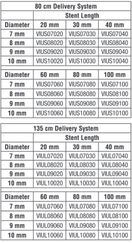
The Bard® LifeStar™ Vascular Stent System is
available in the sizes indicated as follows, listing all item codes for the 80 cm and 135 cm long stent delivery system:
80 cm Delivery System Stent Length Diameter 20 mm 30 mm 40 mm
7 mm VIUS07020 VIUS07030 VIUS07040
8 mm VIUS08020 VIUS08030 VIUS08040
9 mm VIUS09020 VIUS09030 VIUS09040
10 mm VIUS10020 VIUS10030 VIUS10040
Diameter 60 mm 80 mm 100 mm
7 mm VIUS07060 VIUS07080 VIUS07100
8 mm VIUS08060 VIUS08080 VIUS08100
9 mm VIUS09060 VIUS09080 VIUS09100
10 mm VIUS10060 VIUS10080 VIUS10100
135 cm Delivery System Stent Length Diameter 20 mm 30 mm 40 mm
7 mm VIUL07020 VIUL07030 VIUL07040
8 mm VIUL08020 VIUL08030 VIUL08040
9 mm VIUL09020 VIUL09030 VIUL09040
10 mm VIUL10020 VIUL10030 VIUL10040
Diameter 60 mm 80 mm 100 mm
7 mm VIUL07060 VIUL07080 VIUL07100
8 mm VIUL08060 VIUL08080 VIUL08100
9 mm VIUL09060 VIUL09080 VIUL09100
10 mm VIUL10060 VIUL10080 VIUL10100 Each end of the stent has four highly visible
radiopaque Tantalum Markers to facilitate accurate
stent placement. Before deployment, the stent is compressed between the inner catheter and outer catheter at the distal end of the delivery system. In this compressed configuration, the stent struts lie close together and the radiopaque markers appear as a contiguous band at each end of the stent. The
stent MUST NOT be balloon expanded beyond its
2
3
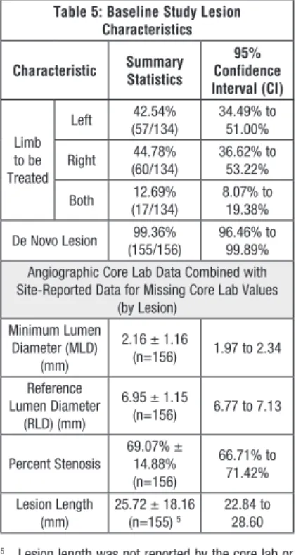
protocol requirements. Table 5 provides pre-treatment lesion characteristics. Antiplatlet/anticoagulant therapy and pre-dilation/post-dilation were left to physician discretion. Overlapping stent placement was permitted and twelve stents in six lesions were placed in an overlapping configuration.
Table 5: Baseline Study Lesion Characteristics
Characteristic Summary Statistics Confidence 95% Interval (CI) Limb to be Treated Left (57/134)42.54% 34.49% to 51.00% Right (60/134)44.78% 36.62% to 53.22% Both (17/134)12.69% 8.07% to 19.38% De Novo Lesion (155/156)99.36% 96.46% to 99.89%
Angiographic Core Lab Data Combined with Site-Reported Data for Missing Core Lab Values
(by Lesion) Minimum Lumen Diameter (MLD) (mm) 2.16 ± 1.16 (n=156) 1.97 to 2.34 Reference Lumen Diameter (RLD) (mm) 6.95 ± 1.15 (n=156) 6.77 to 7.13 Percent Stenosis 69.07% ± 14.88% (n=156) 66.71% to 71.42% Lesion Length (mm) 25.72 ± 18.16 (n=155) 5 22.84 to 28.60
5 Lesion length was not reported by the core lab or
the site for one patient.
At 30 days post-procedure, a telephone contact was made to assess any potential adverse events since the time of the procedure. At nine months post-procedure, a clinic visit was required and the primary and secondary endpoints were assessed. The nine-month follow-up evaluation included a clinical examination, an assessment of adverse events, and a duplex ultrasound evaluation.
8.4 Results:
Results of the Luminexx®Clinical Study are presented
in Table 6.
Thirty-day follow-up compliance was 97.76% (131/134 patients). The percentage of in-office follow-up at nine months post-procedure was 82.09% (110/134 patients); three additional patients were contacted by telephone and one patient’s medical chart was reviewed. Ninety-seven of 134 patients had evaluable ultrasounds that were included in the nine-month assessment interval.
Primary Effectiveness and Safety Endpoint: Using Bayesian statistical models, the study was considered a success if there was at least a 96% probability that the nine-month MACE rate was less than a maximum threshold of 25%. The model was developed on a time-to-event basis within various subintervals of the follow-up period.* At final analysis, the posterior probability was 99.24% that the nine-month MACE
rate was less than 25%. Therefore, the Luminexx®
Clinical Study successfully achieved the primary endpoint outlined in the protocol and demonstrated
that the Luminexx® Stent was safe and effective for
its intended use.
Table 6: The Luminexx® Clinical Study
Endpoints Primary Endpoint:
Posterior Probability: 99.24% that the nine-month 6
MACE rate was < 25% 7
6 Nine months post-procedure (defined as 240-365
days)
7 Using per protocol Bayesian model
* A three-piece piece-wise exponential model was employed for the time until MACE event. The first and last months of exposure were assumed to have different risks than the middle seven months. The
three parameters, λ1, λ2, and λ3 were used within
the model to characterize the efficacy of the Bard®
Luminexx® Iliac Stent. The probability conditional on
λ1, λ2, and λ3 that a patient is free of MACE at 9
months is exp(-λ1 -7λ2 - λ3). Non-informative priors
were used in the model.
Additional collected data:
• Primary Patency: Primary patency was defined as continuous flow through the treated segment without revascularization at nine months post-procedure (i.e., the patient did not have a revascularization procedure, amputation, or bypass surgery). The primary patency rate at nine months post-procedure was 94.03% (95% CI: 88.66% to 96.94%).
• Stent Deployment Success: The stent deployment success rate, defined as the ability of the stent to be successfully delivered and deployed at the target lesion without device malfunction or local arterial complication, was 95.12%.
8.1 Study Endpoints and additional data:
The rate of Major Adverse Clinical Events (MACE) was the primary combined safety and effectiveness endpoint for the study. MACE was defined as peri- procedural death (death during the procedure or prior to hospital discharge), target lesion re- vascularization (any treatment to bypass or increase lumen diameter within the stented segment or within 5mm of its margins), or stented segment restenosis (> 50% stenosis as determined by duplex ultrasound) at nine months post-procedure. Bayesian statistical models, using non-informative prior probabilities for the parameters of interest, were used to evaluate whether there was a 96% probability that the MACE rate would be less than a maximum threshold of 25% at nine months post-procedure.
Additionally for informational purposes, including anatomic success (i.e., achievement of < 30% final residual diameter stenosis) and primary patency (continuous flow through the treated segment without revascularization at nine months post-procedure) were also evaluated.
Evaluations and definitions were adapted from standards established by the Society of Interventional Radiology (SIR), the Society for Vascular Surgery (SVS), the International Society of Cardiovascular Surgery (ISCVS), and described by the SIR Technology Assessment Committee.
To ensure impartiality, all adverse events were submitted for review by an independent Medical
Monitor (i.e., a physician independent of the Luminexx®
Clinical Study and Sponsor). All available information, either from the source documents or summarized on the case report forms was used to adjudicate an event.
8.2 Patient Population:
The protocol allowed for a broad spectrum of patients with iliac artery occlusive disease to be treated with
the Luminexx® Stent, including patients with poor
distal runoff, concomitant or recent distal bypass surgery, and/or restenotic lesions. The intent was to test the device in a non-select population that would more closely represent the clinical population following device commercialization. Patients diagnosed with preoperative coagulation disorders, contraindi-cations to antiplatelet therapy, or who demonstrated the presence of soft, thrombotic, or embolic material within or adjacent to the lesion(s) being treated with the study device were excluded. Characteristics of patients enrolled in the study including age, gender, medical history, and previous vascular procedures are presented in Table 4.
Table 4: Baseline Medical History / Demographics
Characteristic Statistics Summary 1
95% Confidence Interval (CI) 2 Age (Years) 3 67.31 ± 10.31 65.55% to 69.07% Percent Male (73/134)54.48% 46.04% to 62.67% History of Myocardial Infarction (MI) (31/134)23.13% 16.80% to 30.96% History of Percutaneous Trans-luminal Coronary Angioplasty (PTCA) 40.30% (54/134) 32.38% to 48.76% History of Coronary Artery Bypass Graft
(CABG) 25.37% (34/134) 18.76% to 33.36% History of Cardio-vascular Accident (CVA) or Transient Ischemic Attack (TIA)
14.18% (19/134) 9.27% to 21.09% History of Diabetes Mellitus (36/134)26.87% 20.08% to 34.94% History of Hyperlipidemia (98/133 4)73.68% 65.61% to 80.43% History of Hypertension (120/134)89.55% 83.23% to 93.67% History of Peripheral Vascular Disease (PVD)/Claudication 97.76% (131/134) 93.62% to 99.24%
1 All tables: Mean ± Standard Deviation for all
quan-titative variables, Percent (# with characteristic / sample size)
2 All tables: the Score Interval Method was used for
confidence interval percentages
3 Number of patients reporting = 134
4 One patient did not have a value recorded for
History of Hyperlipedemia
8.3 Methods:
Baseline patient assessments included a clinical examination and clinical history targeting the extent of peripheral vascular disease, a clinical category determination, and a thigh/brachial index measurement. At the time of the procedure, lesions were assessed angiographically to determine whether they fit the
• The Bard® LifeStar™ Vascular Stent System is
only compatible with a 0.035“ (0.89 mm) guidewire. • When catheters are in the body, they should be
manipulated only under fluoroscopy with radiographic equipment that produces high quality images. • Read and understand the IFU for any interventional
device to be used in conjunction with the Bard®
LifeStar™ Vascular Stent System.
• During system flushing, DO NOT use the system if fluid
is not observed exiting the catheter at the distal tip. • The delivery system is not designed for use with
power injection systems.
• Faulty placement techniques could lead to stent deployment failure.
• Do not kink the delivery system.
• The delivery system will not function properly until the
Removable Safety Clip (G) is removed. As a precaution
against accidental stent deployment, the Safety Clip should not be removed until the stent is ready to be deployed.
• Administration of adjunctive drug therapy before and after the procedure is left to the discretion of the treating physician (e.g. antiplatelet or anticoagulation). • This product has been designed for single patient use
only. DO NOT reuse. DO NOT resterilize.
• After use, the stent delivery system is a potential biohazard. Handle and dispose of this product in accordance with accepted medical practice and with applicable local, state and federal laws and regulations.
• Store in a cool, dry, dark place. 7.2 Stent Placement Precautions:
• The stent experiences minimal length changes during deployment. (See Table 2)
• Prior to stent deployment, remove all slack from the catheter delivery system to avoid stent misplacement. • DO NOT remove the Removable Safety Clip (G) until
you are ready to deploy the stent.
• DO NOT hold the delivery system catheter during stent deployment.
• DO NOT overlap more than two stents.
• As with all self-expanding nitinol stents, careful attention during stent deployment is warranted to mitigate the potential for movement of the stent. • If more than one stent is required to cover the lesion,
the distal lesion, considered from point of access, should be stented first, followed by stenting of the proximal lesion. Stenting in this order obviates the need to cross the proximal stent for placement of the distal stent, and reduces the potential to dislodge stents that have already been placed.
• To maximize stent placement accuracy, slowly and deliberately deploy the distal portion of the stent until you have visual confirmation of wall apposition before steadily deploying the remaining length of the stent.
7.3 Post-Implant Precautions:
• Caution should be used when crossing a deployed stent with any adjunctive device.
• In the event of thrombosis of the expanded stent, thrombolysis and PTA may be attempted.
• In the event of complications such as infection, pseudoaneurysm or fistualization, surgical removal of the stent may be required.
• The safety and effectiveness of the Bard® LifeStar™
Vascular StentSystem has not been established in patients beyond 9 months of follow-up.
8.0 SUMMARy OF CLINICAL
INVESTIGATIONS
The purpose of the clinical study was to provide the human clinical trial experience to support the safety
and effectiveness of the Bard® LifeStar™ Vascular
Stent System. The U.S. clinical trial proved the device to be safe and effective for its intended use. Data gathered from the clinical study were
collected on both the Bard® Luminexx® Iliac Stent
and the Bard® Luminexx® 6F Iliac Stent (referred to
collectively as the Luminexx® Stent). The stent in
each of these devices was the same; however,
the delivery systems were different. The Bard®
Luminexx® Iliac Stent had a 7F profile and the Bard®
Luminexx® 6F Iliac Stent had a 6F profile. The
commercial device, the Bard® LifeStar™ Vascular
Stent System, uses essentially an electropolished
version of the Luminexx® Stent and includes a Grip
on the 6F delivery system. The clinical data collected
with both the Bard® Luminexx® Iliac Stent and the
Bard® Luminexx® 6F Iliac Stent support the safety
and effectiveness of the Bard® LifeStar™ Vascular
Stent System.
A prospective, multi-center, non-randomized clinical study was conducted at nine sites in the United States
using the Luminexx® Stent. A total of 156 lesions were
treated in 151 limbs using 164 devices. The study objective was to determine the safety and effectiveness
of the Luminexx® Stent for the treatment of common
4
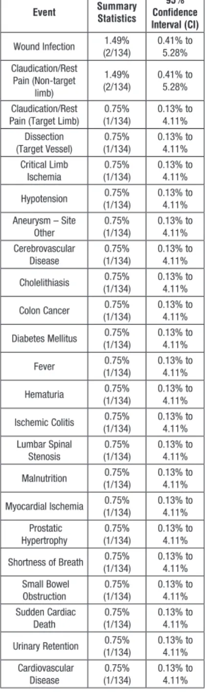
Event Summary Statistics Confidence 95% Interval (CI) Wound Infection (2/134)1.49% 0.41% to 5.28% Claudication/Rest Pain (Non-target limb) 1.49% (2/134) 0.41% to 5.28% Claudication/Rest
Pain (Target Limb) (1/134)0.75% 0.13% to 4.11%
Dissection (Target Vessel) (1/134)0.75% 0.13% to 4.11% Critical Limb Ischemia (1/134)0.75% 0.13% to 4.11% Hypotension (1/134)0.75% 0.13% to 4.11% Aneurysm – Site Other (1/134)0.75% 0.13% to 4.11% Cerebrovascular Disease (1/134)0.75% 0.13% to 4.11% Cholelithiasis (1/134)0.75% 0.13% to 4.11% Colon Cancer (1/134)0.75% 0.13% to 4.11% Diabetes Mellitus (1/134)0.75% 0.13% to 4.11% Fever (1/134)0.75% 0.13% to 4.11% Hematuria (1/134)0.75% 0.13% to 4.11% Ischemic Colitis (1/134)0.75% 0.13% to 4.11% Lumbar Spinal Stenosis (1/134)0.75% 0.13% to 4.11% Malnutrition (1/134)0.75% 0.13% to 4.11% Myocardial Ischemia (1/134)0.75% 0.13% to 4.11% Prostatic Hypertrophy (1/134)0.75% 0.13% to 4.11% Shortness of Breath (1/134)0.75% 0.13% to 4.11% Small Bowel Obstruction (1/134)0.75% 0.13% to 4.11% Sudden Cardiac Death (1/134)0.75% 0.13% to 4.11% Urinary Retention (1/134)0.75% 0.13% to 4.11% Cardiovascular Disease (1/134)0.75% 0.13% to 4.11%
10.0 POTENTIAL COMPLICATIONS
Potential adverse events associated with the use
of the Bard® LifeStar™ Vascular Stent System
include, but may not be limited to: • Abrupt stent closure
• Allergic reaction to nitinol • Amputation
• Aneurysm
• Angina/coronary ischemia • Arterial aneurysm
• Arterial occlusion/thrombus, near the puncture site • Arterial occlusion/thrombus, remote from puncture site • Arterial occlusion/restenosis of the treated vessel • Arterial rupture
• Arteriovenous fistula • Arrhythmia • Atheroembolization • Death related to procedure • Death unrelated to procedure • Embolization, arterial • Embolization, stent • Fever
• Hematoma bleed, remote site
• Hematoma bleed at needle, device path: nonvascular procedure
• Hematoma bleed, puncture site: vascular procedure • Hypersensitivity reactions
• Hypotension/hypertension • Intimal injury/dissection • Ischemia/infarction of tissue/organ
• Ischemia requiring intervention (bypass or amputation of toe, foot or leg)
• Local infection
• Malposition (failure to deliver the stent to the intended site)
• Myocardial infarction • Pseudoaneurysm formation • Pulmonary embolism • Renal failure
• Restenosis of the stented artery • Septicemia/bacteremia
Table 7: In-Hospital Serious Adverse Events Events per Total Patient Population
Event Summary Statistics Confidence 95% Interval (CI) Distal Revascularization (Target Limb) 4.48% (6/134) 2.07% to 9.42% Revascularization (Non-target Limb) (6/134)4.48% 2.07% to 9.42% Major Bleed (2/134)1.49% 0.41% to 5.28% Arterial Thrombosis (2/134)1.49% 0.41% to 5.28% False Aneurysm (2/134)1.49% 0.41% to 5.28% Respiratory Failure (2/134)1.49% 0.41% to 5.28% Amputation on
Study Side Limb (1/134)0.75% 0.13% to 4.11%
Arrhythmia (1/134)0.75% 0.13% to 4.11% Hypertension (1/134)0.75% 0.13% to 4.11% AV Fistula Stenosis (1/134)0.75% 0.13% to 4.11% Dissection (Target Vessel) (1/134)0.75% 0.13% to 4.11% Myocardial Infarction (1/134)0.75% 0.13% to 4.11% Cerebrovascular Disease (1/134)0.75% 0.13% to 4.11% Claudication/Rest Pain (Non-target limb) 0.75% (1/134) 0.13% to 4.11% Claudication/Rest
Pain (Target Limb) 0% (0/134) 2.79%0% to
Critical Limb Ischemia 0% (0/134) 2.79%0% to Sepsis 0% (0/134) 2.79%0% to Target Lesion Revascularization 0% (0/134) 2.79%0% to Death 0% (0/134) 2.79%0% to
• Anatomic Success: Anatomic success was defined as achievement of < 30% final residual diameter stenosis measured at th narrowest point of the stented lumen. The rate of anatomic success based on core lab measurements was 87.50%, while the rate reported by the investigative sites was 98.72%.
Table 7: Additional Collected Data
Primary Patency (88.66% to 96.94%)94.03% Stent Deployment Success (90.67% to 97.51%)95.12% Anatomic Success (Core Lab) (81.11% to 91.94%)87.50% Anatomic Success (Site Reported) (95.45% to 99.65%)98.72% 8.5 Gender Bias:
Males accounted for 54.48% of patients in the study. A comparison between gender and MACE demons-trated a slightly higher incidence of MACE in females than males, but the difference was not significant (Fisher’s Exact Test, P = 0.184).
8.6 Clinical Study Conclusions:
The U.S. multi-center study of the Luminexx® Stent
achieved its primary safety and effectiveness endpoint. The posterior probability was 99.24% that the MACE rate was less than 25% at nine months post-procedure. This probability along with observed rates for other clinical outcomes demonstrated that
the Luminexx® Stent is safe and effective for use in
the treatment of iliac artery occlusive disease.
9.0 SUMMARy OF ADVERSE EVENTS
All adverse events through the nine-month follow-up window were submitted for adjudication by an independent Medical Monitor. The incidence of adverse events was presented descriptively as a percentage of events (i.e., patients could have more than one event) per the total patient population (with 95% CI). No unanticipated adverse device effects
(UADE) were reported in the Luminexx® Clinical Study.
Adverse events were summarized as serious or non-serious and attributed to the stent, procedure, or pre-existing or concomitant condition. Seven patients died through the nine-month follow-up interval (5.2%). None of the deaths occurred within the peri-procedural (< 30 days post-index procedure) timeframe. One patient death (0.75%) was related to complications of thrombectomy of the target lesion and a subsequent chain of revascularization procedures and systemic events. The remaining deaths were the result of pre-existing and/or concomitant conditions, and were not related to the study procedure or the study device.
Table 7 provides a summary of in-hospital serious adverse events (SAEs) and Table 8 provides a cumulative summary of all reported SAEs < nine months post-procedure (< 365 days). The more prevalent SAEs observed through the nine-month follow-up interval are summarized below: • Target Limb Revascularization: Target limb
revascularization was defined as a revascularization procedure outside the margins of the treatment area (i.e., > 5 mm from the proximal or distal end of the stent), but in the same limb. Target limb revascularization was noted in 15 patients (11.19%) through the nine-month follow-up. Revascularization procedures were performed to treat progression of disease or conditions that were not present or did not need treatment at baseline. None of the revascularization
events were attributed to either the Luminexx® Stent
or the study procedure.
• Non-Target Limb Revascularization: Non-target limb revascularizations were noted in 12 patients (8.96%) through the nine-month follow-up period. As with target limb revascularization, these non-target limb procedures represent a progression of the peripheral disease process.
• Amputation: Four amputations were reported (2.24%) through the nine- month interval. All four amputations were performed on the study-limb and were associated with distal-disease progression. Two amputations were performed below-the-knee, one above-the-knee, and one amputation involved a toe. • Major Bleeding Event: Eight patients (5.97%)
experienced major bleeding events throughout the course of the study. Six of these events were unrelated to the study device or procedure. Two patients experienced major bleeding events attributed to the index procedure (1.49%).
• Sepsis: Six patients (eight incidences) experienced sepsis during the course of the study; five patients (3.73%) and six incidences occurred through the nine-month follow-up interval (< 365 days). No incidents of sepsis were attributable to either the device or the iliac stenting procedure.
Table 8: Cumulative Serious Adverse Events through “9 Months” (< 365 days) Events per Total Patient Population
Event Summary Statistics Confidence 95% Interval (CI) Distal Revascularization (Target Limb) 11.19% (15/134) 6.90% to 17.65% Revascularization (Non-target Limb) (12/134)8.96% 5.2% to 15.0% Major Bleed (8/134)5.97% 3.06% to 11.34% Death (7/134)5.22% 2.55% to 10.39% Angina/Coronary Ischemia (7/134)5.22% 2.55% to 10.39% Sepsis/Infection (6/134)4.48% 2.07% to 9.42% Arterial Thrombosis (5/134)3.73% 1.60% to 8.44% Target Lesion Revascularization (5/134)3.73% 1.60% to 8.44% False Aneurysm (4/134)2.99% 1.17% to 7.42% Amputation on
Study Side Limb (4/134)2.99% 1.17% to 7.42%
Arrhythmia (4/134)2.99% 1.17% to 7.42% Stroke (3/134)2.24% 0.76% to 6.38% Myocardial Infarction (3/134)2.24% 0.76% to 6.38% Carotid Artery Disease (3/134)2.24% 0.76% to 6.38% Congestive Heart Failure (2/134)1.49% 0.41% to 5.28% Hypertension (2/134)1.49% 0.41% to 5.28% Renal Complications (2/134)1.49% 0.41% to 5.28% Respiratory Failure (2/134)1.49% 0.41% to 5.28% Anemia (2/134)1.49% 0.41% to 5.28% AV Fistula Stenosis (2/134)1.49% 0.41% to 5.28%
4
Patient
IMPLANT
Information Card
Carry
this
card
with
you.
Prior
to
any
treatment,
please
show
it
to
all
medical
personnel
caring
for
you.
Patient
IMPLANT
Information
Card
MR Conditional
Non-clinical testing has demonstrated the B ar d ® L if e S ta r ™Vascular Stent System
is MR Conditional. It can be scanned safely, immediately after placement of this implant, under the following conditions: • Static magnetic field of 3.0 Tesla or less • Spatial gradient field of 720 Gauss/cm or less • Normal operating mode of the MR system and use of whole body transmit coil. • Maximum whole-body-averaged specific absorption rate (WB-SAR) of 2 W/kg for 15 min. of scanning for patient landmarks above the umbilicus. • Maximum WB-SAR of 1 W/kg for 15 min. of scanning for patient landmarks below the umbilicus. Bard and L ife S tar ™ are trademarks and/or registered trademarks of C. R. Bard, Inc.
Distributed in the USA by: Bard Peripheral Vascular, Inc. Subsidiary
of C. R. Bard, Inc. 1625 West 3 rd Street Tempe, AZ 85281 USA
Manufacturer: Angiomed GmbH & Co. Medizintechnik KG Subsidiary
of C. R. Bard, Inc. TEL: 1-480-894-9515 1-800-321-4254 FAX: 1-480-966-7062 1-800-440-5376 www.bardpv.com Wachhausstraße 6 Tel: ++ 49 721 9445 0 76227 Karlsruhe Fax: ++ 49 721 9445 111 Germany
B
ard ®L
ifeS
tar™
Vascular Stent System
Patient Data:
Name:
Address:
Date
of
birth:
Implant Data:
Product:
Implant
Material:
Implantation
site:
Date
of
implantation:
Follow
up:
Hospital Data:
Name:
Address:
Physician:
Phone:
Apply
“Patient/Inv.
chart”
sticker
here
5
13.0 MAGNETIC RESONANCE IMAGING
(MRI) INFORMATION
Non-clinical testing demonstrated that the Bard®
LifeStar™ Vascular Stent is MR Conditional. A
pati-ent with the Bard® LifeStar™ Vascular Stent can be
scanned safely, immediately after placement of this implant, under the following conditions:
• Static magnetic field of 3.0 Tesla or less
• Normal operating mode of the MR system and use of whole body transmit coil.
• Spatial gradient field of 720 Gauss/cm or less • Maximum whole-body-averaged specific absorption
rate (SAR) of 2-W/kg for 15 minutes of scanning for patient landmarks above the umbilicus.
• Maximum WB-SAR of 1 W/kg for 15 min. of scanning for patient landmarks below the umbilicus.
3.0 Tesla Temperature Rise
Non-clinical testing of RF-induced heating was performed at 128 MHz in a GE Signa HDx 3.0T MR system,software version 4\LX\MR. The testing was according to ASTM F2182 and the stents were in a location and orientation in the phantom that produced the worst case heating. RF power was applied for 15 minutes and the conductivity of the phantom material was about 0.5 S/m. The phantom average SAR calculated using calorimetry was 2.6 W/kg. For scans performed on landmarks above the umbilicus, the maximal temperature rise was 2.3°C when the local SAR was scaled to 2 W/kg for a stent length of 80 mm. The maximal temperature rise was 1.15°C when the local SAR was scaled to 1 W/kg for a stent length of 80 mm. Other stent lengths exhibited a lower rise.
Predicted in-vivo heating based on these non-clincal tests and computer simulation of the patient exposure to the electromagnetic fields in MRI yielded a maximal in-vivo rise of 5°C for the maximal SAR values specified above and a scan time of 15 minutes. The actual in-vivo rise is expected to be less as this calculation did not include the cooling due to blood flood in the lumen of the stent and blood perfusion in the tissue outside the stent.
1.5 Tesla Temperature Rise
Non-clinical testing of RF-induced heating was performed at 64 MHz in a GE Signa whole body coil. The testing was according to ASTM F2182 and the stents were in a location and orientation in the phantom that produced the worst case heating. RF power was applied for 15 minutes and the conductivity of the phantom material was about 0.5 S/m. The phantom average SAR calculated using calorimetry was 1.8 W/kg. For scans performed on landmarks above the umbilicus, the maximal temperature rise was 3.4°C when the local SAR was scaled to 2 W/kg for a stent length of 150 mm. The maximal temperature rise was 1.7°C when the local SAR was scaled to 1 W/ kg for a stent length of 150 mm. Other stent lengths exhibited a lower rise.
Predicted in-vivo heating based on these non-clinical tests and computer simulation of the patient exposure to the electromagnetic fields in MRI yielded a maximal in-vivo rise of 6.1°C for the maximal SAR values specified above and a scan time of 15 minutes. The actual in-vivo rise is expected to be less as this calculation did not include the cooling due to blood flood in the lumen of the stent and blood perfusion in the tissue outside the stent.
Image Artifact
The image artifacts appear as localized signal loss and extend approximately 1.7 mm from the device in the parallel direction and 1.2 mm perpendicular to the stent’s longitudinal axis, both inside and outside the stent lumen when scanned in non-clinical testing using a Gradient echo (GRE) pulse sequence with 100 msec repetition time, 15 msec echo time, 30 degrees flip angle, 256 x 256 matrix size, 10 mm section thickness, 22 cm field of view, number of excitations: 2 and 16 kHz bandwidth, in a 3T Excite General Electric Healthcare (Milwaukee, WI), Software G3.0-052B, with whole body send/receive RF coil.
14.0 HOW SUPPLIED
The Bard® LifeStar™ Vascular Stent System is
supplied sterile (by ethylene oxide gas) unless the package has been opened or damaged. This product has been designed for single patient use only.
DO NOT reuse. DO NOT resterilize. Store in a cool, dry, dark place.
• PRECAUTION: Prior to stent deployment, remove all slack from the catheter delivery system to avoid stent misplacement.
• PRECAUTION:DO NOT hold the delivery system catheter during stent deployment.
11.6 Stent Placement:
• During stent deployment, the entire length of the catheter system should be kept as straight as possible. Maintaining a straight catheter under slight tension during stent deployment is recommended to improve placement accuracy.
• Center the proximal stent markers and both over-lapping distal markers stent markers and marker band on the outer catheter across the stricture. The radiopaque markers on the stent indicate the ends of the compressed stent and the length of the expanded stent.
• By initially advancing the catheter beyond the stricture, micro-adjustments of the stent can be made by pulling the entire system back toward the stricture to improve placement accuracy.
• WARNING: Once the stent is partially or fully deployed, micro-adjustments are no longer possible and the
stent should NOT be dragged or repositioned in the
lumen.
• WARNING: Once stent deployment has been
initiated, the stent CANNOT be recaptured using the
stent delivery system.
• Once the moving marker has passed the proximal end of the stent by approximately 2 cm, the stent is completely deployed.
• Complete stent deployment can be fluoroscopically visualized when the radiopaque markers at the proximal and distal ends of the stent are fully expanded.
11.7 Stent Deployment
• PRECAUTION: DO NOT remove the Removable
Safety Clip (G) until you are ready to deploy the stent.
• Just prior to stent deployment, remove the Safety
Clip. (G)
• Under fluoroscopic visualization, deploy the stent using the conventional “pin & pull-back” technique
by slowly pulling back the Distal T-Luer Adapter (F)
towards the hand that is pinned in place. Pulling back
on the Distal T-Luer Adapter (F) directly retracts the
outer catheter and deploys a corresponding portion of the stent.
• Full stent deployment is ensured when the Distal
T-Luer Adapter (F) reaches the Grip.
• During stent deployment the moving single radio-
paque marker on the outer catheter (D) on the
outer catheter moves backwards toward the proximal markers on the stent. The radiopaque markers on
the stent MUST NOT move during stent deployment.
• After stent deployment, carefully withdraw the delivery system from the patient over the guidewire. After removing the delivery system, visually confirm that the entire stent delivery system has been removed. (a) Inner Catheter
(b) Coaxial Outer Catheter
• Final radiological evaluation of the implanted stent should be conducted by angiogram.
11.8 Post-Stent Placement:
• Post-dilatation of the stent with an appropriately sized balloon dilatation catheter is left to the discretion of the treating physician.
• WARNING: The Bard® LifeStar™ Vascular Stent
System is a self-expanding, nitinol stent that MUST
NOT be expanded beyond its labeled diameter by
dilatation with a PTA balloon.
• PRECAUTION: This product has been designed for
single patient use only. DO NOT reuse. DO NOT
resterilize.
• PRECAUTION: After use, the stent delivery system is a potential biohazard. Handle and dispose of this product in accordance with accepted medical practice and with applicable local, state and federal laws and regulations.
12.0 PATIENT IMPLANT INFORMATION
CARDS:
• A Patient IMPLANT Information Card is provided in the IFU for your convenience.
• The Patient IMPLANT Information Card should be carefully folded along the perforations and removed from the IFU after the completion of the procedure. • The Patient Data, Implant Data, and Hospital Data
should be carefully recorded on the card and given to the patient.
• The patient should carry this card with them and provide to any medical personnel caring for the patient in the future.
• Stent malapposition • Stent migration • Stent strut fracture • Stroke • Vasospasm
• Worsened claudication/rest pain • Tissue necrosis
• Venous occlusion/thrombus, remote from puncture site • Venous occlusion/thrombus, near the puncture site
11.0 DIRECTIONS FOR USE
11.1 Procedural Access:
• Gain access to the treatment site utilizing appropriate
accessory equipment compatible with the 6F Bard®
LifeStar™ Vascular Stent System.
• The working lengths of the 6F Delivery System are indicated on the labels and on the device itself. In
order to allow complete stent deployment, DO NOT
use an introducer sheath or guiding catheter longer than the indicated working length.
• The 6F Delivery System requires a minimum 8F guiding catheter, or a minimum 6F introducer sheath. • Via the femoral route, insert a 0.035“ (0.89 mm)
guidewire under fluoroscopic guidance through the appropriate introducer sheath or guiding catheter and pass the lesion.
11.2 Stent Selection:
• WARNING: Appropriate diameter sizing of the stent to the target lesion is required to reduce the possibility of stent migration.
• Evaluate and mark the stricture. Measure the length of the stricture and the diameter of the target lumen to assist in stent selection.
• The stent should be approximately 1 mm larger in diameter than the target lumen.
• Select the appropriate length of stent to traverse the stricture.
• Allow approximately 5 – 10 mm of the stent to extend beyond each end of the stricture. This will allow for adequate stent coverage at either end of the stenosis. • WARNING: If placing two overlapping stents, both
stents must have identical diameters.
• Stents should overlap by at least 5 mm to include the flared ends.
• PRECAUTION: DO NOT overlap more than two stents.
11.3 General Directions:
• PRECAUTION: Administration of adjunctive drug therapy before and after the procedure is left to the discretion of the treating physician.
• Pre-dilatation of the stricture with an appropriately sized balloon dilatation catheter is left to the discretion of the treating physician.
11.4 Preparation of the Stent Delivery System:
• PRECAUTION: Visually inspect the packaging to
verify that the sterile barrier is intact. DO NOT use if
the sterile barrier is open or damaged.
• PRECAUTION: DO NOT use the device after the “Use By” date specified on the label.
• PRECAUTION: Visually inspect the Bard® LifeStar™
Vascular Stent System to verify that the device has not been damaged due to shipping or improper
storage. DO NOT use damaged equipment.
• WARNING: The delivery system catheter is intended for stent deployment only and not for any other use. • Flush the stent delivery system with sterile saline
using a small volume (e.g., 5 – 10 cc) syringe. Attach the saline filled syringe to the two female Luer ports, the first of which is located at the proximal end of the
device (I) and the second of which is found within
the Distal T-Luer Adapter (F). Continue flushing until
saline drips from the distal tip of the Flexible Catheter
Tip (C) after flushing each Luer port.
• PRECAUTION: During system flushing, DO NOT use the system if fluid is not observed exiting the catheter
at the Flexible Catheter Tip (C) after each Luer port is
flushed.
• During delivery system preparation, ensure that the safety clip remains in place until the stent is ready to be deployed.
• WARNING: If the safety clip has been removed or becomes inadvertently detached from the Grip,
DO NOT use the device.
11.5 Introduction of the Stent Delivery System:
• Insert the guidewire into the distal tip of the catheter until it exits the catheter at the proximal end of the device.
• Advance the delivery catheter over the guidewire into the target lumen.
• Under fluoroscopic visualization, advance the stent delivery system across the stricture using the radiopaque markers to center the stent across the lesion.
• It is recommended to advance the delivery system past the stricture and then to pull back slightly on the entire system to achieve the correct positioning of the markers and to help insure that slack has been remo-ved and that the delivery catheter is straight.
6
7
Symbols used on labelling
mm
Consult Instructions For Use
Keep Away From Sunlight
Keep Dry
Do Not Use If Package Is Damaged
Single Use
Do Not Resterilize
Contents: (1)
MR Conditional
Does Not Contain Natural Rubber Latex
Catalogue Number
Lot Number
Sterilized Using Ethylene Oxide
Use By
Manufacturer
Minimum Introducer Size
Non Pyrogenic
Guidewire Compatibility
Stent Length
Stent Diameter
Working Length
System Length
cm cm6
7
B
ard
®
L
ife
S
tar
™ Vascular Stent System
For the USA only
C. R. BARD, INC. EXCLUDES ALL WARRANTIES, WHETHER EXPRESS OR IMPLIED, By OPERATION OF LAW
OR OTHERWISE, RELATED TO THE BARD
®LIFESTAR™ VASCULAR STENT SySTEM, INCLUDING, BUT NOT
LIMITED TO, ANy IMPLIED WARRANTIES OF MERCHANTABILITy OR FITNESS FOR A PARTICULAR PURPOSE.
IN NO EVENT SHALL C. R. BARD, INC. BE LIABLE FOR ANy INCIDENTAL OR CONSEQUENTIAL LOSS, DAMAGE
OR EXPENSE, DIRECTLy OR INDIRECTLy ARISING FROM USE OF THIS SySTEM. C. R. BARD, INC. NEITHER
ASSUMES NOR AUTHORIZES ANy OTHER PERSON TO ASSUME FOR IT ANy OTHER OR ADDITIONAL LIABILITy
OR RESPONSIBILITy IN CONNECTION WITH THIS SySTEM.
Label Issue Date 08/2011
In the event 2 years have elapsed between this date and product use, the user should contact Bard to see if
additional product information is available.
Telephone Number Inside The US: 1-800-526-4455.
Caution:
Distributed in the USA by:
Bard Peripheral Vascular, Inc.
Subsidiary of C. R. Bard, Inc.
1625 West 3
rdStreet
Tempe, AZ 85281
USA
TEL:
1-480-894-9515
1-800-321-4254
FAX:
1-480-966-7062
1-800-440-5376
www.bardpv.com
Manufacturer:
Angiomed GmbH & Co.
Medizintechnik KG
Subsidiary of C. R. Bard, Inc.
Wachhausstraße 6
76227 Karlsruhe
Germany
Tel.: ++49 721 9445 - 0
Fax: ++49 721 9445 - 111
B05685 Vers.1/08-11
B
ard
®
L
ife
S
tar
™
Vascular Stent System
Bard and L
ife
S
tar
™ are trademarks and/or registered trademarks of C. R. Bard, Inc.
Copyright © 2011 C. R. Bard, Inc.
All Rights Reserved