pain that starts with the first step on arising in the morning or after prolonged sitting. Pain onset is usually insidious but also may com-mence after a traumatic injury. Diagnosis is made by eliciting pain with palpation in the region of the origin of the plantar fascia. Pain may be worsened by passive dorsiflexion of the foot. Overpronation, pes cavus, and re-stricted foot dorsiflexion are common with this condition, although foot pronation itself has not been demonstrated to be a predis-posing factor.10
TIMING AND OTHER CONSIDERATIONS
In plantar fasciitis, corticosteroid injection is a treatment option, usually after other ther-apeutic modalities have failed. These therapies include active stretching, and use of non-steroidal anti-inflammatory drugs (NSAIDs), cushioning heel cups, nighttime plantar fascia splints, and foot orthoses.11-13Corticosteroid
injection effectively provides pain relief,14
although it carries the risk of plantar fascia rupture15and fat pad atrophy.
TECHNIQUE
Pharmaceuticals and equipment are listed in Table 1. The patient is placed in the lateral recumbent position with the affected side down. The physician identifies the medial
T
his article, the final in a series on diagnostic and therapeutic in-jections, covers the ankle and foot. The rationale, indications, contraindications, and general approach to this procedure are covered in the first article in this series.1Subsequent articleshave covered injections of the shoulder, elbow, wrist and hand, hip, and knee regions.2-5
The ankle and foot are susceptible to multi-ple injuries and inflammatory conditions6 that
are amenable to diagnostic and therapeutic injection.7 This article covers the anatomy,
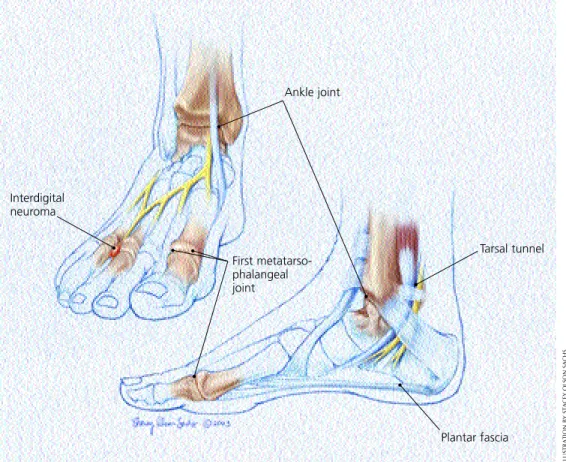
pathology, diagnosis, and injection technique at common sites for which these procedures are applicable. These areas include the plantar fas-cia, ankle joint, tarsal tunnel, interdigital space, and first metatarsophalangeal joint (Figure 1).
Plantar Fascia
The plantar fascia, a band of connective tis-sue deep to the fat layer of the base (plantar aspect) of the foot, spans from the medial plantar tuberosity of the calcaneus to the base of the digits. It helps support the medial lon-gitudinal arch of the foot.
INDICATIONS AND DIAGNOSIS
The plantar fascia is frequently a site of chronic pain.8,9Patients typically complain of
Joint and soft tissue injection of the ankle and foot region is a useful diagnostic and therapeutic tool for the family physician. This article reviews the injection procedure for the plantar fascia, ankle joint, tarsal tunnel, interdigital space, and first metatarsophalangeal joint. Indications for plantar fascia injection include degeneration secondary to repetitive use and traumatic injuries that are unresponsive to con-servative treatment. Diagnostic aspiration or therapeutic injection of the ankle or first metatarsopha-langeal joints can be performed for management of advanced osteoarthritis, rheumatoid arthritis, and other inflammatory arthritides such as gout, or synovitis or an arthrosis such as “turf toe.” Persistent pain and disability resulting from tarsal tunnel syndrome, an analog of carpal tunnel syndrome of the wrist, respond to local injection therapy. A painful interdigital space, such as that occurring in patients with Morton’s neuroma, is commonly relieved with corticosteroid injection. The proper technique, choice and quantity of pharmaceuticals, and appropriate follow-up are essential for effective outcomes. (Am Fam Physician 2003;68:1356-62. Copyright© 2003 American Academy of Family Physicians.)
Diagnostic and Therapeutic Injection
of the Ankle and Foot
ALFRED F. TALLIA, M.D., M.P.H., and DENNIS A. CARDONE, D.O., C.A.Q.S.M.
University of Medicine and Dentistry of New Jersey–Robert Wood Johnson Medical School, New Brunswick, New Jersey
OFFICE PROCEDURES
This article is one in a series of “Office Proce-dures” articles coordi-nated by Dennis A. Cardone, D.O., C.A.Q.S.M., associate professor, and Alfred F. Tallia, M.D., M.P.H., associate professor, Department of Family Medicine, UMDNJ– Robert Wood Johnson Medical School, New Brunswick, New Jersey.
aspect of the foot and palpates the soft tissue just distal to the calcaneus, locating the point of maximal tenderness or swelling.
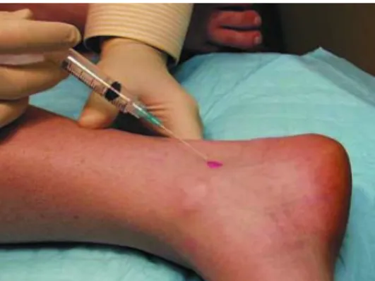
APPROACH AND NEEDLE ENTRY
General technique, including premedica-tion, is discussed in the first article in this series. At the defined soft tissue area, a 25-gauge, 1.5-inch needle is inserted perpen-dicular to the skin (Figure 2). The needle should be inserted directly down past the midline of the width of the foot. The physi-cian should not inject into the fat pad at the base of the foot.
FIGURE 1. Injection target points.
FIGURE 2. Plantar fasciitis injection.
ILLUSTRATION BY STACEY OLSON SACHS
Interdigital neuroma Ankle joint First metatarso- phalangeal joint Tarsal tunnel Plantar fascia
.
.
.
.
.
.
.
.
The pharmaceutical material is injected slowly and evenly through the middle one third of the width of the foot while the needle is being withdrawn. The physician should avoid injecting through the base of the foot, because this approach can result in the com-plications of pharmaceutical leakage and fat pad atrophy.
The patient should remain in the supine position for several minutes after the injec-tion. The physician may put the injected region through passive range of motion. The patient should remain in the office for 30 minutes after the injection to be monitored for adverse reactions. In general, patients
should avoid any strenuous activity involving the injected region for at least 48 hours. Patients should be cautioned that they may experience worsening symptoms during the first 24 to 48 hours. This is related to a possi-ble steroid flair, which can be treated with ice and NSAIDs (e.g., ibuprofen, naproxen). A follow-up examination within three weeks should be arranged.
Ankle Joint
The ankle joint is formed by the articulation of the talus with the tibia and fibula. The medial and lateral malleoli of the tibia and fibula stabilize the talus.
TABLE 1
Equipment and Pharmaceuticals
Hydrocortisone equivalents per
Site Syringe Needle Anesthetic Corticosteroid injection (mg)
Plantar fascia 5 mL 25 gauge, 2 mL of 1% lidocaine 1 mL of Celestone* 150 1.5 inch (Xylocaine) or 0.25% or or
0.5% bupivacaine (Marcaine) 1 mL of 40 mg per mL 200 of methylprednisolone (Solumedrol)
Ankle joint 10 mL (30 to 25 gauge, 1 to 3 to 5 mL of 1% lidocaine or 1 mL of Celestone 150 60 mL if 1.5 inch 0.25% or 0.5% bupivacaine or
aspirating) (18 gauge, 1 mL of Solumedrol 200
1.5 inch if aspirating)†
Tarsal tunnel 3 to 5 mL 25 gauge, 1 or 1 to 2 mL of 1% lidocaine or 0.5 mL of Celestone 75 1.5 inch 0.25% or 0.5% bupivacaine or
0.5 mL of Solumedrol 100 Interdigital 3 to 5 mL 25 gauge, 2 mL of 1% lidocaine or 0.5 mL of Celestone 75
space 1.5 inch 0.25% or 0.5% bupivacaine or
0.5 mL of Solumedrol 100
First metatarso- 3 mL (10 mL if 25 to 27 gauge, 1 mL of 1% lidocaine or 0.25 to 0.5 mL 37.5 to 57.5 phalangeal joint aspirating) 1 or 1.5 inch† 0.25% or 0.5% bupivacaine of Celestone
or
0.25 to 0.5 mL of 50 to 100 Solumedrol
*—1 mL of Celestone is 3 mg each of betamethasone sodium phosphate and betamethasone acetate. †—A hemostat is needed to immobilize the needle when injection follows aspiration.
INDICATIONS AND DIAGNOSIS
Arthritis of the ankle joint may occur in athletes with a history of trauma to the area, and in older patients, and can be an indica-tion for corticosteroid joint injecindica-tion. Besides osteoarthritis, rheumatoid arthritis, and acute traumatic arthritides, other indications for joint injection include crystalloid deposi-tion disease, mixed connective tissue disease, and synovitis.16,17Pain and disability are the
usual presenting complaints, and examina-tion can reveal pain with limitaexamina-tion of motion, tenderness, swelling, crepitus, and deformity. Gait disturbance, erythema, and warmth to palpation also may be present. Radiographs may be helpful to support the diagnosis.
TIMING AND OTHER CONSIDERATIONS
Aspiration of the joint must be performed if infection is suspected. Infection is an absolute contraindication to corticosteroid joint injec-tion. Aspiration also can be useful for con-firming certain arthropathies such as crystal-loid deposition disease and Lyme arthritis.
TECHNIQUE
Pharmaceuticals and equipment are listed in Table 1. The patient is placed in the supine position with the ankle relaxed. The physician identifies the space between the anterior bor-der of the medial malleolus and the medial border of the tibialis anterior tendon and pal-pates this space for the articulation of the talus and tibia.
APPROACH AND NEEDLE ENTRY
As with any joint aspiration or injection procedure, sterile technique must be followed. The needle is inserted into the identified space and directed posterolaterally (Figure 3). Reduced resistance will be felt on entering the joint space, making aspiration and the free flow of pharmaceuticals possible. When aspi-ration precedes injection, the needle is held with a hemostat while the syringe is changed.
Follow-up care is the same as that described for injection of the plantar fascia.
Tarsal Tunnel
The tarsal tunnel is formed by the medial malleolus and a fibrous ligament, the flexor retinaculum. The posterior tibial nerve passes through the tunnel and can be compressed by any condition that reduces the space of the tunnel. The medial plantar, lateral plantar, and calcaneal branches of the posterior tibial nerve innervate the base of the foot.
INDICATIONS AND DIAGNOSIS
Patients with arthritis of the tarsal tunnel may complain of a burning sensation, pain, and paresthesias over the distribution of the poste-rior tibial nerve and its branches that worsen with weight bearing.18 Symptoms are often
related to chronic conditions such as impinge-ment syndromes and hyperpronation, or may be secondary to acute trauma.19,20 Eliciting a
positive Tinel’s sign by tapping over the tarsal tunnel typically causes discomfort in the medial one third of the distal plantar foot, although the entire plantar foot surface may be affected.
Ankle and Foot Injection
FIGURE 3. Ankle joint injection.
Eliciting a positive Tinel’s sign by tapping over the tarsal tun-nel typically will cause discomfort in the medial one third of the distal plantar foot.
TIMING AND OTHER CONSIDERATIONS
Injection is a modality that is performed after a treatment program that can include stretching, rest, and the use of shoe inserts or orthoses, and NSAIDs.21
TECHNIQUE
Pharmaceuticals and equipment are listed in Table 1. The patient is placed in the lateral recumbent position with the affected foot down. Behind the medial malleolus, the point over the posterior tibial nerve where percus-sion elicits the symptoms is identified. Having the patient actively invert the foot against resistance will help the physician identify the
posterior tibial tendon. The nerve lies poste-rior to the tendon.
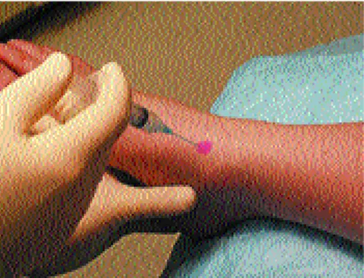
APPROACH AND NEEDLE ENTRY
At approximately 2 cm proximal to the identified location, the needle is inserted at an angle of 30 degrees to the surface of the skin and directed distally (Figure 4). This injection is relatively superficial. The final needle depth will be determined by the amount of subcutaneous tissue. The physi-cian should aspirate before injecting to ensure that the needle is not in an artery or a vein. The pharmaceutical agent is injected slowly. Follow-up care is the same as that previously described.
Interdigital Space
The interdigital spaces of the foot are sites for the occurrence of painful neuromas, a con-dition termed Morton’s neuroma. The second and third common digital branches of the medial plantar nerve are the most frequent sites for development of interdigital neuromas.
INDICATIONS AND DIAGNOSIS
Morton’s neuromas develop secondary to chronic trauma and repetitive stress, as occurs in persons wearing tight-fitting or high-heeled shoes.22Pain and paresthesias are
usu-ally insidious at onset and are located in the interdigital space of the affected nerve. In some cases, the interdigital space between the affected toes may be widened as a result of an associated ganglion or synovial cyst. Pain is elicited in the affected interdigital space when the metatarsal heads of the foot are squeezed together. Injection with 1 percent lidocaine (Xylocaine) can be helpful in confirming the diagnosis.
TIMING AND OTHER CONSIDERATIONS
Treatment for Morton’s neuroma can include the use of NSAIDs, metatarsal pads, orthoses, proper footwear, and injection. Injection may be considered as an early thera-peutic option.23Surgery is a last resort.
FIGURE 4. Tarsal tunnel injection.
The Authors
ALFRED F. TALLIA, M.D., M.P.H., is associate professor and vice chair in the Depart-ment of Family Medicine at the University of Medicine and Dentistry of New Jer-sey (UMDNJ)–Robert Wood Johnson Medical School, New Brunswick, N.J. Dr. Tal-lia is a graduate of the UMDNJ–Robert Wood Johnson Medical School and completed his residency at the Thomas Jefferson University Family Medicine Resi-dency, Philadelphia. He received his public health degree at Rutgers University, New Brunswick, N.J.
DENNIS A. CARDONE, D.O., C.A.Q.S.M., is associate professor and director of sports medicine and the sports medicine fellowship in the Department of Family Medicine at UMDNJ–Robert Wood Johnson Medical School. Dr. Cardone is a graduate of New York College of Osteopathic Medicine, Old Westbury, N.Y., and completed his resi-dency at the UMDNJ–Robert Wood Johnson Medical School of Family Medicine. He completed a sports medicine fellowship at UMDNJ.
Address correspondence to Alfred F. Tallia, M.D., M.P.H., Dept. of Family Medicine, UMDNJ, 1 Robert Wood Johnson Pl., MEB288, New Brunswick, NJ 08903 (e-mail: tallia@ umdnj.edu). Reprints are not available from the authors.
TECHNIQUE
Pharmaceuticals and equipment are listed in Table 1. The patient is placed in a supine position with the knee in a supported flexed position (e.g., with a pillow beneath it) and the foot in a relaxed neutral position. The physician palpates the area of tenderness and fullness on the dorsum of the foot between the affected metatarsal heads.
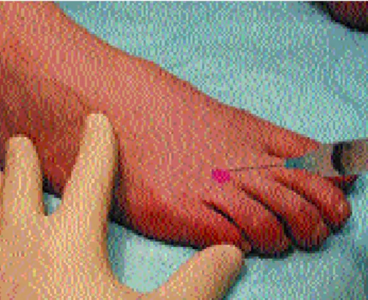
APPROACH AND NEEDLE ENTRY
Injection is performed by inserting the nee-dle on the dorsal foot surface in the distal to proximal direction, at an angle of 45 degrees, and down to the area of fullness between the metatarsal heads (Figure 5). Position is key, because plantar fat pad atrophy can occur if the fat pad is injected.24Follow-up care is the
same as that previously described.
First Metatarsophalangeal Joint
The first metatarsophalangeal joint varies in size and shape, and it may be difficult to pal-pate in patients with conditions such as advanced degenerative arthritis.
INDICATIONS AND DIAGNOSIS
Diagnostic aspiration or therapeutic injec-tion of the first metatarsophalangeal joint can be performed in the management of advanced osteoarthritis, rheumatoid arthritis, and other inflammatory arthritides such as gout, or for synovitis or an arthrosis such as “turf toe.”25-27
Turf toe, a painful ligamentous injury result-ing from hyperextension of the first metatar-sophalangeal joint, often occurs in football linemen. Diagnosis of the specific underlying condition entails eliciting supporting historic and physical findings and, possibly, diagnostic laboratory tests and imaging studies.
TIMING AND OTHER CONSIDERATIONS
Treatment is specific to the underlying con-dition. Injection may be considered as a diag-nostic or therapeutic adjunct. Aside from diagnostic aspiration, therapeutic injection
may be used early in the course of certain inflammatory arthritides, such as gout.
TECHNIQUE
Pharmaceuticals and equipment are listed in Table 1. The patient is placed in a supine position with the knee in a supported flexed position (e.g., with a pillow beneath the knee), and the foot is firmly supported by the table. The physician palpates the joint line on the dorsum of the foot and passively flexes and extends the toe to locate the joint line.
APPROACH AND NEEDLE ENTRY
Distal traction may be applied to the great toe to open the joint space. The needle is inserted on the dorsomedial or dorsolateral surface (Figure 6). The needle should be angled
Ankle and Foot Injection
FIGURE 5. Interdigital neuroma injection.
FIGURE 6. First metatarsophalangeal joint injection.
Ankle and Foot Injection
60 to 70 degrees to the plane of the foot and pointed distally to match the slope of the joint. The joint space is not deep below the skin sur-face. The physician should aspirate before injecting; the injectable agent should flow with-out major resistance when the needle is posi-tioned properly in the joint space. Follow-up care is the same as that previously described. The authors indicate that they do not have any con-flicts of interests. Sources of funding: none reported.
REFERENCES
1. Cardone DA, Tallia AF. Joint and soft tissue injec-tion. Am Fam Physician 2002;66:283-8.
2. Tallia AF, Cardone DA. Diagnostic and therapeutic injection of the shoulder region. Am Fam Physician 2003;67:1271-8.
3. Cardone DA, Tallia AF. Diagnostic and therapeutic injection of the elbow region. Am Fam Physician 2002;66:2097-100.
4. Tallia AF, Cardone DA. Diagnostic and therapeutic injection of the wrist and hand region. Am Fam Physician 2003;67:745-50.
5. Cardone DA, Tallia AF. Diagnostic and therapeutic injection of the hip and knee region. Am Fam Physician 2003;67:2147-52.
6. Omey ML, Micheli LJ. Foot and ankle problems in the young athlete. Med Sci Sports Exerc 1999;31(7 suppl):S470-86.
7. Kerlan RK, Glousman RE. Injections and techniques in athletic medicine. Clin Sports Med 1989;8:541-60. 8. Barrett SJ, O’Malley R. Plantar fasciitis and other causes of heel pain. Am Fam Physician 1999;59: 2200-6.
9. Silko GJ, Cullen PT. Indoor racquet sports injuries. Am Fam Physician 1994;50:374-80,383-4. 10. Cornwall MW, McPoil TG. Plantar fasciitis: etiology
and treatment. J Orthop Sports Phys Ther 1999;29:756-60.
11. Bedinghaus JM, Niedfeldt MW. Over-the-counter foot remedies. Am Fam Physician 2001;64:791-6. 12. Young CC, Rutherford DS, Niedfeldt MW.
Treat-ment of plantar fasciitis. Am Fam Physician 2001; 63:467-74,477-8.
13. Ryan J. Use of posterior night splints in the treat-ment of plantar fasciitis. Am Fam Physician 1995;52:891-8,901-2.
14. Furey JG. Plantar fasciitis. The painful heel syn-drome. J Bone Joint Surg [Am] 1975;57:672-3. 15. Beals TC, Pomeroy GC, Manoli A 2d. Posterior
ten-don insufficiency: diagnosis and treatment. J Am Acad Orthop Surg 1999;7:112-8.
16. Padeh S, Passwell JH. Intraarticular corticosteroid injection in the management of children with chronic arthritis. Arthritis Rheum 1998;41:1210-4. 17. Khoury NJ, el-Khoury GY, Saltzman CL, Brandser EA. Intraarticular foot and ankle injections to iden-tify source of pain before arthrodesis. AJR Am J Roentgenol 1996;167:669-73.
18. Lau JT, Daniels TR. Tarsal tunnel syndrome: a review of the literature. Foot Ankle Int 1999;20:201-9. 19. Oh SJ, Meyer RD. Entrapment neuropathies of the
tibial (posterior tibial) nerve. Neurol Clin 1999;17: 593-615,vii.
20. Schwartzman RJ, Maleki J. Postinjury neuropathic pain syndromes. Med Clin North Am 1999;83:597-626.
21. Malusky LP. Podiatric procedures. In: Roberts JR, Hedges JR, eds. Clinical procedures in emergency medicine. 3d ed. Philadelphia: Saunders, 1998: 879-80.
22. Wu KK. Morton’s interdigital neuroma: a clinical review of its etiology, treatment, and results. J Foot Ankle Surg 1996;35:112-9.
23. Greenfield J, Rea J Jr, Ilfeld FW. Morton’s interdigi-tal neuroma. Indications for treatment by local injections versus surgery. Clin Orthop 1984;185: 142-4.
24. Basadonna PT, Rucco V, Gasparini D, Onorato A. Plantar fat pad atrophy after corticosteroid injec-tion for an interdigital neuroma: a case report. Am J Phys Med Rehabil 1999;78:283-5.
25. Boxer MC. Osteoarthritis involving the sophalangeal joints and management of metatar-sophalangeal joint pain via injection therapy. Clin Podiatr Med Surg 1994;11:125-32.
26. Solan MC, Calder JD, Bendall SP. Manipulation and injection for hallux rigidus. Is it worthwhile? J Bone Joint Surg [Br] 2001;83:706-8.
27. Mizel MS, Michelson JD. Nonsurgical treatment of monarticular nontraumatic synovitis of the second metatarsophalangeal joint. Foot Ankle Int 1997; 18:424-6.