Babak Mokhlesi MD MSc
Historical Perspective Definition
Epidemiology
Clinical Presentation and Diagnosis Morbidity and Mortality
Quality of Life Morbidity Mortality Pathophysiology
The Excessive Load on the Respiratory System Blunted Central Respiratory Drive
Predictors of Hypercapnia in Obese Patients With OSA Treatment
Treatment of Sleep-Disordered Breathing Surgical Interventions
Pharmacologic Respiratory Stimulation Summary
Obesity hyoventilation syndrome (OHS) is defined as the triad of obesity, daytime hypoventilation, and sleep-disordered breathing in the absence of an alternative neuromuscular, mechanical or metabolic explanation for hypoventilation. During the last 3 decades the prevalence of extreme obesity has markedly increased in the United States and other countries. With such a global epidemic of obesity, the prevalence of OHS is bound to increase. Patients with OHS have a lower quality of life, with increased healthcare expenses, and are at higher risk of developing pulmonary hypertension and early mortality, compared to eucapnic patients with sleep-disordered breathing. OHS often remains undiagnosed until late in the course of the disease. Early recognition is impor-tant, as these patients have significant morbidity and mortality. Effective treatment can lead to significant improvement in patient outcomes, underscoring the importance of early diagnosis. This review will include disease definition and epidemiology, clinical characteristics of the syndrome, pathophysiology, and morbidity and mortality associated with it. Lastly, treatment modalities will be discussed in detail.Key words: obesity hypoventilation syndrome; Pickwickian syndrome; hypercap-nia; hypoventilation; sleep apnea; sleep-disordered breathing; CPAP; bi-level PAP. [Respir Care 2010; 55(10):1347–1362. © 2010 Daedalus Enterprises]
Babak Mokhlesi MD MSc is affiliated with the Section of Pulmonary and Critical Care Medicine, the Sleep Disorders Center, and the Sleep Med-icine Fellowship, University of Chicago Pritzker School of MedMed-icine, Chicago, Illinois.
Dr Mokhlesi presented a version of this paper at the 45th RESPIRATORY CAREJournal Conference, “Sleep Disorders: Diagnosis and Treatment,” held December 10-12, 2009, in San Antonio, Texas.
Dr Mokhlesi has disclosed a relationship with Philips/Respironics.
Correspondence: Babak Mokhlesi MD MSc, Section of Pulmonary and Critical Care Medicine, University of Chicago Pritzker School of Med-icine, 5841 S Maryland Avenue, MC 0999, Room L11B, Chicago IL 60637. E-mail: bmokhles@medicine.bsd.uchicago.edu.
Historical Perspective
The association between obesity and hypersomnolence has long been recognized. Of historical interest, OHS was described well before OSA was recognized as a true clin-ical entity in 1969.1,2 The first published report of the
association between obesity and hypersomnolence may have been as early as 1889.3In fact, in 1909 after losing
approximately 90 pounds, President Howard Taft stated, “I have lost that tendency to sleepiness which made me think of the fat boy in Pickwick. My color is very much better and my ability to work is greater.”4 But it was not
until 1955 that Auchincloss described in detail a case of obesity and hypersomnolence paired with alveolar hy-poventilation.5One year later, Burwell described a similar
patient who finally sought treatment after his symptoms caused him to fall asleep during a hand of poker, despite having been dealt a full house of aces over kings.6
Al-though other clinicians had made the comparison some 50 years earlier,3Burwell popularized the term
“Pickwick-ian syndrome” in his case report by noting the similarities between his patient and the boy Joe (Fig. 1), Mr Wardle’s servant in Charles Dickens’s The Posthumous Papers of the Pickwick Club.7Since then our knowledge about the
epidemiology, pathophysiology, treatment, and outcomes of OHS has improved significantly.
Definition
OHS is defined as daytime hypercapnia and hypoxemia (PaCO2⬎45 mm Hg and PaO2⬍ 70 mm Hg at sea level) in an obese patient (body mass index [BMI]ⱖ30 kg/m2)
with sleep-disordered breathing in the absence of any other cause of hypoventilation.8It is important to recognize that
OHS is a diagnosis of exclusion and should be distin-guished from other conditions that are commonly associ-ated with hypercapnia (Table 1). Hypercapnia is very un-likely to develop in OSA patients with BMI below 30 kg/m2
(Fig. 2). In 90% of patients with OHS, the sleep-disor-dered breathing is simply OSA. The remaining 10% have sleep hypoventilation, which is defined as an increase in PaCO2of ⬎10 mm Hg above that of wakefulness or sig-nificant oxygen desaturations, neither of which is the re-sult of obstructive apneas or hypopneas. Therefore, in these patients non-obstructive hypoventilation characterized by an apnea-hypopnea index (AHI)⬍ 5 per hour is noted to be present. There is no accurate way to know into which category a patient with OHS will fall without performing an overnight polysomnogram.
Epidemiology
In the United States, a third of the adult population is obese, and the prevalence of extreme obesity (BMIⱖ40 kg/
m2) has increased dramatically. From 1986 to 2005 the
prevalence of BMIⱖ 40 kg/m2has increased by 5-fold,
from affecting 1 in every 200 adults to 1 in every 33 adults. Similarly, the prevalence of BMIⱖ50 kg/m2has
increased by 10-fold, from affecting 1 in every 2,000 adults to 1 in every 230 adults.9The obesity epidemic is not only
impacting adults in the United States, it is a global phe-nomenon affecting children and adolescents.10-13With such
a global epidemic of obesity the prevalence of OHS is likely to increase.
Numerous studies have reported a prevalence of OHS between 10 –20% in obese patients with OSA (Table 2).14-20
The prevalence of OHS is higher in the subgroup of pa-tients with OSA with extreme obesity (Fig. 3). A recent meta-analysis of 4,250 patients with obesity and OSA— who did not have COPD—reported a 19% prevalence of hypercapnia.21 The prevalence of OHS among
hospital-ized adult patients with BMI ⬎ 35 kg/m2 has been
re-ported at 31%.22
Although the prevalence of OHS tends to be higher in men, the male predominance is not as clear as in OSA. In fact, 3 studies had a higher proportion of women with OHS.22-24Similarly, there is no clear racial or ethnic
pre-Fig. 1. Joe the “Fat Boy,” the character in Dickens’sThe Posthu-mous Papers of the Pickwick Club, from which the term “Pick-wickian syndrome” was derived. (From Reference 7.)
dominance. However, due to higher prevalence of extreme obesity in African-Americans compared to other races, the prevalence of OHS might be higher in African Ameri-cans.25,26 Because of cephalometric differences, such as
narrowing of the bony oropharynx and inferior displace-ment of the hyoid bone, OHS associated with OSA occurs at a lower BMI in Asians, compared to whites.19,27,28
The prevalence of OHS in a community-based cohort is unknown, as it has not been studied, but its prevalence can be estimated among the general adult population in the United States. If approximately 3% of the general United States population has severe obesity (BMI ⱖ40 kg/m2)9
and half of patients with severe obesity have OSA,29and
10 –20% of the severely obese patients with OSA have OHS, then a conservative estimated prevalence of OHS in the general adult population is anywhere between 0.15– 0.3% (approximately 1 in 300 to 1 in 600 adults in the general population).30OHS may be more prevalent in the
United States than in other nations because of its obesity epidemic.
Taken together, with such an epidemic of extreme obe-sity the prevalence of OHS is likely to increase and a high index of suspicion on the part of clinicians may lead to early recognition and treatment of this syndrome.
Clinical Presentation and Diagnosis
While most patients with OHS have had prior hospital-izations and have high healthcare resources utilization, the formal diagnosis of OHS is established late in the fifth or sixth decade of life. The 2 most common presentations are an acute-on-chronic exacerbation with acute respiratory acidosis leading to admission to an intensive care unit, or during a routine out-patient evaluation by a sleep specialist or a pulmonologist.16,22,31,32Patients with OHS tend to be
severely obese (defined as a BMIⱖ 40 kg/m2), have an
AHI in the severe range, and are usually hypersomnolent. The vast majority of patients have the classic symptoms of OSA, including loud snoring, nocturnal choking episodes with witnessed apneas, excessive daytime sleepiness, and morning headaches. In contrast to eucapnic OSA, patients with stable OHS frequently complain of dyspnea and may have signs of cor pulmonale. Physical examination find-ings can include a plethoric obese patient with an enlarged neck circumference, crowded oropharynx, a prominent pul-monic component of the second heart sound on cardiac auscultation (this is often hard to hear, due to obesity), and lower extremity edema. Table 3 summarizes the clinical features of 757 patients with OHS reported in the litera-ture.14-20,22,33-38
Several laboratory findings are supportive of OHS, yet the definitive test for alveolar hypoventilation is an arterial blood gas performed on room air. Elevated serum bicar-bonate level due to metabolic compensation of respiratory acidosis is common in patients with OHS and points to-ward the chronic nature of hypercapnia.22,35,39Therefore,
serum bicarbonate from venous blood could be used as a sensitive test to screen for chronic hypercapnia.20Figure 4
shows the prevalence of OHS in obese patients with OSA (BMIⱖ30 kg/m2and AHIⱖ5), using serum bicarbonate
level combined with other readily available measures such
Table 1. Definition of Obesity Hypoventilation Syndrome
Required
Conditions Description Obesity Body mass indexⱖ30 kg/m2
Chronic hypoventilation Awake daytime hypercapnia (PaCO2ⱖ45 mm Hg and PaO2
⬍70 mm Hg)
Sleep-disordered breathing Obstructive sleep apnea (apnea-hypopnea indexⱖ5 events/h, with or without sleep hypoventilation) present in 90% of cases
Non-obstructive sleep hypoventilation (apnea-hypopnea index⬍5 events/ h) in 10% of cases
Exclusion of other causes of hypercapnia
Severe obstructive airways disease Severe interstitial lung disease Severe chest-wall disorders (eg,
kyphoscoliosis) Severe hypothyroidism Neuromuscular disease
Congenital central hypoventilation syndrome
Fig. 2. Summary of 19 case series of patients with obesity hy-poventilation syndrome, in which the authors reported the mean body mass index (BMI) and arterial blood gases. The mean PaCO2
(in solid) and PaO2(in hollow) are plotted against the BMI. Although
there were no patients with BMI below 30 kg/m2in these series, if
the regression line for PaCO2is continued to a BMI of 30 kg/m
2the
PaCO2would be 45.7 mm Hg. Therefore, hypercapnia with OSA is
as severity of obesity, and severity of OSA. Therefore, serum bicarbonate level is a reasonable test to screen for hypercapnia, because it is readily available, physiologi-cally sensible, and less invasive than an arterial puncture to measure blood gases.
Additionally, hypoxemia during wakefulness is not com-mon in patients with simple OSA. Therefore, abnormal arterial oxygen saturation detected via finger pulse oxim-etry (SpO2) during wakefulness should also lead clinicians to exclude OHS in patients with OSA.20,40A useful tool
for a sleep physician interpreting the polysomnogram of a patient they have not seen in clinic may be the percent of total sleep time with SpO2spent below 90%.
In a recent meta-analysis, the mean difference of per-cent of total sleep time with SpO2 spent below 90% was 37% (56% for hypercapnic OSA patients, 19% for eucap-nic OSA patients), with very little overlap in the 95% confidence intervals (Table 4).21In another study,
Baner-jee and colleagues prospectively compared sleep parame-ters in 23 patients with OHS (mean PaCO254 mm Hg) to 23 patients with eucapnic OSA, by performing overnight in-laboratory polysomnography.41 These 2 groups were Table 3. Clinical Features of Patients With Obesity Hypoventilation
Syndrome*
Mean (range)
Age (y) 52 (42–61)
Male (%) 60 (49–90)
Body mass index (kg/m2) 44 (35–56)
Neck circumference (cm) 46.5 (45–47)
pH 7.38 (7.34–7.40)
PaCO2(mm Hg) 53 (47–61)
PaO2(mm Hg) 56 (46–74)
Serum bicarbonate (mEq/L) 32 (31–33) Hemoglobin (g/dL) 15 (ND) Apnea-hypopnea index (events/h) 66 (20–100) Oxygen nadir during sleep (%) 65 (59–76) Percent time SpO2⬍90% 50 (46–56)
FVC (% predicted) 68 (57–102) FEV1(% predicted) 64 (53–92)
FEV1/FVC 0.77 (0.74–0.88)
Medical Research Council dyspnea class 3 or 4 (%)
69 (ND)
Epworth sleepiness scale score 14 (12–16)
* The studies included a total of 757 patients with obesity hypoventilation syndrome.14-20,22,24,33-38
ND⫽nonsufficient data available to calculate a range (Adapted from Reference 8.)
Table 2. Prevalence of Obesity Hypoventilation Syndrome in Patients With Obstructive Sleep Apnea
First
Author N Design Country
Age* (mean) BMI* (mean) AHI* (mean) OHS (%)
Verin14 218 Retrospective France 55 34 51 10
Laaban15 1,141 Retrospective France 56 34 55 11
Kessler16 254 Prospective France 54 33 76 13
Resta17 219 Prospective Italy 51 40 42 17
Golpe18 175 Retrospective Spain ND 32 42 14
Akashiba19 611 Retrospective Japan 48 29 52 9
Mokhlesi20 359 Prospective United States 48 43 62 20
* Age, body mass index (BMI), and apnea-hypopnea index (AHI) are mean values for all patients, calculated from the data provided by the authors in the article. ND⫽no data available. (Adapted from Reference 8.)
Fig. 3. Prevalence of obesity hypoventilation syndrome (OHS) in patients with obstructive sleep apnea (OSA), by categories of body mass index (BMI) in the United States,20France,15and Italy. The
data from Italy was provided by Professor Onofrio Resta from the University of Bari, Italy. In the study from the United States the mean BMI was 43 kg/m2and 60% of the subjects had a BMI
above 40 kg/m2. In contrast, the mean BMI in the French study
was 34 kg/m2and 15% of the subjects had BMI above 40 kg/m2.
Consequently, OHS may be more prevalent in the United States, compared to other nations, because of its more exuberant obesity epidemic. (Adapted from Reference 8, with permission.)
matched for age (mean 45 y vs 43 y), BMI (58.7 kg/m2vs
59.9 kg/m2), AHI (49.6 events/h vs 39.4 events/h), and
forced vital capacity (FVC) percentage of predicted (69% vs 74%). The only significant polysomnographic differ-ence between these 2 well matched groups was the sever-ity of nocturnal hypoxemia (Fig. 5).
If hypercapnia is present and confirmed with a measure-ment of arterial blood gases, pulmonary function testing and chest imaging should be performed to exclude other causes of hypercapnia. In patients with OHS, pulmonary function tests can be normal but typically reveal a mild to moderate restrictive defect due to body habitus, without significant evidence of airways obstruction (normal or near normal FEV1/FVC). The expiratory reserve volume is
sig-nificantly reduced in these patients with significant obe-sity. Patients with OHS may also have mild reductions in maximum expiratory and inspiratory pressures related to the combination of abnormal respiratory mechanics and relative weak respiratory muscles.42In general, lung
func-tion is better preserved in OHS, compared to other chronic diseases in which patients develop hypercapnia (Fig. 6).43
Other laboratory testing should include a complete blood count to rule out secondary erythrocytosis and severe hy-pothyroidism.
Morbidity and Mortality
The majority of patients with OHS are severely obese and have severe OSA.8Severe obesity44and severe OSA
(defined as an AHI ⬎ 30 events/h),45-47 independent of
hypercapnia, are known to negatively affect quality of life, morbidity, and mortality. OHS seems to present an addi-tional burden on these patients above and beyond that of severe obesity and severe OSA.
Quality of Life
Hida et al matched patients with OHS to patients with eucapnic OSA by age, BMI, and lung function, and as-sessed quality of life with the Short Form-36 [36-item version of the Medical Outcomes Study Short-Form ques-tionnaire].48There was no significant difference between
the 2 groups, with the exception of social functioning, those with OHS being worse (P⬍ .01). The authors hy-pothesized that this was because the patients with OHS were sleepier (Epworth sleepiness score 14.6 ⫾ 4.9 vs 12.5⫾4.6,P⬍.05). Quality of life improved after 6 months of treatment with continuous positive airway pressure (CPAP) in both groups, but the authors did not examine whether the patients with OHS had a significantly greater improvement. Patients with OHS have a lower quality of life than those with other hypercapnic respiratory diseases, despite having a significantly lower PaCO2.
49A
confound-ing factor is that the patients with OHS were, predictably, more obese than those with other causes of hypercapnia.
Morbidity
It is also unclear whether patients with OHS experience higher morbidity than patients who are similarly obese and have OSA, as no studies have been performed to date. Berg et al performed a study where 26 patients with OHS were matched with patients of similar BMI, age, gender, and postal code (to control for socioeconomic factors).36
The group with OHS was significantly more obese, al-though both groups were severely obese. The group with OHS was found to be more likely to carry a diagnosis of congestive heart failure (odds ratio 9, 95% CI 2.3–35), angina pectoris (odds ratio 9, 95% CI 1.4 –57.1), and cor pul-monale (odds ratio 9, 95% CI 1.4 –57.1). Pulmonary hy-pertension is more common (50% vs 15%) and more se-vere in patients with OHS than in patients with OSA.16,50-52
Patients with OHS were more likely to be hospitalized and more likely to be admitted to the intensive care unit. Rates of hospital admission decreased and were equivalent with the control group 2 years after treatment was instituted. In another prospective study, 47 patients with OHS had higher rates of admission to the intensive care unit (40% vs 6%) and need for invasive mechanical ventilation (6% vs 0%), when compared to 103 patients with similar degree of obesity but without hypoventilation.22
Fig. 4. Decision tree to screen for obesity hypoventilation syn-drome (OHS) in 522 obese patients with obstructive sleep apnea (OSA) (body mass index [BMI]ⱖ30 kg/m2and apnea-hypopnea
index (AHI)ⱖ5 events/h). Among those with a serum bicarbonate
⬎27 mEq/L, obesity hypoventilation syndrome (OHS) was present in 50% of the patients. Very severe OSA (AHI⬎100 events/h or SpO2nadir during sleep less than 60%) increased the prevalence of
Mortality
Patients with untreated OHS have a significant risk of death. A retrospective study reported that 7 out of 15 patients with OHS (46%) who refused long-term nonin-vasive positive airway pressure (PAP) therapy died during an average 50-month follow-up period.35 A prospective
study by Nowbar et al followed a group of 47 severely obese patients after hospital discharge.22 The 18-month
mortality rate for patients with untreated OHS was higher than the control cohort of 103 patients with obesity alone (23% vs 9%), despite the fact that the groups were well
matched for BMI, age, and a number of comorbid condi-tions. When adjusted for age, sex, BMI, and renal func-tion, the hazard ratio of death in the OHS group was 4.0 in the 18-month period. Only 13% of the 47 patients were treated for OHS after hospital discharge. The difference in survival was evident as early as 3 months after hospital discharge. Budweiser and colleagues conducted a retro-spective analysis of 126 patients with OHS and found the 1, 2, and 5-year survival rates to be 97%, 92%, and 70%, respectively.37
Together, these 2 studies suggest that adherence to PAP therapy may lower the short-term mortality of patients with OHS (Fig. 7).8,37 Accordingly, identifying patients
with OHS in a timely manner is important. Treatment should be initiated without delay to avoid adverse out-comes such as readmission to the hospital, acute-on-chronic respiratory failure requiring intensive care monitoring, or
Table 4. Weighted Averages of Individual Determinants of Hypercapnia Between 788 Hypercapnic Obese Patients With OSA and 3,462 Eucapnic Obese Patients With OSA
Weighted Mean (95% CI)
Hypercapnic Eucapnic Mean Difference
(95% CI) P BMI (kg/m2) 39 (34 to 44) 36 (31 to 41) 3 (2 to 4) ⬍.001 AHI (events/h) 64 (52 to 76) 51 (42 to 60) 12 (7 to 18) ⬍.001 FEV1(% predicted) 71 (63 to 79) 82 (75 to 89) –11 (–16 to 7) ⬍.001 FVC (% predicted) 85 (72 to 98) 93 (82 to 104) –8 (11 to 5) ⬍.001 FEV1/FVC 78 (74 to 83) 80 (77 to 84) –2 (–5 to 1) .02
Total lung capacity (% predicted) 77 (70 to 85) 84 (79 to 89) –6 (10 to –3) ⬍.001 Percent total sleep time with SpO2⬍90% 56 (42 to 70) 19 (–10 to 47) 37 (30 to 45) ⬍.001
OSA⫽obstructive sleep apnea BMI⫽body mass index AHI⫽apnea-hypopnea index FVC⫽forced vital capacity (Data from Reference 21.)
Fig. 5. Polysomnographic differences between patients with obe-sity hypoventilation syndrome (OHS) (n⫽23) and patients with obstructive sleep apnea (OSA) (n⫽23) matched for age, body mass index (BMI), apnea-hypopnea index, and lung function (forced vital capacity and FEV1percent of predicted). Patients with OHS
have severe nocturnal hypoxemia and spend a significant percent of total sleep time with oxygen saturation (SpO2) below 90% and
80%. In fact, none of the patients with eucapnic OSA had a sus-tained oxygen saturation below 80%. Therefore, the severity of nocturnal hypoxemia is a useful tool for a sleep physician to sus-pect OHS when interpreting the polysomnogram of a patient for whom they have no other clinical background information. (Data from Reference 41.)
Fig. 6. Spirometric results from patients receiving chronic nonin-vasive ventilation at home for hypoventilation (n ⫽ 211). Lung function is better preserved in obesity hypoventilation syndrome (OHS), compared to other chronic diseases associated with hy-percapnia. Data presented as mean and standard deviation. FVC⫽ forced vital capacity. TB⫽post-tuberculosis. Kyphosis⫽ kypho-scoliosis. NMD ⫽ neuromuscular disease. (Data from Refer-ence 43.)
death. More importantly, adherence to therapy should be emphasized and monitored objectively.24It is also
impor-tant to note that the data on morbidity and mortality are limited because they are driven by sleep-laboratory-de-rived cohorts, retrospective studies, and small sample sizes. Larger studies with community cohorts are needed to con-firm these findings.
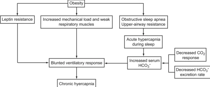
Pathophysiology
The PaCO2 is determined by the balance between CO2 production and elimination (minute ventilation and the frac-tion of dead-space ventilafrac-tion). The hypercapnia in OHS is entirely due to hypoventilation, as short-term treatment with PAP improves hypercapnia without any significant changes in body weight, CO2production, or the volume of
dead space.39However, the exact mechanisms that lead to
hypoventilation in obese individuals are complex and prob-ably multifactorial (Fig. 8). There have been a variety of physiologic differences between patients with OHS and those with obesity and/or OSA described to date: increased upper-airway resistance;53 an excessive mechanical load
imposed on the respiratory system by excess weight, ven-tilation-perfusion mismatching secondary to pulmonary edema,54 or low lung volumes/atelectasis;55 an impaired
central response to hypoxemia and hypercapnia; the pres-ence of sleep-disordered breathing; and impaired neuro-hormonal responses (leptin resistance). Although these are undoubtedly present, the most convincing evidence for the pathogenesis lies behind the universal presence of
sleep-disordered breathing and a blunted central response to hy-percapnia and hypoxia. Recently, Norman and colleagues proposed a mathematical model that combines sleep-dis-ordered breathing, central respiratory dive, and renal buff-ering to explain the development of this condition.56
The Excessive Load on the Respiratory System Upper-Airway Obstruction. Patient with OHS have a higher upper-airway resistance both in the sitting and su-pine position, when compared to patients with eucapnic OSA with similar degrees of obesity and control subjects.53
However, it remains unclear if an increased upper-airway resistance plays a role in the development of daytime hy-percapnia in this subset of patients.
Respiratory System Mechanics. In OHS there is an increase in the work of breathing in order to move the excess weight on the thoracic wall and abdomen during breathing.57However, it is unclear what contribution, if
any, these altered mechanics have in the pathogenesis of OHS. The lung compliance of OHS patients is less than an equally obese control group (0.122 L/cm H2O vs 0.157 L/
cm H2O). This can be explained by the lower functional
residual capacity of the OHS group (1.71 L vs 2.20 L). There is an even greater difference in chest-wall compli-ance between the 2 groups (OHS 0.079 L/cm H2O vs
obese controls 0.196 L/cm H2O).57Patients with OHS also
have a 3-fold increase in lung resistance that has been attributed to a low functional residual capacity.57,58 The
changes in lung mechanics are frequently demonstrated on spirometry by a low FVC and FEV1and a normal FEV1/
FVC ratio. The spirometric abnormalities may be related to the combination of abnormal respiratory mechanics and weak respiratory muscles.17,34,59,60The changes in
respi-ratory system mechanics in subjects with OHS impose a significant load on the respiratory muscles and lead to a 3-fold increase in the work of breathing.57 As a result,
morbidly obese patients dedicate 15% of their oxygen con-sumption to the work of breathing compared to 3% in non-obese individuals.61
Respiratory Muscles. The maximal inspiratory and ex-piratory pressures are normal in eucapnic morbidly obese patients but are reduced in patients with OHS.62-64Patients
with mild OHS, however, might have normal inspiratory and expiratory pressures.65A more accurate assessment of
the diaphragmatic strength by cervical magnetic stimula-tion has not been performed in patients with obesity hy-poventilation.66The role of diaphragmatic weakness in the
pathogenesis of this disorder remains uncertain because patients with OHS can generate similar transdiaphragmatic pressures at any level of diaphragmatic activation, com-pared to eucapnic obese subjects.63
Fig. 7. Survival curves for patients with untreated obesity hypoven-tilation syndrome (OHS) (n⫽47, mean age 55⫾14 y, mean body mass index [BMI] 45⫾9 kg/m2, mean P
aCO252⫾7 mm Hg) and
eucapnic morbidly obese patients (n⫽103, mean age 53⫾13 y, mean BMI 42⫾8 kg/m2), as reported by Nowbar et al,22
com-pared to patients with OHS treated with noninvasive ventilation (NIV) therapy (n ⫽ 126, mean age 55.6 ⫾ 10.6 y, mean BMI 44.6⫾7.8 kg/m2, mean baseline P
aCO255.5⫾7.7 mm Hg, mean
adherence to NIV of 6.5⫾2.3 h/d). Data for OHS patients treated with NIV was provided courtesy of Dr Stephan Budweiser and colleagues from the University of Regensburg, Germany.37
In a study by Sampson, patients with OHS were able to generate transdiaphragmatic pressure equivalent to that of eucapnic obese patients during hypercapnia-induced hy-perventilation, suggesting that respiratory muscle weak-ness may not play a role in the development of OHS. In addition, the OHS group showed no evidence of acute diaphragmatic fatigue (or neuromuscular uncoupling) throughout the hypercapnic trial when measured by the ratio of peak electrical activity of the diaphragm to peak transdiaphragmatic pressure, which should theoretically eliminate the variable of inadequate patient cooperation.63
Hypercapnia is also known to have deleterious effects on diaphragmatic function, so it will be difficult to determine whether respiratory muscle fatigue is a cause of or an effect of OHS.67
Taken together, the data suggest that obesity imposes a significant load on the respiratory system in patients with OHS. Obesity is not, however, the only determinant of hypoventilation, since less than a third of morbidly obese individuals develop hypercapnia.15,20,59
Blunted Central Respiratory Drive
Patients with OHS are able to voluntarily hyperventilate to eucapnia.68This is probably the simplest evidence for a
defective central respiratory drive, although there is plenty of additional evidence. Patients with OHS do not hyper-ventilate to the same degree as morbidly obese patients when rebreathing CO2.60,63,65This deficit corrects in most
patients after therapy with PAP.65,69,70 In patients with
severe OSA but without hypercapnia, the hypercapnic ven-tilatory response does not change with PAP therapy.70In
addition, patients with OHS do not augment their minute ventilation to the same degree as when forced to breathe a
hypoxic gas mixture.65,70,71 This blunted hypoxic drive
also corrects with PAP therapy.65,70 The reversibility of
the blunted central drive suggests that they are secondary effects of the syndrome (and necessary for its persistence), but not the origin of it.
There are a few hypotheses as to the origin of these defects. Obesity, genetic predisposition, sleep-disordered breathing, and leptin resistance have been proposed as mechanisms for the blunted response to hypercapnia. The weight load was suggested as a mechanism behind the blunted respiratory drive, because weight loss improves PaCO2level in patients with OHS. But this is unlikely to be related directly to weight, because, if anything, weight loss blunts the response of eucapnic morbidly obese subjects to hypercapnia.72The blunted respiratory response to
hyper-capnia is also unlikely to be familial, because the ventila-tory response to hypercapnia is similar between first-de-gree relatives of patients with OHS and control subjects.73
Treatment of sleep-disordered breathing with PAP ther-apy might improve the response to hypercapnia.65,69,70The
airway-occlusion pressure 0.1 s after the start of inspira-tory flow (P0.1) response to hypercapnia improves as early
as 2 weeks and reaches normal levels after 6 weeks of therapy with PAP in patients with mild OHS (PaCO2 be-tween 46 –50 mm Hg). The response of minute ventilation to hypercapnia improves by the sixth week of therapy, but does not normalize.65These finding, however, are not
uni-versal.39,69,74
Leptin. Leptin, a satiety hormone produced by adipo-cytes, stimulates ventilation.75-78 Obesity leads to an
in-crease in the CO2production and load.75Therefore, with
increasing obesity the excess adipose tissue leads to in-creasing levels of leptin in order to increase ventilation to Fig. 8. Mechanisms by which obesity can lead to chronic daytime hypercapnia.
compensate for the additional CO2load. This is the reason
as to why the vast majority of severely obese individuals do not develop hypercapnia. Patients with OHS and OSA have significantly higher leptin levels, compared to lean or BMI matched subjects without OSA. Although the inde-pendent contribution of OSA or OHS to leptin production remains unclear, the data suggest that excess adiposity is a much more significant contributor to elevated serum leptin levels than OSA or OHS.79-82 Patients with OHS,
how-ever, have a higher serum leptin level than eucapnic sub-jects with OSA matched for percent body fat and AHI, and their serum leptin level drops after treatment with PAP.81,83,84These observations suggest that patients with
OHS might be resistant to leptin. For leptin to affect the respiratory center and increase minute ventilation it has to penetrate into the cerebrospinal fluid. The leptin cerebro-spinal fluid-to-serum ratio is 4-fold higher in lean individ-uals, compared to obese subjects (0.045 ⫾ 0.01 vs 0.011⫾0.002,P⬍.05).85Individual differences in leptin
cerebrospinal fluid penetration may explain why some obese patients with severe OSA develop OHS and others do not.
Sleep-Disordered Breathing. Sleep-disordered breath-ing is considered necessary for the diagnosis of OHS and can take 2 forms. The first and by far the most common type is OSA, and the second is central hypoventilation. OSA is well established in the pathophysiology of OHS by the resolution of hypercapnia in most (but not all) patients by treatment with either tracheostomy or PAP thera-py.23,24,33,35,65,86,87Norman and colleagues have proposed
an elegant mathematical model that explains the transition from acute hypercapnia during sleep-disordered breathing to chronic daytime hypercapnia.56 In most patients with
OSA, the hyperventilation after an apnea eliminates all CO2accumulated during the apnea.88But if the
inter-ap-nea hyperventilation is inadequate or the ventilatory re-sponse to the accumulated CO2is blunted, it could lead to
an increase in PaCO2during sleep (Fig. 9).
89Even in this
acute setting during sleep the kidneys can retain small amounts of bicarbonate to buffer the decrease in pH. If the time constant for the excretion of the small amount of accumulated bicarbonate is slow, then the patient will have a net gain of bicarbonate and will retain some CO2during
wakefulness to compensate for the retained bicarbonate.56
Therefore, the combination of a decreased response to CO2and a slow rate of bicarbonate excretion rate will lead
to a blunted respiratory drive for the next sleep cycle.
Predictors of Hypercapnia in Obese Patients With OSA
Many studies have tried to find risk factors or predictors of hypercapnia (OHS) in cohorts of patients with OSA, but
the results have been mixed.14-20,34,90 In a recent large
meta-analysis from 15 studies of obese patients with OSA— but without COPD—Kaw et al were able to identify 3 significant predictors of chronic hypercapnia: severity of obesity, as measured by the BMI; severity of OSA, mea-sured by either AHI or hypoxia during sleep; and degree of restrictive chest physiology. The mean AHI in the hyper-capnic group was 64 events/h (95% CI 52–76 events/h) versus 51 events/h in the eucapnic group (95% CI 42– 60 events/h, difference between groupsP⬍.001).21In 2
studies the authors found the prevalence of OHS in pa-tients with an AHI⬎60 events/h to be 25–30%.30,90
Like-wise, Kaw found the mean BMI in the hypercapnic group to be 39 kg/m2(95% CI 34 – 44 kg/m2) versus 36 kg/m2
(95% CI 31– 41 kg/m2, difference between groups P ⬍
.001) (see Table 4).21
Treatment
Although there are no established guidelines on treat-ment of OHS, treattreat-ment modalities are each based on dif-ferent perspectives of the underlying pathophysiology of the condition: reversal of sleep-disordered-breathing, weight reduction, and pharmacotherapy.
Treatment of Sleep-Disordered Breathing
Positive Airway Pressure Therapy. PAP (in the form of CPAP therapy) was first described in the treatment of OHS in 1982.86Although subsequent studies confirmed its
Fig. 9. Schematic demonstrating how CO2excretion is dependent
upon inter-event hyperventilation. In the first cycle, the inter-event hyperpnea is sufficient to excrete the CO2accumulated during the
hypopnea. In the second cycle, much more CO2is accumulated
during the apnea than is excreted after the event. Repetitive cycles of inability to eliminate CO2accumulation during the apneic period
leads to CO2retention and increase in PaCO2level. (Adapted from
efficacy, failure of CPAP in some cases has led to uncer-tainty whether CPAP should be attempted initially or if bi-level PAP therapy (more commonly known as nonin-vasive ventilation [NIV]) is a better modality.15,24,39,86,91In
a recent prospective study of ambulatory patients with severe OHS— based on the severity of obesity, OSA and the degree of hypercapnia—57% of patients were titrated successfully with CPAP alone, and the mean pressure re-quired was 13.9 cm H2O.41The remaining 43% of patients
with OHS failed CPAP titration because of persistent hy-poxemia at therapeutic or near therapeutic pressures. In these patients the oxygen saturation remained below 90% for more than 20% of total sleep time. However, these patients who “failed CPAP” had a residual AHI of 25 events/h, which suggests that in some patients a ther-apeutic pressure was not achieved. Although both groups were extremely obese, the CPAP-failure group was more
obese (mean ⫾ SEM BMI 61.6 ⫾ 1.7 kg/m2 vs
56.5⫾1.2 kg/m2,P⫽.02). Since this was a single-night
titration study, the question of whether residual hypoxemia would resolve with long-term treatment was left unan-swered.92
A recent prospective randomized study performed by Piper et al93compared the long-term efficacy of bi-level
PAP versus CPAP. In this study 45 consecutive patients with OHS underwent a full night of CPAP titration. Nine patients (20%) were excluded because of persistent hy-poxemia—arbitrarily defined as 10 continuous minutes of SpO2⬍ 80% without frank apneas— during the CPAP ti-tration. The remaining 36 patients who had a successful CPAP titration night were subsequently randomized to ei-ther CPAP (n ⫽ 18) or bi-level PAP (n ⫽ 18). Those randomized to bi-level PAP underwent an additional titra-tion night to establish the effective inspiratory and expi-ratory pressures. Supplemental oxygen administration was necessary in 3 patients in the CPAP group and 4 in the bi-level PAP group. After 3 months, there was no signif-icant difference between the groups in adherence to PAP therapy or in improvement in daytime sleepiness, hypox-emia, or hypercapnia. This study confirms that the major-ity of patients with OHS (80%) can be successfully titrated with CPAP. These findings also suggest that, as long as OSA and nocturnal hypoxemia are effectively treated with CPAP, it makes no significant difference at 3 months if patients are given bi-level PAP or CPAP therapy. There-fore, bi-level PAP is not superior to CPAP a priori; rather, treatment should be individualized to each patient.
Bi-level PAP should be instituted if the patient is intol-erant of higher CPAP pressure (⬎15 cm H2O) that may be
required to resolve apneas and hypopneas or if hypoxemia is persistent despite adequate resolution of obstructive re-spiratory events during the titration study.94 During
bi-level PAP titration, the inspiratory PAP (IPAP) should be at least 8 to 10 cm H2O above the expiratory PAP (EPAP)
in order to effectively increase ventilation.33,35,95,96In the
minority of patients with OHS who do not have OSA, EPAP can be set at 5 cm H2O and IPAP can be titrated to
improve ventilation.95,96Bi-level PAP should also be
con-sidered if the PaCO2does not normalize after 3 months of therapy with CPAP.
Adherence to PAP Therapy. Adherence to PAP ther-apy, measured as average hours of daily use in the last 30 days, is directly correlated with improvement in day-time arterial blood gas values. In a retrospective study of 75 out-patients with stable OHS, the PaCO2 decreased by 1.8 mm Hg and the PaO2increased by 3 mm Hg per hour of daily CPAP or bi-level PAP use during the last 30 days before a repeated measurement of arterial blood gases. Patients who used PAP therapy for⬎4.5 h/d had a con-siderably greater improvement in blood gases than less adherent patients (⌬PaCO27.7⫾5 mm Hg vs 2.4⫾4 mm Hg,
P ⬍ .001; ⌬PaO29.2⫾ 11 mm Hg vs 1.8 ⫾ 9 mm Hg,
P⬍.001). In addition, the need for daytime oxygen ther-apy decreased from 30% of patients to 6%.24There was no
significant difference in improvement of hypercapnia and hypoxemia between patients on CPAP (n⫽ 48) and pa-tients on bi-level PAP therapy (n⫽27). Improvement in blood gas values may be seen as early as one month after the institution of PAP therapy.24,65,97
The impact of long-term NIV on vital capacity and lung volumes is contradictory. Several studies have reported no change in lung volumes or FVC after successful treatment of OHS with bi-level PAP.23,35,74In contrast, 2 studies of
patients with OHS reported significant improvements in vital capacity and expiratory reserve volume after 12 months of NIV, without any significant changes in BMI or in FEV1/FVC ratio.96,98
Lack of Improvement in Hypercapnia With PAP Ther-apy. The most common reason for persistent hypercap-nia in patients with OHS is lack of adherence to PAP therapy. However, if there is documented evidence of ad-equate adherence by objective monitoring of PAP devices, other possibilities need to be entertained, such as inade-quate PAP titration, CPAP failure, other causes of hyper-capnia such as COPD, or metabolic alkalosis due to high doses of loop diuretics.
The improvement in chronic daytime hypercapnia in patients who are adherent to PAP therapy is neither uni-versal nor complete. In 2 studies,24,93 the P
aCO2 did not improve significantly in approximately a quarter of pa-tients who had undergone successful PAP titration in the laboratory and were highly adherent (⬎6 h/night) to either CPAP or bi-level PAP therapy. In one study, 8 patients (23%) among the 34 patients who used PAP for at least 4.5 h/d, did not have a significant improvement in their PaCO2— decrease in PaCO2 of less than 4 mm Hg. These
non-responders had lower AHI, compared to responders (44⫾ 45 events/h vs 86⫾ 47 events/h,P⫽ .03). Mean adherence to PAP therapy was 7.2 ⫾ 2.1 h/d for non-responders versus 6.0⫾1.7 h/d for responders (P⫽.1).24
This lack of response to PAP therapy, combined with reports of persistent hypoventilation after tracheostomy,33
suggests that in a subset of patients with OHS, factors other than sleep-disordered breathing are the driving force behind the pathogenesis of hypoventilation. These patients will most likely need more aggressive nocturnal mechan-ical ventilation, with or without respiratory stimulants (see below).
Average Volume-Assured Pressure-Support Ventila-tion. Average volume-assured pressure-support ventila-tion is a hybrid mode of pressure-support and volume-controlled ventilation that delivers a more consistent tidal volume with the comfort of pressure-support ventilation. Average volume-assured pressure-support ventilation en-sures a preset tidal volume during bi-level-S/T mode, and the expiratory tidal volume is estimated based on pneu-motachographic inspiratory and expiratory flows. The IPAP support is then titrated in steps of 1 cm H2O/min in order
to achieve the preset tidal volume. As a result, the IPAP is variable. The EPAP on the other hand is set between 4 – 8 cm H2O and the respiratory backup rate can be set at
12–18 breaths/min with an inspiratory/expiratory ratio of 1:2. The role of a backup rate remains unclear, since pa-tients with OHS are typically tachypneic during sleep, with respiratory rates ranging between 15–30 breaths/min. However, it is conceivable that during titration central apneas could develop with pressure-support ventilation, and in those instances a backup rate would be useful.99
Although more costly than CPAP or bi-level PAP therapy, it has been shown effective in a randomized controlled study of OHS patients with milder degrees of hypercap-nia.100
Oxygen Therapy. In up to 50% of patients with OHS, oxygen therapy (in addition to PAP therapy) is necessary to keep SpO2 ⬎ 90% in the absence of hypopneas and apneas.41The need for nocturnal oxygen may abate with
regular PAP usage. One retrospective cohort study found the need for daytime supplemental oxygen decreased from 30% to 6% in patients who were adherent to PAP thera-py.24 Therefore, patients should be reassessed for both
diurnal and nocturnal oxygen requirements a few weeks to months after PAP therapy is instituted, since oxygen ther-apy is costly.
Phlebotomy. Phlebotomy has not been systematically studied in patients with OHS who develop secondary eryth-rocytosis. Secondary erythrocytosis is a physiological re-sponse to tissue hypoxia in order to enhance oxygen
car-rying capacity. However, hyperviscosity impairs oxygen delivery and can counteract the beneficial effects of eryth-rocytosis. In adult patients with congenital cyanotic heart disease, phlebotomy has been recommended if the hemat-ocrit is above 65% only if symptoms of hyperviscosity are present.101However, it is difficult to extrapolate this
rec-ommendation to patients with OHS, because many symp-toms of hyperviscosity are similar to the sympsymp-toms of OHS. Reversing hypoventilation and hypoxemia with PAP therapy eventually improves secondary erythrocytosis, and phlebotomy is rarely needed in patients with OHS.98
In-Laboratory PAP and Oxygen Titration. Figure 10 provides a therapeutic algorithm during polysomnography in order to address the variety of respiratory events ob-served in patients with OHS.33Even though auto-adjusting
PAP technology can be used in patients with simple OSA to bypass laboratory-based titration studies, this technol-ogy cannot be recommended in patients with OHS, be-cause it does not have the ability to recognize hypoventi-lation and hypoxemia. As a result, patients with OHS require a laboratory-based PAP and oxygen titration.
Fig. 10. Suggested therapeutic algorithm during continuous pos-itive airway pressure (CPAP) titration in patients with obesity hy-poventilation syndrome. IPAP⫽inspiratory positive airway pres-sure. EPAP ⫽ expiratory positive airway pressure. AVAPS ⫽ average volume-assured pressure-support ventilation.
Taken together, the data suggest that CPAP is effective in the majority of patients with stable OHS, particularly in the subgroup that have severe OSA. Bi-level PAP should be strongly considered in patients who fail CPAP, patients with OHS who experience acute-on-chronic respiratory failure, and in patients who have OHS without OSA. Whether average volume-assured pressure-support venti-lation has long-term benefits over bi-level PAP remains uncertain.
Treatment of OHS with PAP improves blood gases, morning headaches, excessive daytime sleepiness and vig-ilance, dyspnea, pulmonary hypertension, and leg ede-ma.23,35,74 Improvement in symptoms and blood gases is
directly related to adherence to therapy, and maximum improvement in blood gases can be achieved as early as 2 to 4 weeks. Therefore, early follow-up is imperative and should include repeat measurement of arterial blood gases and objective assessment of adherence to PAP, as patients frequently overestimate adherence.102-104 Changes in
se-rum bicarbonate level and pulse oximetry could be used as a less invasive measure of ventilation if the patient is reluctant to undergo a repeated measurement of arterial blood gases. Discontinuing oxygen therapy when no longer indicated can decrease the cost of therapy in patients with OHS.
Surgical Interventions
Weight-Reduction Surgery. Bariatric surgery has vari-able long-term efficacy in treating OSA. One study of patients undergoing Roux-en-Y showed that those with severe OSA had a reduction in AHI of 80 events/h to 20 events/h an average of 11 months after surgery.105
Al-though this drastic reduction in sleep-disordered breathing would probably be enough to normalize daytime blood gases, some of these patients still have moderate OSA and would benefit from continued PAP therapy. In another study, approximately half of the patients who had mild OSA after bariatric surgery had developed severe OSA 7 years postoperatively, despite no significant change in their weight.106 A recently published meta-analysis that
included 12 studies with 342 patients who underwent poly-somnography before bariatric surgery and after maximum weight loss reported that there was a 71% reduction in the AHI, with a reduction from baseline of 55 events/h (95% CI 49 – 60 events/h) to 16 events/h (95% CI 13–19 events/h). Only 38% achieved cure, defined as AHI⬍5 events/h. In contrast, 62% of patients had residual disease, with the mean residual AHI of 16 events/h. Many of these patients had persistent moderate OSA, defined as AHIⱖ15 events/ h.107It is also known that in the 6 – 8 years after
weight-reduction surgery, patients experience 7% weight gain.108
Therefore, patients with OHS who undergo bariatric
sur-gery should be monitored closely for recurrence of sleep-disordered breathing.
Only one study has examined the impact of bariatric surgery on OHS. Initially, blood gases improved. In 31 patients, preoperative PaO2increased from 53 mm Hg to 73 mm Hg one year after surgery, and PaCO2 decreased from 53 mm Hg to 44 mm Hg. In the 12 patients from whom arterial blood gas measurements were available 5 years after surgery, values had worsened, with the mean PaO2 dropping to 68 mm Hg and PaCO2 increasing to 47 mm Hg.109 In these 12 patients, BMI had hardly
in-creased from 1 to 5 years postoperatively (38 kg/m2 to
40 kg/m2). The worsening in daytime blood gases is
prob-ably from the redevelopment of sleep-disordered breath-ing.
Bariatric surgery is associated with significant risk. The perioperative mortality is between 0.5% and 1.5%. OSA and OHS may be associated with higher operative mortal-ity.110,111The independent risk factors associated with
mor-tality are intestinal leak, pulmonary embolism, preopera-tive weight, and hypertension. Depending on the type of the surgery, intestinal leak occurs in 2– 4% of patients and pulmonary embolism occurs in 1% of patients.111Ideally,
patients with OHS should be treated with PAP therapy— or tracheostomy in cases of PAP failure— before undergoing surgical intervention, in order to decrease perioperative morbidity and mortality. Moreover, PAP therapy should be initiated immediately after extubation to avoid postop-erative respiratory failure,112-114particularly in that there is
no evidence that PAP therapy initiated postoperatively leads to anastomotic disruption or leakage.113,115
Tracheostomy. Tracheostomy was the first therapy de-scribed for the treatment of OHS.116 In a retrospective
study of 13 patients with OHS, tracheostomy was associ-ated with significant improvement in OSA. With the tra-cheostomy closed, the mean non-rapid-eye-movement (non-REM) AHI and REM AHI were 64 events/h and 46 events/h, respectively; with the tracheostomy open, the non-REM AHI and REM AHI decreased to 31 events/h and 39 events/h, respectively. In 7 patients the AHI re-mained above 20 events/h. These residual respiratory events were associated with persistent respiratory effort, suggest-ing that disordered breathsuggest-ing was caused by hypoventila-tion through an open tracheostomy, rather than central apneas. However, the overall improvement in the severity of sleep-disordered breathing after tracheostomy led to the resolution of hypercapnia in the majority of the patients.117
Today tracheostomy is generally reserved for patients who are intolerant of, or not adherent to, PAP therapy. It is also an option for that minority of patients who do not have a significant improvement in daytime blood gases despite adherence to PAP therapy, especially those patients who have signs or symptoms of cor pulmonale. Patients with
tracheostomy may require nocturnal ventilation, as it does not treat any central hypoventilation that may be present.118
A polysomnogram with the tracheostomy open is neces-sary to determine whether nocturnal ventilation is re-quired.33
Pharmacologic Respiratory Stimulation
Respiratory stimulants can theoretically increase respi-ratory drive and improve daytime hypercapnia, but the data are extremely limited.
Medroxyprogesterone. Medroxyprogesterone acts as a respiratory stimulant at the hypothalamic level.119The
re-sults of treatment in patients with OHS have been contra-dictory. In a series of 10 men with OHS treated with high doses of oral medroxyprogesterone (60 mg/d) for one month, the PaCO2decreased from 51 mm Hg to 38 mm Hg and the PaO2increased from 49 mm Hg to 62 mm Hg.
120
All these patients were able to normalize their PaCO2with 1–2 min of voluntary hyperventilation, suggesting that there was no limitation to ventilation. Of note, polysomnographic data were not available for these 10 men with OHS, so it remains unclear whether they had concomitant OSA as well. In contrast, medroxyprogesterone did not improve PaCO2, minute ventilation, or ventilatory response to hy-percapnia in 3 OHS patients who remained hypercapnic after tracheostomy.39Administration of a medication that
may increase the risk of venous thromboembolism to a population whose mobility is limited may be unwise.121,122
In addition, high doses of medroxyprogesterone can lead to breakthrough uterine bleeding in women and to de-creased libido and erectile dysfunction in men.
Most but not all patients with OHS can normalize their PaCO2 with voluntary hyperventilation.
68 The inability to
eliminate CO2with voluntary hyperventilation may be due
to mechanical impairment. In one study, the ability to drop the PaCO2by at least 5 mm Hg with voluntary hyperven-tilation was the main predictor of a favorable response to medroxyprogesterone.123 Therefore, a respiratory
stimu-lant in patients who cannot normalize their PaCO2 with voluntary hyperventilation— due to limited ventilation and/or mechanical impairment— can lead to an increase in dyspnea or even worsening of acidosis with acetazolamide.
Acetazolamide. Acetazolamide induces metabolic aci-dosis through carbonic anhydrase inhibition, which in-creases minute ventilation in normal subjects. There is only one published case report describing normalization of blood gases after tracheostomy,39although, interestingly,
the agent reduces the AHI in patients with moderate to severe OSA.124,125
In summary, the treatment options other than PAP are poorly studied. PAP therapy is the mainstay of treatment,
but the best approach for those who do not respond to this modality is unknown and may include a combination of PAP therapy and pharmacotherapy with respiratory stim-ulants or tracheostomy, with or without nocturnal ventila-tion.
Summary
With such a global epidemic of obesity, the prevalence of OHS is likely to increase. Despite the significant mor-bidity and mortality associated with the syndrome, it is often unrecognized and treatment is frequently delayed. A high index of suspicion can lead to early recognition of the syndrome and initiation of appropriate therapy. The treat-ment options other than PAP have been poorly studied, and further research is needed to better understand the long-term treatment outcomes of patients with OHS. For the time being, clinicians should encourage adherence to PAP therapy in order to prevent the serious adverse out-comes of untreated OHS. Weight-reduction surgery or tra-cheostomy, with or without pharmacotherapy with respi-ratory stimulants, should be considered in cases of PAP failure. Further research is needed to better understand the pathophysiology, discover newer PAP modalities, explore non-PAP treatment options, and improve long-term treat-ment outcomes of patients with OHS.
REFERENCES
1. Lugaresi E, Coccagna G, Tassinari CA, Ambrosetto C. [Particular-ite´s cliniques et polygraphiques du syndrome d’impatience des mem-bres infe´rieurs]. Rev Neurol 1965;115:545.Article in French.
2. Gastaut H, Tassinari CA, Duron B. Polygraphic study of the epi-sodic diurnal and nocturnal (hypnic and respiratory) manifestations of the Pickwick syndrome. Brain Res 1966;1(2):167-186. 3. Lavie P. Who was the first to use the term Pickwickian in
connec-tion with sleepy patients? History of sleep apnoea syndrome. Sleep Med Rev 2008;12(1):5-17.
4. Sotos JG. Taft and Pickwick: sleep apnea in the White House. Chest 2003;124(3):1133-1142.
5. Auchincloss JH Jr, Cook E, Renzetti AD. Clinical and physiolog-ical aspects of a case of obesity, polycythemia and alveolar hy-poventilation. J Clin Invest 1955;34(10):1537-1545.
6. Burwell CS, Robin ED, Whaley RD, Bickelmann AG. Extreme obesity associated with alveolar hypoventilation: a Pickwickian syn-drome. Am J Med 1956;21(5):811-818.
7. Dickens, C. The posthumous papers of the Pickwick club. Boston: Ticknor and Fields; 1867.
8. Mokhlesi B, Kryger MH, Grunstein RR. Assessment and manage-ment of patients with obesity hypoventilation syndrome. Proc Am Thorac Soc 2008;5(2):218-225.
9. Sturm R. Increases in morbid obesity in the USA: 2000-2005. Pub-lic Health 2007;121(7):492-496.
10. Prentice A, Webb F. Obesity amidst poverty. Int J Epidemiol 2006; 35(1):24-30.
11. Skidmore PM, Yarnell JW. The obesity epidemic: prospects for prevention. QJM 2004;97(12):817-825.
12. Spritzer DA. Obesity epidemic migrates east. CMAJ 2004;171(10): 1159.
13. Miech RA, Kumanyika SK, Stettler N, Link BG, Phelan JC, Chang VW. Trends in the association of poverty with overweight among US adolescents, 1971-2004. JAMA 2006;295(20):2385-2393. 14. Verin E, Tardif C, Pasquis P. Prevalence of daytime hypercapnia or
hypoxia in patients with OSAS and normal lung function. Respir Med 2001;95(8):693-696.
15. Laaban JP, Chailleux E. Daytime hypercapnia in adult patients with obstructive sleep apnea syndrome in France, before initiating noc-turnal nasal continuous positive airway pressure therapy. Chest 2005;127(3):710-715.
16. Kessler R, Chaouat A, Schinkewitch P, Faller M, Casel S, Krieger J, et al. The obesity-hypoventilation syndrome revisited: a prospec-tive study of 34 consecuprospec-tive cases. Chest 2001;120(2):369-376. 17. Resta O, Foschino Barbaro MP, Bonfitto P, Talamo S,
Mastrosi-mone V, Stefano A, et al. Hypercapnia in obstructive sleep apnoea syndrome. Neth J Med 2000;56(6):215-222.
18. Golpe R, Jimenez A, Carpizo R. Diurnal hypercapnia in patients with obstructive sleep apnea syndrome. Chest 2002;122(3):1100-1101.
19. Akashiba T, Akahoshi T, Kawahara S, Uematsu A, Katsura K, Sakurai S, et al. Clinical characteristics of obesity-hypoventilation syndrome in Japan: a multi-center study. Intern Med 2006;45(20): 1121-1125.
20. Mokhlesi B, Tulaimat A, Faibussowitsch I, Wang Y, Evans AT. Obesity hypoventilation syndrome: prevalence and predictors in patients with obstructive sleep apnea. Sleep Breath 2007;11(2):117-124.
21. Kaw R, Hernandez AV, Walker E, Aboussouan L, Mokhlesi B. Determinants of hypercapnia in obese patients with obstructive sleep apnea: a systematic review and meta-analysis of cohort studies. Chest 2009;136(3):787-796.
22. Nowbar S, Burkart KM, Gonzales R, Fedorowicz A, Gozansky WS, Gaudio JC, et al. Obesity-associated hypoventilation in hospitalized patients: prevalence, effects, and outcome. Am J Med 2004;116(1): 1-7.
23. Masa JF, Celli BR, Riesco JA, Hernandez M, Sanchez De Cos J, Disdier C. The obesity hypoventilation syndrome can be treated with noninvasive mechanical ventilation. Chest 2001;119(4):1102-1107.
24. Mokhlesi B, Tulaimat A, Evans AT, Wang Y, Itani A, Hassaballa HA, et al. Impact of adherence with positive airway pressure ther-apy on hypercapnia in obstructive sleep apnea. J Clin Sleep Med 2006;2(1):57-62.
25. Freedman DS, Khan LK, Serdula MK, Galuska DA, Dietz WH. Trends and correlates of class 3 obesity in the United States from 1990 through 2000. JAMA 2002;288(14):1758-1761.
26. McTigue K, Larson JC, Valoski A, Burke G, Kotchen J, Lewis CE, et al. Mortality and cardiac and vascular outcomes in extremely obese women. JAMA 2006;296(1):79-86.
27. Sakakibara H, Tong M, Matsushita K, Hirata M, Konishi Y, Suet-sugu S. Cephalometric abnormalities in non-obese and obese pa-tients with obstructive sleep apnoea. Eur Respir J 1999;13(2):403-410.
28. Yu X, Fujimoto K, Urushibata K, Matsuzawa Y, Kubo K. Cepha-lometric analysis in obese and nonobese patients with obstructive sleep apnea syndrome. Chest 2003;124(1):212-218.
29. Lee W, Nagubadi S, Kryger MH, Mokhlesi B. Epidemiology of obstructive sleep apnea: a population-based perspective. Expert Rev Respir Med 2008;2(3):349-364.
30. Littleton SW, Mokhlesi B. The Pickwickian syndrome: obesity hy-poventilation syndrome. Clin Chest Med 2009;30(3):467-478. 31. Lee WY, Mokhlesi B. Diagnosis and management of obesity
hy-poventilation syndrome in the ICU. Crit Care Clin 2008;24(3):533-549.
32. Quint JK, Ward L, Davison AG. Previously undiagnosed obesity hypoventilation syndrome. Thorax 2007;62(5):462-463.
33. Berger KI, Ayappa I, Chatr-Amontri B, Marfatia A, Sorkin IB, Rapoport DM, et al. Obesity hypoventilation syndrome as a spec-trum of respiratory disturbances during sleep. Chest 2001;120(4): 1231-1238.
34. Leech JA, Onal E, Baer P, Lopata M. Determinants of hypercapnia in occlusive sleep apnea syndrome. Chest 1987;92(5):807-813. 35. Perez de Llano LA, Golpe R, Ortiz Piquer M, Veres Racamonde A,
Vazquez Caruncho M, Caballero Muinelos O, et al. Short-term and long-term effects of nasal intermittent positive pressure ventilation in patients with obesity-hypoventilation syndrome. Chest 2005; 128(2):587-594.
36. Berg G, Delaive K, Manfreda J, Walld R, Kryger MH. The use of health-care resources in obesity-hypoventilation syndrome. Chest 2001;120(2):377-383.
37. Budweiser S, Riedl SG, Jorres RA, Heinemann F, Pfeifer M. Mor-tality and prognostic factors in patients with obesity-hypoventila-tion syndrome undergoing noninvasive ventilaobesity-hypoventila-tion. J Intern Med 2007;261(4):375-383.
38. Resta O, Foschino-Barbaro MP, Bonfitto P, Talamo S, Legari G, De Pergola G, et al. Prevalence and mechanisms of diurnal hyper-capnia in a sample of morbidly obese subjects with obstructive sleep apnoea. Respir Med 2000;94(3):240-246.
39. Rapoport DM, Garay SM, Epstein H, Goldring RM. Hypercapnia in the obstructive sleep apnea syndrome: a reevaluation of the Pick-wickian syndrome. Chest 1986;89(5):627-635.
40. Olson AL, Zwillich C. The obesity hypoventilation syndrome. Am J Med 2005;118(9):948-956.
41. Banerjee D, Yee BJ, Piper AJ, Zwillich CW, Grunstein RR. Obesity hypoventilation syndrome: hypoxemia during continuous positive airway pressure. Chest 2007;131(6):1678-1684.
42. Koenig SM. Pulmonary complications of obesity. Am J Med Sci 2001;321(4):249-279.
43. Janssens JP, Derivaz S, Breitenstein E, De Muralt B, Fitting JW, Chevrolet JC, et al. Changing patterns in long-term noninvasive ventilation: a 7-year prospective study in the Geneva Lake area. Chest 2003;123(1):67-79.
44. Flegal KM, Graubard BI, Williamson DF, Gail MH. Excess deaths associated with underweight, overweight, and obesity. JAMA 2005; 293(15):1861-1867.
45. Young T, Finn L, Peppard PE, Szklo-Coxe M, Austin D, Nieto FJ, et al. Sleep disordered breathing and mortality: eighteen-year fol-low-up of the Wisconsin Sleep Cohort. Sleep 2008;31(8):1071-1078.
46. Punjabi NM, Caffo BS, Goodwin JL, Gottlieb DJ, Newman AB, O’Connor GT, et al. Sleep-disordered breathing and mortality: a prospective cohort study. PLoS Med 2009;6(8):e1000132. 47. Marin JM, Carrizo SJ, Vicente E, Agusti AG. Long-term
cardio-vascular outcomes in men with obstructive sleep apnoea-hypop-noea with or without treatment with continuous positive airway pressure: an observational study. Lancet 2005;365(9464):1046-1053.
48. Hida W. Quality of life in obesity hypoventilation syndrome. Sleep Breath 2003;7(1):1-2.
49. Budweiser S, Hitzl AP, Jo¨rres RA, Schmidbauer K, Heinemann F, Pfeifer M. Health-related quality of life and long-term prognosis in chronic hypercapnic respiratory failure: a prospective survival anal-ysis. Respir Res 2007;8:92.
50. Atwood CW Jr, McCrory D, Garcia JG, Abman SH, Ahearn GS. Pulmonary artery hypertension and sleep-disordered breathing: ACCP evidence-based clinical practice guidelines. Chest 2004;126(1 Suppl):72S-77S.
51. Kessler R, Chaouat A, Weitzenblum E, Oswald M, Ehrhart M, Apprill M, et al. Pulmonary hypertension in the obstructive sleep apnoea syndrome: prevalence, causes and therapeutic consequences. Eur Respir J 1996;9(4):787-794.
52. Sugerman HJ, Baron PL, Fairman RP, Evans CR, Vetrovec GW. Hemodynamic dysfunction in obesity hypoventilation syndrome and the effects of treatment with surgically induced weight loss. Ann Surg 1988;207(5):604-613.
53. Lin CC, Wu KM, Chou CS, Liaw SF. Oral airway resistance during wakefulness in eucapnic and hypercapnic sleep apnea syndrome. Respir Physiol Neurobiol 2004;139(2):215-224.
54. Kaltman AJ, Goldring RM. Role of circulatory congestion in the cardiorespiratory failure of obesity. Am J Med 1976;60(5):645-653. 55. Piper AJ, Grunstein RR. Big breathing: the complex interaction of obesity, hypoventilation, weight loss and respiratory function. J Appl Physiol 2010;101(1):199-205.
56. Norman RG, Goldring RM, Clain JM, Oppenheimer BW, Charney AN, Rapoport DM, et al. Transition from acute to chronic hyper-capnia in patients with periodic breathing: predictions from a com-puter model. J Appl Physiol 2006;100(5):1733-1741.
57. Sharp JT, Henry JP, Sweany SK, Meadows WR, Pietras RJ. The total work of breathing in normal and obese men. J Clin Invest 1964;43:728-739.
58. Rubinstein I, Zamel N, DuBarry L, Hoffstein V. Airflow limitation in morbidly obese nonsmoking men. Ann Intern Med 1990;112(11): 828-832.
59. Javaheri S, Colangelo G, Lacey W, Gartside PS. Chronic hyper-capnia in obstructive sleep apnea-hypopnea syndrome. Sleep 1994; 17(5):416-423.
60. Lopata M, Freilich RA, Onal E, Pearle J, Lourenco RV. Ventilatory control and the obesity hypoventilation syndrome. Am Rev Respir Dis 1979;119(2 Pt 2):165-168.
61. Kress JP, Pohlman AS, Alverdy J, Hall JB. The impact of morbid obesity on oxygen cost of breathing (VO2RESP) at rest. Am J Respir
Crit Care Med 1999;160(3):883-886.
62. Kelly TM, Jensen RL, Elliott CG, Crapo RO. Maximum respiratory pressures in morbidly obese subjects. Respiration 1988;54(2):73-77.
63. Sampson MG, Grassino K. Neuromechanical properties in obese patients during carbon dioxide rebreathing. Am J Med 1983;75(1): 81-90.
64. Becker HF, Piper AJ, Flynn WE, McNamara SG, Grunstein RR, Peter JH, et al. Breathing during sleep in patients with nocturnal desaturation. Am J Respir Crit Care Med 1999;159(1):112-118. 65. Han F, Chen E, Wei H, He Q, Ding D, Strohl KP. Treatment effects
on carbon dioxide retention in patients with obstructive sleep ap-nea-hypopnea syndrome. Chest 2001;119(6):1814-1819. 66. American Thoracic Society, European Respiratory Society. ATS/
ERS Statement on respiratory muscle testing. Am J Respir Crit Care Med 2002;166(4):518-624.
67. Laffey J, Kavanagh B. Permissive hypercapnia. In: Tobin MJ, ed-itor. Principles and practice of mechanical ventilation. New York: McGraw-Hill Professional; 2006:379.
68. Leech J, Onal E, Aronson R, Lopata M. Voluntary hyperventilation in obesity hypoventilation. Chest 1991;100(5):1334-1338. 69. Berthon-Jones M, Sullivan CE. Time course of change in
ventila-tory response to CO2with long-term CPAP therapy for obstructive
sleep apnea. Am Rev Respir Dis 1987;135(1):144-147.
70. Lin CC. Effect of nasal CPAP on ventilatory drive in normocapnic and hypercapnic patients with obstructive sleep apnoea syndrome. Eur Respir J 1994;7(11):2005-2010.
71. Zwillich CW, Sutton FD, Pierson DJ, Greagh EM, Weil JV. De-creased hypoxic ventilatory drive in the obesity-hypoventilation syndrome. Am J Med 1975;59(3):343-348.
72. Emirgil C, Sobol BJ. The effects of weight reduction on pulmonary function and the sensitivity of the respiratory center in obesity. Am Rev Respir Dis 1973;108(4):831-842.
73. Jokic R, Zintel T, Sridhar G, Gallagher CG, Fitzpatrick MF. Ven-tilatory responses to hypercapnia and hypoxia in relatives of pa-tients with the obesity hypoventilation syndrome. Thorax 2000; 55(11):940-945.
74. Chouri-Pontarollo N, Borel JC, Tamisier R, Wuyam B, Levy P, Pepin JL. Impaired objective daytime vigilance in obesity-hypoventilation syndrome: impact of noninvasive ventilation. Chest 2007;131(1):148-155.
75. Kalra SP. Central leptin insufficiency syndrome: an interactive eti-ology for obesity, metabolic and neural diseases and for designing new therapeutic interventions. Peptides 2008;29(1):127-138. 76. Tankersley C, Kleeberger S, Russ B, Schwartz A, Smith P.
Mod-ified control of breathing in genetically obese (ob/ob) mice. J Appl Physiol 1996;81(2):716-723.
77. Tankersley CG, O’Donnell C, Daood MJ, Watchko JF, Mitzner W, Schwartz A, et al. Leptin attenuates respiratory complications as-sociated with the obese phenotype. J Appl Physiol 1998;85(6): 2261-2269.
78. Considine RV, Sinha MK, Heiman ML, Kriauciunas A, Stephens TW, Nyce MR, et al. Serum immunoreactive-leptin concentrations in normal-weight and obese humans. N Engl J Med 1996;334(5): 292-295.
79. Ip MS, Lam KS, Ho C, Tsang KW, Lam W. Serum leptin and vascular risk factors in obstructive sleep apnea. Chest 2000;118(3): 580-586.
80. Barcelo A, Barbe F, Llompart E, de la Pena M, Duran-Cantolla J, Ladaria A, et al. Neuropeptide Y and leptin in patients with ob-structive sleep apnea syndrome: role of obesity. Am J Respir Crit Care Med 2005;171(2):183-187.
81. Shimura R, Tatsumi K, Nakamura A, Kasahara Y, Tanabe N, Takigu-chi Y, et al. Fat accumulation, leptin, and hypercapnia in obstruc-tive sleep apnea-hypopnea syndrome. Chest 2005;127(2):543-549. 82. Makinodan K, Yoshikawa M, Fukuoka A, Tamaki S, Koyama N, Yamauchi M, et al. Effect of serum leptin levels on hypercapnic ventilatory response in obstructive sleep apnea. Respiration 2008; 75(3):257-264.
83. Phipps PR, Starritt E, Caterson I, Grunstein RR. Association of serum leptin with hypoventilation in human obesity. Thorax 2002; 57(1):75-76.
84. Yee BJ, Cheung J, Phipps P, Banerjee D, Piper AJ, Grunstein RR. Treatment of obesity hypoventilation syndrome and serum leptin. Respiration 2006;73(2):209-212.
85. Caro JF, Kolaczynski JW, Nyce MR, Ohannesian JP, Opentanova I, Goldman WH, et al. Decreased cerebrospinal-fluid/serum leptin ratio in obesity: a possible mechanism for leptin resistance. Lancet 1996;348(9021):159-161.
86. Rapoport DM, Sorkin B, Garay SM, Goldring RM. Reversal of the Pickwickian syndrome by long-term use of nocturnal nasal-airway pressure. N Engl J Med 1982;307(15):931-933.
87. Leech JA, Onal E, Lopata M. Nasal CPAP continues to improve sleep-disordered breathing and daytime oxygenation over long-term follow-up of occlusive sleep apnea syndrome. Chest 1992;102(6): 1651-1655.
88. Ayappa I, Berger KI, Norman RG, Oppenheimer BW, Rapoport DM, Goldring RM. Hypercapnia and ventilatory periodicity in ob-structive sleep apnea syndrome. Am J Respir Crit Care Med 2002; 166(8):1112-1115.
89. Berger KI, Ayappa I, Sorkin IB, Norman RG, Rapoport DM, Gold-ring RM. CO2homeostasis during periodic breathing in obstructive


![Fig. 4. Decision tree to screen for obesity hypoventilation syn- syn-drome (OHS) in 522 obese patients with obstructive sleep apnea (OSA) (body mass index [BMI] ⱖ 30 kg/m 2 and apnea-hypopnea index (AHI) ⱖ 5 events/h)](https://thumb-us.123doks.com/thumbv2/123dok_us/1160857.2655564/5.904.87.436.112.403/decision-screen-obesity-hypoventilation-patients-obstructive-hypopnea-events.webp)

![Fig. 7. Survival curves for patients with untreated obesity hypoven- hypoven-tilation syndrome (OHS) (n ⫽ 47, mean age 55 ⫾ 14 y, mean body mass index [BMI] 45 ⫾ 9 kg/m 2 , mean P aCO](https://thumb-us.123doks.com/thumbv2/123dok_us/1160857.2655564/7.904.87.437.112.325/survival-patients-untreated-obesity-hypoven-hypoven-tilation-syndrome.webp)