Epstein-Barr Virus Lymphoproliferative Disease following
Allogeneic Hematopoietic Stem Cell Transplantation:
Prediction and early Intervention
Cover design : Job van Esser
Lay-out cover : Inge Enneking-Leijten
Lay-out : Edwin Bosma
Printed by : Optima grafische communicatie, Rotterdam
ISBN : 90-6734-096-0
Publication of this thesis was financially supported by Roche Nederland
Copyright : J.W.J. van Esser, Rotterdam, 2002
© All rights reserved. No part of this publication may be reproduced, stored in a retrieval
system, or transmitted in any form or by any means, mechanically, by photocopying, by recording or otherwise without the prior permission of the author.
Allogeneic Hematopoietic Stem Cell Transplantation:
Prediction and early Intervention
Epstein-Barr virus geïnduceerde lymfoproliferatieve ziekte na
allogene hematopoëtische stam cel transplantatie:
voorspelling en vroegtijdige interventie
Proefschrift
ter verkrijging van de graad van doctor aan de
Erasmus Universiteit Rotterdam
op gezag van de
Rector Magnificus
Prof.dr.ir. J.H. van Bemmel
en volgens besluit van het College voor Promoties.
De openbare verdediging zal plaatsvinden op
vrijdag 17 januari 2003 om 13.30 uur
door
Joseph Willem Jan van Esser
geboren te Roermond
Promotor: Prof.dr. B. Löwenberg
Overige leden: Prof.dr. J.M. Middeldorp
Prof.dr. A.D.M.E. Osterhaus
Prof.dr. W. Weimar
De wereld gaat en gaat, als lang na dezen mijn roem verging, mijn kennis hooggeprezen. Wij werden vóór ons komen niet gemist, na ons vertrek zal het niet anders wezen. Uit de Rubaijat J.H. Leopold
Chapter 1. Introduction 11 Chapter 2. Development of a real-time quantitative assay for detection of
Epstein-Barr Virus.
(Journal of Clinical Microbiology. 2000;38:712-715) 53
Chapter 3. Epstein-Barr virus (EBV) reactivation is a frequent event after allogeneic hematopoietic stem cell transplantation and quantitatively predicts EBV-lymphoproliferative disease following T-cell-depleted stem cell transplantation.
(Blood. 2001;98:972-978) 67
Chapter 4. Molecular quantification of viral load in plasma allows for fast and accurate prediction of response to therapy of Epstein-Barr virus-associated lymphoproliferative disease after allogeneic hematopoietic stem cell transplantation.
(British Journal of Haematology. 2001;113:814-821) 91
Chapter 5. Prevention of Epstein-Barr virus-lymphoproliferative disease by molecular monitoring and pre-emptive rituximab in high-risk patients after allogeneic hematopoietic stem cell transplantation.
(Blood. 2002;99:4364-4369)
111
Chapter 6. Recipients of an allogeneic hematopoietic stem cell graft not recovering Epstein-Barr virus (EBV) specific immunity are at risk to develop high-level EBV load reactivation and
EBV-lymphoproliferative disease. 131
Chapter 7. General discussion 153
Chapter 8. Summary / Samenvatting 167
List of publications 174
Dankwoord 176
ATG Anti-Thymocyte Globulin
Allo-SCT Allogeneic hematopoietic Stem Cell Transplantation
BM Bone Marrow
Bu Busulphan
CFU-GM Granulocyte-Macrophage Colony Forming Units
CMV Cytomegalovirus
COD Cause of Death
CR Complete Remission Ct Cycle threshold CTL Cytotoxic T-cells Cy Cyclophosphamide CyA/CsA Cyclosporin A D Donor
DNA Deoxyribonucleic Acid
DLI Donor Lymphocyte Infusion
EBV Epstein-Barr Virus
EBNA Epstein-Barr virus Nuclear Antigen
EBER Epstein-Barr virus Encoding RNA
EM Electron Microscopy
h/hrs hour(s)
HLA Human Leucocyte Antigen
HIV Human Immunodeficiency Virus
IEA Immediate Early Antigens
IM Infectious Mononucleosis
LMP Latent Membrane Protein
LP Leader Protein
LPD Lymphoproliferative Disease
MNC Mononuclear Cells
MUD Matched Unrelated Donor
NK-cell Natural Killer cell
NHL Non-Hodgkin’s Lymphoma
OKT3 Anti-T-cell antibody Ortho-Klone
PB Peripheral Blood
PCR Polymerase Chain Reaction
PD Progressive Disease
PR Partial Remission
PTLD Post-Transplant Lymphoproliferative Disease
PV+/- Positive/negative predictive value
RNA Ribonucleic Acid
SCT Stem Cell Transplantation
Sib HLA-identical family donor
TRM Treatment-Related mortality
TCD T-Cell Depleted
TBI Total Body Irradiation
1. Introduction 13 2. Infection, immunity, and malignancy
EBV-infection
Immortalization of B-cells Latency and reactivation
Immune response to EBV-infection EBV-induced infectious syndromes Malignancies associated with EBV
13 13 14 14 16 17 18 3. Post-transplant lymphoproliferative disease
Introduction
Clinical presentation Diagnosis of PTLD
Risk factors for developing PTLD Therapeutic approaches
Prevention of PTLD following solid organ transplantation and allogeneic hematopoietic stem cell transplantation
20 20 20 21 23 27 32
1. Introduction
Since its discovery, Epstein-Barr virus (EBV) has been associated with a variety of both infectious and malignant human diseases. EBV was first detected by electron microscopy
in cultured Burkitt’s lymphoma cells in 1964 by Epstein, Achong and Barr.1 EBV is a
DNA virus with a linear genome of 172 kb and belongs to the genus lymphocryptovirus within the family of gamma herpesviruses. These viruses are characterized by (B-cell) lymphotropism, their ability to establish latent infection in host cells and to induce
proliferation of these latently infected cells.2 EBV was distinguished from other, at the
time known human Herpes viruses, because of its strong association with Burkitt’s lymphoma and its potent growth-transforming ability for B-lymphocytes in vitro. Approximately 90% of humans will become infected with EBV, generally without clinical evidence of disease. Primary infection usually occurs asymptomatically in childhood and results in a lifetime carrier state with periodic release of infectious virus into saliva which
may cause infection of naive individuals.2 Sometimes later, e.g. during adolescence, the
latter type of infection may be referred to as kissing disease or Pfeiffer’s disease.3 EBV
causes various benign syndromes, such as infectious mononucleosis, chronic active EBV infection, X-linked lymphoproliferative disease and oral hairy leukoplakia. EBV has also been associated with malignant diseases including nasopharyngeal carcinoma, Burkitt’s lymphoma, Hodgkin’s lymphoma, and post-transplant lymphoproliferative disease
(PTLD).4-7 This thesis deals with the development of molecular monitoring of EBV-DNA
in plasma of recipients of allogeneic hematopoietic stem cell grafts and the introduction of such monitoring in therapeutic and preventive approaches. In this chapter we shall briefly review the pathogenetic role of EBV in infectious and malignant diseases. Subsequently the clinical presentation, diagnosis, incidence, risk factors and treatment of EBV associated lymphoproliferative disease (LPD) in recipients of hematopoietic stem cell transplants is discussed followed by an outline of this thesis.
2. Infection, immunity, and malignancy
EBV-infectionEBV shedding in the oropharynx occurs intermittently in EBV-seropositive individuals, during such periods of EBV shedding other persons may become infected. When EBV enters the oropharyngeal cavity it penetrates local lymphoid tissue (lingual-, palatine-, and pharyngeal tonsils). Squamous epithelium covering the lymphoid tissue dips into the tonsillar crypts where cells are densely situated. Following primary infection these
B-cells are the first to be infected. 8 Although earlier studies have discussed elaborately
whether EBV replicates in epithelium 9-14, a recent study identified EBV replication in
epithelial cells in vitro.15 Following contact between virus and cells, EBV enters the
(receptor for complement C3d) on the surface of the B-cell. Binding of glycoprotein 42 to major-histocompatibility-complex class II (co-receptor) and binding of glycoprotein
350/220 to its receptor facilitate fusion of the viral envelope with the B-cell during
endocytosis.15-18 Receptorbinding results in activation of the B-cell, which may further
favor penetration of the virus into the cell. The de-enveloped capsid subsequently travels into the nucleus, degrades and releases viral DNA.
Immortalization of B-cells
Linear EBV-DNA is being transported to the cell-nucleus, where transcription and translation of Epstein-Barr virus nuclear antigen (EBNA)-2 and EBNA-leader protein (LP) start within 4 hours following infection. EBNA-2 is essential for B-cell transformation and is an activator of EBV-latent membrane protein (LMP)-1 and LMP-2 and genes involved in growth and transformation of the infected B cell. EBNA-LP augments the ability of
EBNA-2 to up-regulate LMP-1.2,8LMP-1 contributes to the growth and transformation of
B-cells and is the major transforming gene of EBV.19,20 LMP-1 mimics in this respect the
function of CD40, a receptor constitutively present on the surface of B-cells.21 B-cell
activation leads to differentiation of the B-cell to a B-blast, that expresses besides
EBNA-2, LMP-1 and EBNA-3A, 3B, 3C and EBNA-4. The circularisation of linear EBV-DNA to episomal EBV-DNA (circular form of EBV-DNA) in the B-cell nucleus is completed twenty hours after infection, and with the expression of EBNA-1 the transformation of the B-cell to an immortalized B-cell is completed. LMP-2 is only transcribed once the circular
viral episome is formed and prevents B-cell activation stabilising the latent state. 20 The
non-translated types of EBV-encoded RNA (EBER) do not encode proteins but may be
important for oncogenesis and resistance to programmed cell death, or apoptosis. 22
Latency and reactivation
EBNA-1 plays a pivotal role in the maintenance of EBV in dividing latently infected
B-cells.23-25 EBV-infected B-cells in vivo can express four different programmes of gene
usage depending on the location and differentiation stage of the infected B-cell. One of these programmes is used to produce infectious virus, the other three are all associated with latent infection, in which no virus is produced. These latent growth programmes are known as the growth programme, the default programme and the latency programme
(Table 1).26,27 During the growth programme EBV infects resting naive B-cells (CD27-,
sIgD+) in tonsils and drives these cells out of their resting state to become activated
proliferating lymphoblasts.28 Three viral proteins are expressed in the default programme,
of which EBNA-1 ensures replication of the viral genome during cell division.2,27
Physiologically, B-cells that encounter antigen become activated and migrate into the follicle of a lymph node where they form germinal centers. Following proliferation and somatic hypermutation, cells that have mutated their immunoglobulin genes become antibody producing plasma cells or memory B-cells, remaining cells that are not selected
go into apoptosis.27 Signals through the B-cell receptor and from T helper cells are essential for B-cell survival during this period. EBV mimics these pathways to rescue an activated B-lymphoblast into the memory B-cell pool and create a state of latency. The key proteins to achieve that goal are LMP-1 and LMP-2A.
Table 1. Latency transcription Programmes (adapted from 27) Programmes Gene expression Proposed function Growth
programme
EBNA 1-6, LMP-1, LMP-2A, LMP-2B
Activates resting B-cell to become a proliferating lymphoblast
Default programme
EBNA-1, LMP-1, LMP-2A
Provides necessary survival signals for:
1. infected lymphoblasts to differentiate
into memory B-cells,
2. maintenance of persistently infected
memory B-cells Latency
programme
None (LMP-2A) Persistence of virus in resting recirculating
memory B-cells
EBNA indicates Epstein-Barr virus nuclear antigen; LMP, latent membrane protein.
First, LMP-1 interacts with a set of molecules that act as intermediary in signalling by
tumour necrosis-factor-receptor-associated factors (TRAFs).29CD40 is a member of TRAF
and situated on germinal centre B-cells. Following stimulation by CD154, situated on T helper cells, CD40 can deliver a survival signal for the B-cell, rescuing it from apoptosis
and driving proliferation.30 LMP-1 acts as a functional homologue of CD40 and in addition
induces cellular bcl-2 providing apoptosis resistance.21 Secondly, receptors for LMP-2A
and the B-cell receptor contain a common pathway (immunoreceptor tyrosine based
activation motifs, ITAM).31 In the absence of antigen, the B-cell receptor delivers a
non-proliferative signal that is essential for survival of B-cells. This is the signal that LMP-2A
mimics. During this process the immunodominant EBNA-2, EBNA-3A, 3B, 3C are not
expressed. Analogously to normal B-cells encountering an antigen, EBV-infected B-cells enter the follicles and undergo germinal centre differentiation to become memory B-cells. When the EBV-infected B-cell ultimately leaves the tonsil into the peripheral circulation as
resting memory B-cell (CD27+, Ig+, CD23-, CD5-) it will switch off all latent gene
expression.32 In this way the virus can persist in a benign state and will not be recognized
by the immune system as the main targets for cytotoxic T-cell response lack
expression.33,34
In a small proportion of latently infected B-cells, EBV eventually may undergo lytic replication. This lytic replication is accompanied by considerable nuclear and cytoplasmic
changes of infected cells, because new virus particles need to be produced. Production of new virus includes, synthesis of viral DNA, assembly of nucleocapsids, and transport of the virus through the nuclear membrane followed by cytoplasmic transport of tegumented
capsid and envelopment through budding into Golgi vesicles followed by release of
enveloped virions by exocytosis at the plasmamembrane.35-38 The switch from latency to
lytic replication involves the expression of the immediate early genes BZLF1 and BLRF1 followed by the coordinate expression of a cascade of about 80 early and late genes. Early genes are essential for viral replication. BHRF1, most abundantly expressed during this cycle, shows structural and functional homology to host-encoded bcl-2 having
anti-apoptotic activity also.39,40 Other important early genes are, BALF1 which regulates the
function of BHRF1, BALF2 being important in DNA replication, and BALF5, encoding
the DNA polymerase protein.26,39 Lastly, viral genome replication, production of structural
proteins and subsequent virion assembly takes place. These processes finally lead to donor cell death and release of viral progeny. Important genes in this stage are the BLLF1 gene, which encodes for the glycoprotein 350/220 (capsid antigen) that mediates binding to CD21, and BCRF1, a viral homologue of interleukin-10, which may stimulate B cell
proliferation and inhibit the function of cytotoxic T-cells.41,42
Immune response to EBV-infection
Cellular immunity
Initial cell mediated defense against EBV consists of direct cytolysis by non-specific T-cell or natural killer cell responses. These are followed by the development of EBV-specific
CD4+ and CD8+ T-cell responses. Cytotoxic T-cells especially recognize epitopes from the
EBNA-3 family of latent proteins.43,44 Less common, subdominant, reactivities against
EBNA-2, EBNA-LP, LMP-1 and -2 have been described. Cytotoxic T-cells may become memory cells after clearance of infection (1/1000 T-lymphocytes versus 1/100,000 EBV infected B-cells), that persist for live and may respond rapidly upon reactivation of the
virus.45,46 EBNA-1 may escape the immune response by virtue of impaired major
histocompatibility complex presentation, as its Glycine/Alanine repeat domain prevents proteasome-dependent processing for presentation by major histocompatibility complex
class I.47,48 Reactivation of EBV from latently infected B-cells generally results in lytic
antigen expression.35 Several immediate early (BZLF1, BRLF1) and early (BMLF1,
BMRF1, BALF2) antigens serve as targets for specific CD8+ cytotoxic T-cells.49,50 These
lytic-antigen specific T-cells can mount up to 40% of the total CD8+ T-cell population
during acute infection, whereas only 2% is targeted to a latent EBV protein sequence.51,52
Humoral immunity
The antibody response to primary EBV infection is characterized by substantial immunoglobulin (Ig) M titres against viral capsid antigen and rising IgG titres to early antigen. Functionally, antibodies to viral membrane antigen (glycoprotein 350) can both neutralize infective free virus and direct antibody-dependent cellular cytotoxicity against
productively infected cells.53,54 Such neutralising antibodies develop late after primo-infection and persist lifelong, but they are not able to control the proliferation of latently infected cells, and may only limit infection and prevent superinfection by new incoming
strains.55 Anti-EBV antibody titres are characterised by a lifelong persistence at a specific
level in one patient.56 The serologic hallmarks of a non-immuno-compromised EBV
seropositive person are: Ig G antibodies to EBNA-1, Ig G antibodies to viral capsid antigen
and Ig G antibodies to Zebra (BZLF1, lytic gene).16,53,54,56,57
EBV-induced infectious syndromes
Infectious Mononucleosis
Most primary EBV infections occur in infancy and are asymptomatic. In adolescence and in adults however up to 50% of EBV infections may result in symptomatic infectious
mononucleosis.2 A recent study showed that expansion of T-cells differs depending on
whether the infection progresses without clinical symptoms or develops into infectious
mononucleosis.58 The classical triad consists of fever, lymphadenopathy, and pharyngitis.
Most patients have lymphocytosis with cytologically atypical lymphocytes, heterophilic serum antibodies, and elevated serum aminotransferase levels. In general, symptoms subside within a few weeks without sequelae. Complications may include splenic rupture, hepatitis, myocarditis, encephalitis, meningitis, aplastic anemia, and thrombocytopenia. To date no specific therapy has been proven to be effective in infectious mononucleosis. Chronic active EBV infection
Chronic active EBV-infection starts as a primary EBV infection but lasts for more than 6 months and is accompanied by abnormally elevated EBV antibodies titres (anti-viral capsid antigen IgG, anti-early antigen IgG) and low titres to EBNA. In addition, histologic evidence of major organ involvement (pneumonitis, uveitis, hepatitis, splenomegaly, bone
marrow hypoplasia) and increased viral load in affected tissues may be present.59,60
Although the exact pathogenesis of chronic active EBV-infection is not clear, clonal
expansion of EBV-infected T or natural killer (NK) cells may be observed.61,62 The T-cell
type of disease is characterised by fever, and high titres of EBV antibodies, whereas hypersensitivity to mosquito bites and high IgE may prevail in patients with the NK-cell type of chronic active EBV infection. The cytotoxic T-cell response in chronic active EBV
infection is restricted to a few epitopes.63 Furthermore, this limited immune response and
the low antigenicity (expression of latency genes EBER and EBNA-1 only) of EBV infected T- and NK cells in chronic active EBV-infection may explain the aggressive clinical course. Although allogeneic bone marrow transplantation and cytotoxic T-cell infusion have been reported successful, to date, no specific treatment for chronic active
X-linked lymphoproliferative disease
X-linked lymphoproliferative disease is an inherited disease only affecting males, which results from a mutation in the signalling lymphocyte activation molecule (SLAM) gene located on the X-chromosome. The absence of a functional SLAM gene may impair normal T and B cell interaction, which results in unregulated growth of EBV-infected
B-cells.67 Most patients die of infectious mononucleosis (57%), furthermore, frequent other
complications are hypogammaglobulinemia (29%) and malignant lymphomas (24%).68
Oral hairy leukoplakia
Oral hairy leukoplakia especially occurs in immunocompromised patients (transplant recipients, human immunodeficiency virus (HIV)-infected patients). It presents as white wartlike epithelial lesions of the oral mucosa, especially the lateral border of the tongue, which contain EBV-DNA and Herpesvirus like particles. Multiple strains are often present
in one lesion, showing active viral replication and expression of lytic viral proteins.69,70
Aciclovir may be an effective treatment modality, however, frequent recurrences occur
after therapy has been stopped.71
Malignancies associated with EBV
Hodgkin’s Disease
Hodgkin’s disease is a malignant lymphoma characterised by the presence of mononuclear Hodgkin cells and their multinucleated variant, the Sternberg-Reed cell. The background of these cells consists of lymphocytes, plasma cells, histiocytes and eosinophils. Hodgkin
cells and Sternberg-Reed cells are derived from B-cells in most cases.72 EBV-DNA
(BamHI-W region of the EBV genome) can be detected in 40-60% of patients with
Hodgkin’s disease, especially in the lymphocyte depleted subtype and mixed cellularity
subtype. 6 The EBV genome may be present in the Hodgkin’s and Reed-Sternberg cells,
and is monoclonal.6,73,74 Individuals with a history of infectious mononucleosis carry a 2-4
fold increased risk of developing Hodgkin’s disease, the risk being most pronounced
during the first 3 years following primary infection.75 Antibody titres to viral capsid
antigen- and early antigen-proteins may be high at diagnosis of Hodgkin’s disease.76 The
latter 3 observations suggest a causative link between EBV and Hodgkin’s disease. Burkitt’s Lymphoma
Burkitt’s lymphoma is a high-grade lymphoma of small, noncleaved B-cells. The endemic
(African) form is EBV DNA positive in 90% of cases.77 Endemic Burkitt’s lymphoma is
characteristically located in the jaw or abdomen and preferentially occurs during
childhood. 2 In Africa infection with Plasmodium falciparum is thought to play a role in
the pathogenesis of Burkitt’s lymphoma because it stimulates B-cell proliferation and diminishes T-cell control of proliferating EBV-infected B-cells favoring Burkitt’s
lymphoma.78 Children with elevated antibodies to EBV are at risk for developing Burkitt’s
United States, is associated with EBV in only 20% of cases. It most commonly presents with an abdominal mass and is seen at older age. HIV infected individuals are at risk for
Burkitt’s lymphoma. The clinical presentation resembles that of the sporadic form.2
Burkitt’s lymphoma cells often contain chromosomal translocations involving chromosomes 8 (c-myc oncogene), 14 (immunoglobulin heavy chain), or chromosomes 22 and 2 (immunoglobulin light chain). The translocation t(8;14) is apparent in 80% of patients with Burkitt’s lymphoma. The variant translocations, t(2:8) or t(8:22), are evident in 20% of cases. The translocations result in juxtapositioning of the c-myc oncogene to the
constant region of the heavy or light chain.80 Overexpression of c-myc results in increased
tumorigenicity of EBV-immortalized B-cells.81
T-cell non-Hodgkin’s lymphoma
Although EBV is considered a B-lymphotropic virus, EBV may also infect T-cells and NK-cells. To date, 3 distinct categories of T-cell non-Hodgkin’s lymphomas (NHL) have been associated with EBV. These include: 1. virus-associated hemophagocytosis
associated T-cell lymphocytosis/lymphoma, 2. nasal T-NHL, and 3. peripheral T-NHL.2 It
has been postulated that EBV may enter the T-cell during the process of B-cell kill by an
activated cytotoxic T-cell. 82 As T-cells may exhibit a very low expression of CD21, EBV
may enter the T-cell via CD21, if present and functional, or an alternative receptor or
mechanism.83-85 Despite intensive chemotherapy outcome of EBV related T-cell NHL is
poor. Especially, fulminant T-cell NHL occurring in the setting of a chronic active
EBV-infection has a dismal outcome.86
Nasopharyngeal Carcinoma
Nasopharyngeal carcinoma is especially prevalent in southern China, northern Africa and Alaskan Eskimos. The incidence approaches 50 per 100,000 persons per year in southern
China as compared to <1 per 100,000 in other parts of the world.87 Almost 100% of
anaplastic or poorly differentiated nasopharyngeal carcinoma contain EBV and express EBV proteins, whereas the majority of keratinizing squamous-cell and non-keratinizing
nasopharyngeal carcinoma express EBV.88 The EBV genome is present in transformed
epithelial cells but not in the lymphocytes of the tumor. Clonal EBV is found in the early dysplastic lesions or carcinoma in situ, indicating that EBV infection precedes the
development of nasopharyngeal carcinoma.89 The detection of EBV specific IgA
antibodies in nasopharyngeal carcinoma patients may be useful in screening individuals for nasopharyngeal carcinoma and monitoring patients during follow-up after treatment of
established disease.90,91
Gastric Carcinoma
Approximately 10% of gastric carcinomas are associated with EBV worldwide. 92,93 The
expression pattern is exceptional as only EBNA-1, EBER and LMP-2 are detected, and LMP-1 and EBNA-2 are not expressed. Transcription of the transforming and
detected in addition which suggests that it may act as an alternative viral transforming
factor instead of LMP-1.94
3. Post-transplant lymphoproliferative disease
IntroductionMost solid organ and bone marrow recipients/donors carry latent EBV at the time of transplantation, as a result of the widespread distribution of EBV. Uncontrolled outgrowth of EBV infected B-cells may occur in patients with a severe immune deficiency, as may occur following allogeneic hematopoietic stem cell transplantation, solid organ
transplantation, and congenital/acquired immune deficiencies.16,17,59,68,95-97 Uncontrolled
B-cell proliferation may result in B-B-cell lymphoproliferative disease, which is associated with
considerable morbidity and mortality.17 Post-transplantation lymphoproliferative disorders
(PTLD) refer to a range of hyperplastic to neoplastic lymphoid proliferations which occur after solid organ- or bone marrow transplantation, they mostly are of B-cell origin, and
commonly (90%) contain EBV.98 We shall refer to PTLD as EBV-lymphoproliferative
disease (EBV-LPD) only if the presence of EBV was unequivocally demonstrated in malignant lymphocytes, thereby showing the causative role of EBV. Crawford et al were the first to demonstrate the presence of EBNA in a PTLD occurring in a renal transplant
recipient.99 The majority of PTLDs is EBV positive (EBV-LPD), as has been concluded
from immunohistological and molecular studies. EBV-DNA has been found in PTLD
tumor tissue by in situ hybridisation and Southern blotting.17,100-102 Furthermore,
EBV-specific proteins have been detected by immuno-histochemistry and Western blotting.102,103
The incidence of PTLD varies among different transplant groups. The incidence is ± 1% among recipients of renal grafts, ± 2% after liver transplantation, 3-4% after heart
transplantation, 8% after lung transplantation, 28.5% in recipients of intestinal transplants.
The incidence of PTLD after allogeneic hematopoietic stem cell transplantation may vary
between 2-25%.104-112 Differences of incidence may reflect the intensity of
immunosuppressive measures applied to prevent rejection or graft-versus-host disease after transplantation.
Clinical presentation
The clinical presentation of PTLD may vary considerably as B-cell lymphoproliferation may present in nodal or extranodal site(s) and also as a B-cell leukemia. Generally, 3
characteristic clinical presentations may be distinguished.17,98,113-116:
1. A mononucleosis-like syndrome with fever, sore throat, myalgia, tonsillar
hypertrophy and cervical lymphadenopathy.
2. A tumorous form with symptoms secondary to the presence of lymphoid tumours
3. Disseminated disease with monoclonal B-cells in blood and bone marrow, high fever, and/or multiorgan failure.
Most commonly, the first 2 presentations of PTLD are seen following solid organ transplantation, whereas the third form is more frequently encountered following allogeneic hematopoietic stem cell transplantation. The more aggressive clinical presentation of the latter may be explained by an immediate and severe immunopromised condition in the first months following allogeneic hematopoietic stem cell transplantation. PTLD is localised within the allografted organ in 36-100% of recipients of a renal or lung graft. In contrast, patients allografted with a liver or heart rarely experience PTLD at the
site of their donor organ 17,104,116 Both origin (donor versus recipient) of the B-cell and
virus may differ comparing PTLD following allogeneic hematopoietic stem cell transplantation and solid organ transplantation. PTLD generally develops in donor B-cells in the setting of an allogeneic hematopoietic stem cell transplantation, whereas recipient
B-cells usually cause PTLD following solid organ transplantation.117-119 PTLD most
commonly originates from recipient EBV if the recipient has been EBV-seropositive
before transplantation. 119 EBV seronegative recipients of a solid organ transplant may
acquire donor EBV and subsequently be at risk of developing PTLD caused by donor
EBV.118,120-122
Diagnosis of PTLD
A diagnosis of PTLD is usually made on the basis of lymph node histology. However, thorough histological examination, including morphology, immunohistochemistry to assess clonality and presence of LMP, and molecular evaluation to detect EBER or EBV-DNA should be performed in order to assess clonality and the causal relation with EBV (EBV-LPD). Investigations required for staging include whole body computer tomography, flow cytometric or molecular detection of monoclonal B-cells in blood, bone marrow, and cerebrospinal fluid (if indicated), or T-cell receptor rearrangement studies in case of a T cell lymphoma. Serology is of little value in the diagnosis of PTLD, and negative serology
may even be misleading due to the severe immune dysfunction of patients.123
Pathological classification systems of PTLD distinguish 3 subtypes.105,116,124-128
1. Plasmacytic hyperplasia. The underlying architecture of the lymph node is
preserved but is diffusely infiltrated by plasmacytoid lymphocytes with some plasma cells and sporadic immunoblasts. B-cells are preferably polyclonal. It furthermore lacks oncogene and tumor suppressor gene rearrangements, such as
N-ras, c-myc, p-53, bcl-2 or bcl-6.124
2. Polymorphic B-cell hyperplasia/lymphoma. These tumors arise at nodal and/or
extranodal sites and mostly show monoclonal lymhocyte infiltration. Histological examination shows a combination of plasmacytic lymphocytes / plasma cells and immunoblasts without atypia and only mild necrosis. No oncogene and tumor
suppressor gene rearrangements are present. When the transition to lymphoma occurs plasmacytoid differentiation is lacking, and atypical immunoblasts prevail in the presence of necrosis.
3. Immunoblastic lymphoma. Histologically, large monomorphic atypic immunoblasts
are present with abundant necrosis. It may present as a disseminated monoclonal lymphocyte neoplasm, containing only one EBV-strain with alterations in one or more proto-oncogenes or tumor suppressor genes, such as N-ras, c-myc, p-53,
bcl-2 or bcl-6. 124
From a clinical point of view, the distinction between polymorphic hyperplasia and immunoblastic lymphoma has proven to be rather artificial, because both histological subtypes can be present concomitantly and they do not predict a different clinical course and response to therapy. Lesions may contain monoclonal and/or oligoclonal lymphocyte components, and may or may not express oncogene alterations. The term polymorphic PTLD has been introduced to refer to those PTLDs with characteristics of both
immunoblastic and polymorphic B-cell hyperplasia.105,128,129
Immunohistology may include antibody staining with CD19, CD20, LMP-1, EBNA-1, and EBNA-2. In contrast to LMP, both EBNA-1 and –2 are expressed very heterogeneously in
PTLD lesions.126 In order to differentiate between poly- and monoclonal disease staining
with monoclonal antibodies to kappa and lambda can be performed. Alternatively, immunoglobulin gene rearrangement studies can be done. Assessing clonality, however, may be of limited value because both poly- and monoclonal proliferations may coexist in
one PTLD lesion.130,131 Using molecular diagnostic techniques, such as in situ
hybridisation and polymerase chain reaction, EBER and EBV-DNA can be detected.132
These assays are important for a diagnosis of EBV associated disease.100 Karyotypic
analysis of biopsy material from PTLD lesions is usually normal, however, deletions or
translocations of chromosome 6, trisomy 11, and t(8;14) can be found.116 Untill recently,
no systematic analysis has been performed to assess whether the presence of certain oncogenetic mutations in PTLD predicts clinical outcome. In a recent study, the prognostic value of mutations in the proto-oncogene BCL-6 in 33 solid organ recipients with
histologically proven PTLD has been reported.133 Mutations in BCL-6 appeared to predict
failure of response to reduction in immunosuppression. EBV load
EBV serology is the gold standard for diagnosing EBV-infection in immunocompetent
individuals, but is of only limited value in immuno-compromised individuals.134 Therefore,
various authors have explored other ways to monitor EBV-infection in immunodeficient individuals. The diagnosis of EBV infection depends to a large extent on assaying viral nucleic acids (DNA) in conditions of immunodeficiency. Viral nucleic acids can be measured quantitatively in different compartments using various methods. Viral load can
be assessed in plasma, mononuclear cells (MNC), or in whole blood samples.135-147
Comparative polymerase chain reaction (PCR) assays with end-point dilution, quantitative competitive PCR assays as well as the more recent real-time quantitative PCR assays have
been used.144,145,148,149 Real-time PCR monitoring of EBV DNA has emerged as a sensitive
and reproducible test.144,150,151,152 High levels of EBV DNA in peripheral blood may be
associated with a diagnosis of EBV-LPD. However, a high viral load may also be present in patients with infectious mononucleosis and in transplant recipients without definite
EBV-LPD.142,150,153-155 On the other hand, a gradually increasing EBV load is an important
risk factor for impending EBV-LPD.135,138,143,149,155-157 In case of established EBV-LPD,
EBV load may be used to monitor response to therapy.139,151,153,156-159 A recent report
suggested that plasma viral load may be more specific for a diagnosis of EBV-LPD
following renal transplantation as compared with measurement of EBV-DNA in MNC.151
Furthermore, plasma PCR may reflect more closely the response to therapy for EBV-LPD both following solid organ transplantation and allogeneic hematopoietic stem cell
transplantation.139,151,159
Detection of EBV specific immunity
By using tetramers of specific HLA-class I molecules loaded with viral peptides, it has become possible to detect peptide-specific T-cells in the setting of cytomegalovirus
(CMV)-disease and EBV-lymphoproliferative disease.160-163 Marshall et al studied the
development of EBV-specific CD8+ T-cells using the tetramer technique following
unmanipulated- and T-cell depleted allogeneic hematopoietic stem cell transplantation. They showed that EBV-specific T-cells formed a substantial percentage of CD8 T-cells within one month of unmanipulated transplantation. A correlation between T-cell numbers
and viral load was demonstrated.164 The recovery and function of these
EBV-tetramer-binding T-cells following T-cell depleted allogeneic hematopoietic stem cell transplantation is currently unknown, but might greatly add to our understanding of the pathogenesis of EBV-LPD following allogeneic hematopoietic stem cell transplantation. Humoral immunity is of little value in the management of patients with PTLD following
allogeneic hematopoietic stem cell transplantation and will therefore not be discussed.123
Risk factors for developing PTLD
The presence of a sufficient number of EBV-specific T-cells with potent anti-EBV activity
is pivotal for prevention of PTLD.52,98 Therefore, especially factors, which adversely affect
the presence and maturation of T-cell-dependent immune responses and generation of cytotoxic T-cells, may become risk factors for EBV-LPD.
Risk factors in recipients of solid organ transplants.
Table 2 shows results observed in large studies performed in at least 100 recipients. Opelz et al assessed the incidence of PTLD among 52,775 solid organ transplant recipients (kidney and heart). They showed that the risk of PTLD correlated with the cumulative intensity of the immunosuppressive regimen applied for prevention of transplant
Table 2. Risk factors for developing PTLD follow
ing solid organ transplantation *
Reference Type of Tx No of patients Risk factor No of patients wi th r is k factor
No of patients at risk developing PTLD
Relative Risk ( range ) Swinnen 165 Heart 154 OKT3 dose (> 75 m g) 14 5 9.5 Opelz 104 Kidney , heart 52,7 75 ATG/OKT3 Cy A+AZT 17,3 55 27,4 56 117 145 1.8 ( 1.3-2. 5 ) 1.5 ( 1.03-2 .08 ) Walker 16 7
Liver,heart, lung, kidne
y-pancreas 381 EBV-serost at us
recipient (-) OKT3 CMV-serost
atus recipient (-) 18 177 66 9 12 6 24 6 4 Shpilberg 177 Liver, lung , kidne y, heart 303 CD56 + DR + count nr 32 6.9 Swerdlow 174 Heart,lung, heart-lung 1,56 3 EBV-serost at us recipient (-) 127 23 10
Tx indicates transplantation; OKT3, ant
i-T-cell antibody Ortho-Klone; ATG, Anti-Thymocyte
globul
in; CyA, cyclosporin A; AZT,
azathiopri
n;
EBV, Epstein-Barr virus;
CMV, Cytomegalovirus; nr, not reported; (-), negativ
e; PTLD, post-transplant
lymphoprolif
erative disease.
*) only risk factors that appeared significant were included in this table
Patients at highest risk were those, who had received immuno prophylaxis with anti T-cell antibodies (Relative Risk (RR): 1.8) and patients, who received a combination of the
immunosuppressive agents cyclosporin A and azathioprine (RR: 1.5).104,165 Swinnen et al evaluated
the effect of the use of the anti-T-cell antibody Ortho-Klone (OKT3) on the incidence of PTLD in
heart transplant recipients.165 OKT3 is a murine monoclonal antibody reactive with the human
CD3-receptor-T-cell complex and exerts broad anti-T-cell activity.165,166 The incidence of PTLD
was 1.4% in patients without OKT3 treatment as compared to 11.4% in patients who had been treated with OKT3 (RR: 9.5). Successive treatment courses with OKT3 further increased the
cumulative incidence of PTLD to 35%.165,167 The immunosuppressive drug mycofenolate mofetil in
combination with cyclosporin A did not increase the incidence of PTLD in renal allograft recipients
as compared to azathioprine/cyclosporin A or cyclosporin A alone.168,169 Both cyclosporin A and
tacrolimus primarily inhibit T-helper cell activation through inhibition of interleukin-2
production.116 Mycofenolate mofetil exerts reversible inhibition of the enzyme inosine
monophosphate dehydrogenase required for purine synthesis during cell division. Mycofenolate mofetil inhibits proliferation of both T and B cells and thereby the production of antibodies and generation of cytotoxic T-cells. Mycofenolate mofetil may therefore have a beneficial effect on
prevention of proliferation of EBV infected B-cells.170
Apart from the intensity of the immunosuppressive regimen, the serostatus of the recipient also emerged as an important risk factor. The incidence of PTLD in EBV seronegative recipients may increase more than 20-fold as compared to EBV seropositive patients, which may be explained by
the lack of EBV-specific cellular immunity of the recipient. 17,171-175 In addition, CMV serostatus
also proved an adverse risk factor if the recipient had been CMV seronegative prior to
transplantation and the donor CMV seropositive. 167
That association is not entirely clear, but may be explained by reactivation of CMV in the absence
of an adequate immune response and subsequent CMV-induced immune suppression.167,176 A high
absolute count of activated NK-cells (CD56+DR+), as assessed prior to transplantation, may also
increase the risk for PTLD.177 It may be explained by chronic antigenic stimulation, which
especially occurs in patients with prior autoimmune diseases and an immune deficiency prior to transplantation.
Risk factors in recipients of an allogeneic hematopoietic stem cell graft
Table 3 shows results observed in large studies performed in at least 100 recipients. As in recipients of solid organ transplants, recipients of allogeneic hematopoietic stem cell grafts may become severely immunocompromised by immunosuppressive agents needed for prevention of rejection and graft-versus-host disease. In contrast to solid organ recipients, recipients of allogeneic hematopoietic stem cell grafts regenerate a new immune system, which originates from the donor. Well known factors impairing immune reconstitution following allogeneic hematopoietic stem cell transplantation include the degree of human leucocyte antigen (HLA-)mismatching between donor and recipient, higher age of the recipient, more intensive radiotherapy applied before transplantation, presence of graft-versus-host disease (GVHD) and T-cell depletion (TCD) of the donor stem cell graft.
Table 3. Risk factors for developing PTLD follow
ing Allogeneic hematopoietic stem cell transplantation *
Reference
No of Patients
Risk factor
No of Patients with risk factor No of patients at risk developing PTLD
Relative Risk Zutter 12 2 2,47 5
OKT3 TCD aGVHD II-I
V 24 64 2,03 1 4 2 6 nr nr nr Witherspoon 17 9 2,24 6
TCD OKT3 ATG HLA m
ismatched donor nr nr nr 336 2 3 7 8 12.4 15.6 4.9 3.8 Gerritsen 115 142 Cam path+ α CD11a HLA TCD mis m at ched donor 25 65 9 9 nr nr Hale 111 2,58 2 TCD 20 5 15 Micallef 110 428
TCD ATG aGVHD II-I
V TBI+ATG 44 45 221 38 5 5 7 5 30.5 12.7 7.7 nr Curtis 11 2 18,0 14 HLA m ismatched donor
TCD ATG Anti-CD3 aGVHD II-I
V TBI Extensive cG VHD 3,39 0 2,52 1 1,10 1 21 7,06 3 13,0 25 3,87 2 30 42 25 3 42 74 16 4.1 12.7 6.4 43.2 1.9 2.9 4.0 Gross 109 2,39 5 TCD HLA m is m at ch 247 315 13 11 5.4 2.2
TCD indicates T-cell depletion; aGVHD, acute
Graft-versus-Host
Disease;
HL
A, Human Leucocyte Antigen; TBI, total
body
irradiation;
ext cGVHD, extensive
chronic GVHD ; nr, not reported.
All these factors impair reconstitution of a new B-cell and T-cell repertoire following allogeneic hematopoietic stem cell transplantation and thus are also implicated as potential
risk factors for PTLD.109-112,115,122,175,176 Several studies have evaluated whether these
factors would predict for PTLD and compared them to established risk factors in the setting of solid organ transplantation. The use of anti-thymocyte globulin (ATG) and especially OKT3, as has been observed after solid organ transplantation, were also strongly associated with a considerable risk for PTLD after hematopoietic stem cell
transplantation.110,112,122 Furthermore, the degree of T-cell depletion of the donor graft also
emerged as an important risk factor. Stringent depletion enhanced the risk of PTLD in
several studies.109-112,115,122,175 The coexistence of more than 1 risk factor further enhances
the risk. The incidence of PTLD rose to 24% in patients, who received a T-cell depleted
stem cell graft from a HLA-mismatched donor in a study by Gerritsen et al.115 The use of
unrelated donor stem cell grafts, but especially the use of HLA mismatched donor grafts were also associated with an increased risk of PTLD, although the relative weight was less than the risk associated with the use of OKT3 or T-cell depletion.
A greater incidence of PTLD was also observed in patients developing graft-versus-host disease requiring treatment with anti-T-cell immuno-therapy such as anti-thymocyte
globulin. 112
Therapeutic approaches
One of the major problems in evaluating treatment of PTLD is the lack of prospective randomised trials. Furthermore, patients often receive multiple therapy modalities concomitantly with a reduction of immunosuppression. Therefore, it has been difficult to assess the merits of each intervention individually. However, various treatments have been evaluated and reported in literature.
Reduction of immunosuppression
Reduction of immunosuppression may induce complete remissions of PTLD after solid
organ transplantation.177 Especially patients with localized disease may respond, as patients
with disseminated PTLD often show progression despite reduction of
immunosuppression.177,178 Reduction of immuno-suppression as such has not been studied
following allogeneic hematopoietic stem cell transplantation. A retrospective analysis in heart-transplant recipients revealed that the majority of PTLDs occurring in the first year
following transplantation respond favorably to reduction in immunosuppression.179 If
PTLD develops more than 1 year after transplantation, responses tended to be rare.178-181
Histological subtype of PTLD did not affect the response rate, as responses were readily
observed in each subtype and in polyclonal as well as monoclonal PTLDs.129,133 A
significant disadvantage of reducing immunosuppression is the obvious risk of organ graft rejection or acute graft-versus-host disease in case of allogeneic hematopoietic stem cell
transplantation.115,182-185 Therefore, immunosuppression is often interrupted temporarily or
that reduction of immune suppression is an important part in the treatment of patients with PTLD.
Antiviral drugs
To date no randomised studies have been published addressing the role of antiviral drugs in patients with PTLD. Also no clinical studies have shown that aciclovir and/or ganciclovir may induce durable responses in PTLD. In addition, PTLD has been described consistently in patients receiving both aciclovir and ganciclovir prophylaxis, again
indicating the low therapeutic efficacy of these antiviral drugs.153,180 Although EBV
shedding from the oropharynx has been reported to be reduced by both aciclovir and
ganciclovir, numbers of EBV-infected B-cells were not affected by these drugs.186,187 The
lack of efficacy of aciclovir and ganciclovir may be explained by the absence of EBV-induced thymidine kinase in latently infected B-cells, which is needed for both aciclovir
and ganciclovir to exert their cytotoxicity.188 Thymidine kinase is expressed during lytic
cycle of infection, but not during latency.188 At present it is unknown whether or when
B-cells in PTLD lesions express thymidine kinase. It has been suggested to combine ganciclovir with arginine butyrate, as the latter compound may induce the expression of
thymidine kinase.188,189 However, no clinical studies have been reported recently. In
conclusion aciclovir and ganciclovir cannot be recommended as therapy of PTLD. Chemotherapy
In analogy with the treatment of malignant lymphoma in general, chemotherapy may also induce complete remissions in a high proportion (33-100%) of patients with PTLD. However, long term outcome has been rather disappointing due to high toxicity and
sometimes transient duration of responses.190-193 Septic complications in neutropenic
patients accounted for the majority of treatment related mortality.185,190,192,194 In order to
prevent cardiac- and infectious toxicity, low-dose cyclophospamide, doxorubicin, oncovin
and prednisone (CHOP) like regimes have been studied by Gross and Oertel.195,196 They
showed complete remission rates of 75% (6/8) and 100% (3/3), respectively, without severe toxicity in limited numbers of patients. Reduced treatment related mortality was also reported following ProMACE-CytaBOM chemotherapy in cardiac transplant
recipients.194 Complete remission rate measured 75% (6 out of 8 patients) and no patient
relapsed after a median follow up of 64 months.194 Only case-reports, no larger studies
have been published with regard to the use of chemotherapy in PTLD following allogeneic hematopoietic stem cell transplantation. In conclusion, the reduced toxicity profile and high response rates of new chemotherapy schemes may favour the use of chemotherapy in PTLD following solid organ transplantation. Chemotherapy should be applied very cautiously after allogeneic hematopoietic stem cell transplantation as chemotherapy may adversely affect the immune system. Furthermore, the fact that hematopoietic stem cell recipients have often been treated with intensive radio-/chemotherapy prior to transplantation places them at an elevated risk of cumulative toxicity.
Interferon-α
Interferon-α is a cytokine with antiviral- and anti-neoplastic activity.197-199 Several case
reports have described complete remissions in patients with PTLD following solid organ
transplantation treated with a combination of interferon-α and intravenous
immunoglobulin.198,200-203 Liebowitz studied 18 solid organ transplant recipients with
PTLD, who were treated with a reduction of immunosuppression and interferon-α. Fifteen
patients obtained complete or partial remissions, but median survival was short and
measured approximately 6 months.204 Three studies evaluated the use of interferon-α for
PTLD following allogeneic hematopoietic stem cell transplantation. Durable responses
were reported in several patients, however, the specific value of interferon-α could not be
assessed in these studies as most patients received additional treatment modalities.109,205,206
Toxic side effects of interferon-α were noted in a considerable proportion of patients.
Interferon-α may also negatively contribute to induction of graft rejection or induction of
graft-versus-host disease, which may preclude a wider use of interferon-α in PTLD.109,204
In conclusion, although initial reports on interferon-α are promising, larger prospective
trials are needed.
Anti B-cell immunotherapy
Monoclonal antibodies with specificity for the B-cell surface molecules CD21, CD24 and
CD20 have been successfully applied in the treatment of PTLD (Table 4, Table 5).185,207-220
These monoclonal antibodies may exert anti B-cell cytotoxicity by opsonization and antibody-dependent cellular cytotoxicity. Anti-CD21 may also block the receptor by which
EBV intrudes the B-cell.221 Recent reports showed that especially complement mediated
cytotoxicity plays an important role when CD20 is used.222,223 High remission rates
(50-80%) were reported in the first two prospective clinical studies using CD21 and
anti-CD24 in solid organ- and bone marrow recipients with established PTLD.207,208 Relapse
rates in those studies were approximately 10% and survival at 12 months from immunotherapy was ± 50%. Comparable response rates were observed in studies using anti-CD20 immunotherapy using the humanized anti-CD20 monoclonal antibody
rituximab (± 70%).211 Both recipients of solid organ transplants and hematopoietic stem
cell grafts showed high response rates and overall survival probabilities of 65 to 75%. Mortality from progressive PTLD was mainly observed in patients with multivisceral disease, central nervous system involvement, or late onset PTLD (> 1 year following
transplantation). 207,221 No serious adverse effects have been described following anti
B-cell immunotherapy. Mild neutropenia may occur in 40% of patients receiving anti-CD21 and anti-CD24, and hypotensive reactions secondary to cytokine release have been
reported in 10% of patients with bulky disease.221 B-cell lymphopenia of 6-9 months
duration may occur following monoclonal anti-CD20 immuno therapy, which as yet has not been associated with a significant increase in the susceptibility to infections nor with
Table 4. Response of PTLD to anti B-cell immunotherapy following allogeneic hematopoietic stem cell transplantation
Remission Reference No of patients Therapy Complete Partial Relapse Survival (%) at 1 year Fischer 208 10 αCD21+αCD24 9 1 3 58 Benkerrou 207 28 αCD21+αCD24 16 1 2 38 Milpied 211 6 αCD20 5 - 1 66 Kuehnle 215 3 αCD20 3 - 0 nr Faye 225 12 αCD20 8 - 0 88
α CD20/21/24 indicates, monoclonal antibody therapy against CD20/21/24, respectively; nr, not
reported.
In conclusion, anti-B-cell immuno therapy has been established in several phase II studies as an effective and non-toxic therapy for PTLD in both solid organ- and allogeneic stem cell transplantation. Since anti-CD20 monoclonal antibody therapy has been licensed (rituximab), that type of immunotherapy will be applied most frequently. The dose and scheme of administration of rituximab may be subject of further study.
Adoptive transfer of T-cell immunity
Infusion of donor lymphocytes from an EBV-seropositive donor has been proven to be effective in the treatment of established PTLD following allogeneic hematopoietic stem
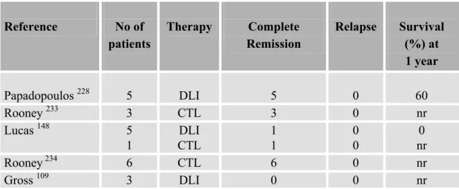
cell transplantation (Table 6). 109,148,228-231 A major drawback, however, is graft-versus-host
disease ensuing following donor lymphocyte infusion (30-60%), often resulting in considerable morbidity and mortality. To avoid graft-versus-host disease induced by allo-reactive T-cells, Rooney et al infused polyclonal EBV-specific cytotoxic T-cells in patients who had evidence of uncontrolled EBV replication, either as an elevated EBV load (> 20,000 EBV genomes per µg mononuclear cell DNA) or frank EBV-LPD, following allogeneic hematopoietic stem cell transplantation. Two of 3 patients obtained complete remission, the third patient had a complete remission of a histologically proven PTLD. No patient developed graft-versus-host disease. Functional EBV-specific cytotoxic T-cells
could be traced by gene-marking until 18 months following initial infusion.232,233 Recently,
these results were confirmed in a larger study. Anti-EBV cytotoxic T-cell infusion were administered prophylactically at a median of 3 months following allogeneic hematopoietic stem cell transplantation in 39 patients.
Table 5. Response of PTLD to anti B-cell immunotherapy follow
ing solid organ transplantation
Remission Reference Tx No of patients Therapy Complete Partial Relapse Survival (%) at 1 year Fischer 208
Heart, heart-lung, liver, kidney, kidney-pancreas
8 α CD21+ α CD24 7 1 0 58 Leblond 18 5
Heart, lung, kidney
10 α CD21+ α CD24 8 1 1 50 Benkerrou 20 7
Heart, kidney, lung, heart-lung
31 α CD21+ α CD24 20 3 2 55 Cook 212 Lung 3 α CD20 2 1 1 nr Zo m pi 216 Liver 3 RI+ α CD20 2 - 0 nr Milpied 21 1
Liver, kidney, heart, lung, kidney-pancreas, live
r-kidn ey 30 α CD20 15 2 0 77 Oertel 213
Heart, Liver, kidney
6 α CD20 4 - 0 nr Cailla rd 22 6 Kidney 13 RI+ α CD20 7 - nr nr Verschuuren 227 Lung 3 α CD20 3 - 1 66 Tx indicates transplanta tio n; α CD20/21/24, monoclonal antibody t herapy agai
nst CD20/21/24; RI, reduction of immune
suppression;
nr, not repor
In these patients no PTLD occurred, whereas 11% of historical controls (7 out of 61)
developed PTLD.234 PTLD following solid organ has also been treated with EBV-specific
cytotoxic T-cells. Complete remissions were observed in several cases without the
occurrence of rejection.235-237 Although the treatment strategy is highly specific and
without major side-effects, preparation of EBV-specific cytotoxic T-cells is time-consuming and technically elaborate. Furthermore, additional drawbacks may include a diminished effectivity during use of steroids, occurrence of exaggerated inflammatory response in case of bulky disease, and unresponsiveness to EBV-specific cytotoxic T-cells
as a result of mutation of EBV epitopes.215,234,238 In conclusion, treatment of PTLD
following allogeneic hematopoietic stem cell transplantation by donor lymphocyte infusion is effective, but may be complicated by severe graft-versus-host disease. The preparation of EBV-specific cytotoxic T-cells seems promising, but requires rather elaborate technical preparations.
Table 6. Response of PTLD to donor lymphocyte infusion or EBV specific CTLs following allogeneic hematopoietic stem cell transplantation
Reference No of patients Therapy Complete Remission Relapse Survival (%) at 1 year Papadopoulos 228 5 DLI 5 0 60 Rooney 233 3 CTL 3 0 nr Lucas 148 5 1 DLI CTL 1 1 0 0 0 nr Rooney 234 6 CTL 6 0 nr Gross 109 3 DLI 0 0 nr
DLI, indicates donor lymphocyte infusion; CTL, cytotoxic T-lymphocyte infusion; nr, not reported.
Prevention of PTLD following solid organ transplantation and allogeneic hematopoietic stem cell transplantation
As morbidity and mortality of established PTLD following solid organ transplantation and allogeneic hematopoietic stem cell transplantation is high, the emphasis should be on prevention of PTLD. The therapeutic approaches described above may also be applied for prevention. To date, no trials have been reported specifically addressing prophylaxis with antiviral agents such as aciclovir or ganciclovir. A reduced incidence of PTLD following solid organ transplantation was suggested in patients treated prophylactically with aciclovir or ganciclovir. However, these studies mostly had a non-randomized design without a
control group or PTLD was a secondary endpoint.239-244 In the setting of allogeneic hematopoietic stem cell transplantation, no benefit has been reported of aciclovir or
ganciclovir prophylaxis with respect to PTLD.122,178 B-cell depletion of the hematopoietic
stem cell graft using monoclonal anti B-cell antibodies was reported very effective in the prevention of PTLD following allogeneic hematopoietic stem cell transplantation as
compared to historical controls.245,246 Furthermore, B-cell depletion did not delay
engraftment, or increase incidence of infection. Rooney et al. evaluated the prophylactic infusion of EBV-specific cytotoxic T-cells in patients with high risk features such as HLA mismatch and T-cell depletion. A significant reduction in the incidence of PTLD was
observed as compared to historical controls (0/39 versus 7/100).234 Gustaffson et al.
prophylactically administered EBV-specific cytotoxic T-cells following allogeneic hematopoietic stem cell transplantation guided by viral load and observed 1 PTLD out of 4
patients treated.246 In contrast to the experience with CMV, prophylaxis guided by viral
load as yet has gained little interest in the prevention of PTLD sofar.
4. Outline of this thesis
Until recently, EBV-LPD could only be diagnosed in recipients of an allogeneic hematopoietic stem cell transplantation with overt established EBV-LPD, usually presenting as a critical illness in patients with generalized lymphadenopathy. The diagnosis was made on the basis of lymph node histology, since early and sensitive markers of EBV infection and reactivation were lacking. Recently the development of PCR-based assays has created the possibility to sensitively and quantitatively monitor EBV-DNA in peripheral blood samples and to evaluate their diagnostic value in immunocompetent patients with EBV infection and in immuocompressed patients with (impending) EBV-LPD.
The development of a real-time quantitative PCR assay for detection of EBV-DNA is described in chapter 2. The assay was evaluated in-vitro using an EBV standard determined by electron microscopy and in-vivo using plasma samples of patients with EBV-infection or EBV-LPD. Using that assay, we then retrospectively assessed the predictive value of the assay using plasma samples from a large cohort of recipients of an allogeneic hematopoietic stem cell transplantation. One hundred and fifty-two patients of whom 2-weekly plasma samples were available, were examined for predictive parameters as regards the incidence of EBV reactivation and EBV-disease. In addition, risk factors for reactivation and disease were assessed and correlated to transplant outcome measures (chapter 3).
In a cohort of 14 patients presenting with EBV-LPD, we next asked the question whether quantitative follow-up levels of EBV-DNA would predict response to therapy and survival. Quantification of EBV-DNA appeared as a very accurate and sensitive marker of response (chapter 4). Moreover, quantification of DNA before the onset of clinical overt
EBV-LPD appeared to accurately predict impending EBV-EBV-LPD. High positive and negative predictive values of EBV-DNA were established. Based on these results a prospective clinical study was designed aiming at the prevention of EBV-LPD and EBV-LPD-mortality. This prospective phase II study included 49 recipients of an allogeneic hematopoietic stem cell transplantation, and 15 patients received pre-emptive treatment when EBV load in plasma exceeded a threshold level of 1,000 EBV genome equivalents per ml. Comparison of prospectively followed patients to a recent historical cohort showed effective prevention of EBV-LPD and EBV-LPD-mortality (chapter 5).
Although the positive predictive value of quantified EBV-DNA appeared relatively high, most patients with viral reactivation were able to mount an immune response and clear their viral reactivation. Sofar, little was known with respect to the recovery of EBV specific cellular immunity following allogeneic hematopoietic stem cell transplantation. The recent introduction of HLA-class I tetramers presenting viral peptides has enabled the
monitoring of peptide specific cytotoxic CD8+ T-cells in peripheral blood samples
following allogeneic hematopoietic stem cell transplantation. We were able to use several
EBV peptide specific tetramers and monitor the recovery of EBV specific CD8+ T-cells.
The question whether impaired recovery of cellular T-cell immunity would identify patients at serious risk of EBV reactivation and progression to EBV-LPD was addressed in 61 recipients of a T-cell depleted allogeneic hematopoietic stem cell transplantation. Results described in chapter 6 suggest a pivotal protective role of T-cell immunity against EBV in recipients of an allogeneic hematopoietic stem cell graft. Finally, the overall results of our studies and future issues with respect to diagnosis, prevention and treatment of EBV-LPD are discussed in the general discussion (chapter 7).
References
1. Epstein MA, Achong BG, Barr YM. Virus particles in cultured lymphoblasts from Burkitt’s lymphoma. Lancet. 1964;i:702-703.
2. Rickinson AB, Kieff E. in: Fields BN, Knipe DM, Howley PM eds. Fields Virology. Philadelphia: Lippincott-Raven. Epstein-Barr virus. 1996:2397-2446.
3. Henle G, Henle W, Diehl V. Relation of Burkitt’s tumor-associated Herpes-type virus to infectious mononucleosis. Proc Natl Acad Sci USA. 1967;59:94-101.
4. Klein G. The Epstein-Barr virus and neoplasia. N Engl J Med. 1975;293:1353-1357.
5. Zur Hausen H, Schulte-Holthausen H, Klein G, Henle W, Henle G, Clifford P, Santesson L. EBV DNA in biopsies of Burkitt tumours and anaplastic carcinomas of the nasopharynx. Nature. 1970;228:1056-1058.
6. Weiss LM, Movahed LA, Warnke RA, Sklar J. Detection of Epstein-Barr viral genomes in Reed-Sternberg cells of Hodgkin’s disease. N Engl J Med. 1989;320:502-506.
7. Matas AJ, Hertel BF, Rosai J, Simmons RL, Najarian JS. Post-transplant malignant lymphoma. Am J Med. 1976;61:716-720.
8. Faulkner GC, Krajewski AS, Crawford DH. The ins and outs of EBV infection. Trends Immunol. 2000;8:185-189.
9. Jones K, Rivera C, Sgadari C, Franklin J, Max EE, Bhatia K, Tosato G. Infection of human endothelial cells with Epstein-Barr virus. J Exp Med. 1995;182:1213-1221.
10. Sixbey JW, Nedrud JG, Raab-Traub N, Hanes RA, Pagano JS. Epstein-Barr virus replication in oropharyngeal cells. N Engl J Med. 1984;310:1225-1230.
11. Niedobitek G, Hamilton-Dutoit S, Herbst H, Finn T, Vetner M, Pallesen G, Stein H. Identification of Epstein-Barr virus-infected cells in tonsils of acute infectious mononucleosis by in situ hybridisation. Hum Pathol. 1989;20:796-799.
12. Niedobitek G, Herbst H, Young LS, Brooks L, Masucci MS, Crocker J, Rickinson AB, Stein H. Patterns of Epstein-Barr virus infection in non-neoplastic lymphoid tissue. Blood. 1992;79:2520-2526.
13. Tao Q, Srivastava G,Chan ACL, Chung LP, Loke SL, Ho FCS. Evidence for lytic infection by Epstein-Barr in mucosal lymphocytes instead of nasopharyngeal epithelial cells in normal individuals. J Med Virol. 1995;45:71-77.
14. Anagnostopaulos I, Hummel M, Kreschel C, Stein H. Morphology, immunophenotype, and distribution of latently and/or productively Epstein-Barr virus-infected cells in acute infectious mononucleosis: implications for the interindividual infection route of Epstein-Barr virus. Blood. 1995;85:744-750.
15. Borza CM, Hutt-Fletcher LM. Alternate replication in B-cells and epithelial cells switches tropism of Epstein-Barr virus. Nat Med. 2002;8:594-599.
16. Gratama JW. Epstein-Barr virus infections in bone marrow transplantation recipients. In: Forman JS, Blume KG, Thomas ED eds. Bone Marrow Transplantation. Boston: Blackwell scientific publications; 1995:429-442.
17. Preiksaitis JK, Cockfield SM. in: Bowden RA, Ljungman P, Paya PV eds. Transplant Infections. Philadelphia: Lippincott-Raven; 1998:245-263.
18. Li Q, Spriggs MK, Kovats S, Turk SM, Comeau MR, Nepom B, Hutt-Fletcher LM. Epstein-Barr virus uses HLA class II as a cofactor for infection of B lymphocytes. J Virol. 1997;71:4657-4662.
19. Johannsen E, Koh E, Mosialos G, Tong X, Kieff E, Grossman SR. Epstein-Barr virus nuclear protein 2 transactivation of the latent membrane protein 1 promotor is mediated by Jκ and PU.1. J Virol. 1995;69:253-262.
20. Middeldorp JM, Brink AATP, van den Brule AJC, Meijer CJLM. Pathogenetic roles for Epstein-Barr virus (EBV) gene products in EBV-associated proliferative disorders. Crit Rev Oncol Hematol. In press.
21. Uchida J, Yasui T, Takaoka-Shichijo Y, Muraoka M, Kulwichit W, Raab-Traub N, Kikutani H. Mimicry of CD40 signals by Epstein-Barr virus LMP1 in B lymphocyte responses. Science. 1999;286:300-303.
22. Komano J, Maruo S, Kurozumi K, Oda T, Takada K. Oncogenic role of Epstein-Barr virus-encoded RNAs in Burkitt’s lymphoma cell line Akata. J Virol. 1999;73:9827-9831.
23. Shaw JE. The circular intracellular form of Epstein-Barr virus DNA is amplified by the virus-associated DNA polymerase. J Virol. 1985;53:1012-1015.
24. Leight ER, Sugden B. EBNA-1: a protein pivotal to latent infection by Epstein-Barr virus. Rev Med Virol. 2000;10:83-100.
25. Kieff E. in: Fields BN, Knipe DM, Howley PM eds. Fields Virology. Philadelphia: Lippincott-Raven; Epstein-Barr virus and its replication. 1996:2343-2396.
26. Thorley-Lawson DA, Miyashita EM, Khan G. Epstein-Barr virus and the B cell: that’s all it takes. Tr Microbiol. 1996;4:204-208.
27. Thorley-Lawson DA. Epstein-Barr virus: exploiting the immune system. Nature Immunol. 2001;1:75-81.
28. Aman P, Ehlin-Henriksson B, Klein G. Epstein-Barr virus susceptibility of normal human B-lymphocyte populations. J Exp Med. 1984;159:208-220.
29. Wang D, Liebowitz D, Kieff E. An EBV membrane protein expressed in immortalized lymhocytes transforms established rodent cells. Cell. 1985;43:831-840.
30. Banchereau J, Bazan F, Blanchard D, Briere F, Galizzi JP, van Kooten C, Liu YJ, Saeland S. The CD40 antigen and its ligand. Annu Rev Immunol. 1994;12:881-922.
31. Beaufils P, Choquet D, Marnoun R, Malissen B. The (YXXL/1)2 signalling motif found in
the cytoplasmic segments of the bovine leukaemia virus envelope protein and Epstein-Barr virus latent membrane protein 2A can elicit early and late lymphocyte activation events. EMBO J. 1993;12:5105-5112.
32. Babcock GJ, Decker LL, Freeman RB, Thorley-Lawson DA. Epstein-Barr virus-infected resting memory B-cells, not proliferating lymphoblasts, accumulate in the peripheral blood of immunosuppressed patients. J Exp Med. 1999;190:567-576.
33. Chen F, Zou J-Z, di Renzo L, Winberg G, Hu L-F, Klein E, Klein G, Ernberg I. A subpopulation of normal B cells latently infected with Epstein-Barr virus resembles Burkitt lymphoma cells in expressing EBNA-1 but not EBNA-2 or LMP-1. J Virol. 1995;69:3752-3758.
34. Tierney RJ, Steven N, Youg LS, Rickinson AB. Epstein-Barr virus latency in blood mononuclear cells: analysis of viral gene transcription during primary infection and in the carrier state. J Virol. 1994;68:7374-7385.
35. Speck SH, Chatila T, Flemington E. Reactivation of Epstein-Barr virus: regulation and function of the BZLF1 gene. Tr Microbiol. 1997;5:399-405.
36. Adamson AL, Darr D, Holley-Guthrie E, Johnson RA, Mauser A, Swenson J, Kenney S. Epstein-Barr virus immediate-early proteins BZLF1 and BRLF1 activate the ATF2