HJC
439
Review Article
Review Article
Manuscript received: August 21, 2010; Accepted: September 15, 2010. Address: Christina-Maria Kotta 14, Paloumbioti St. 114 76 Athens, Greece e-mail: mckotta@hotmail. com Key words: Phenotype, ion channels, genetics, function, SNPs.Effects of Mutations and Genetic Overlap in
Inherited Long-QT and Brugada Arrhythmia
Syndromes
C
hristina-M
ariaK
otta, a
risa
nastasaKis, C
hristodouloss
tefanadisFirst Department of Cardiology, University of Athens Medical School, Hippokration Hospital, Athens, Greece
T
he functional characterisation of
causative mutations behind the
in-herited long-QT and Brugada
ar-rhythmia syndromes has been the focus of
much research that has attempted to link
the genotype to the phenotype. This
arti-cle will examine our current knowledge of
the effects of mutations, as well as the
ge-netic and phenotypic overlap in inherited
arrhythmia syndromes.
Long-QT syndrome
Long-QT syndrome (LQTS) is an
inher-ited cardiac disease that is characterised
by prolongation of the QT interval on the
electrocardiogram and is associated with
syncopal episodes, dangerous
ventricu-lar arrhythmias of the torsades de pointes
type, and a high risk of sudden death on a
substrate of a structurally normal heart.
1Today we know that LQTS is an
inherit-ed autosomal dominant arrhythmogenic
disease that is caused by mutations in the
genes of cardiac ion channels and their
subunits.
2-5According to the genes
in-volved, the type of syndrome is labelled as
LQT1, LQT2, etc. (Table 1).
Brugada syndrome
Brugada syndrome (BrS) is an inherited
cardiac disease that is characterised
elec-trocardiographically by ST-segment
el-evation in the right precordial leads V
1-V
3, with or without right bundle branch
block, and is associated with syncopal
epi-sodes and a high risk of sudden death on a
substrate of a structurally normal heart.
6-9The syndrome is inherited in an
autoso-mal dominant manner and is estimated
to be responsible for 4-12% of total
sud-den deaths, and for up to 20% of sudsud-den
deaths with a “normal” heart.
8-10In
addi-tion, it is estimated to account for 40-60%
of the cases of ventricular fibrillation that
were previously characterised as
idiopath-ic.
11,12Up to now, mutations in the
SC-N5A
gene, which encodes the cardiac Na
+ion channel and constitutes the I
Nacur-rent, appear to form the main genetic
sub-strate of the syndrome and are detected
in about 18-30% of patients, with a higher
prevalence in the purely familial forms of
the syndrome (Table 1).
13-17Structure of the cardiac ion channels
The cardiac ion channels are
macromo-lecular protein complexes of the cellular
membrane, which, by responding to
differ-ences in transmembrane potential, change
their stereochemical structure. These
ste-reochemical changes, though small, lead
to the opening of a pore through which,
within fractions of a second, millions of
ions enter or leave the cell, creating a current of some
picoamperes.
18The process of cellular
depolarisa-tion and repolarisadepolarisa-tion is carried out via four
succes-sive, discrete stages of changes in the stereochemical
condition of the channels, which are specific to each
channel.
19Nevertheless, all voltage-dependent
chan-nels have several structural and functional
character-istics in common, such as a voltage sensor that detects
the changes in potential, the pore, which in response
to the sensor opens and closes, allowing or preventing
the flow of ions, and the ion selection filter.
18,19Functional consequences of the mutations
The functional cardiac currents are the result of the
perfectly coordinated expression of the biophysical,
bio-chemical, and biogenic properties of their channels.
20Obviously, even small changes in the structures of the
channels as a result of mutations are capable of
influ-encing and damaging these complex functional
proper-ties. This influence may result in a partial or complete
loss of functionality, or an additional, greater than
nor-mal functionality. Thus, as a first approximation, the
mutations are classified into those that lead to
gain-of-function
and those that result in loss-of-function.
LQTS
In LQTS we know that mutations in the genes for
the K
+channels (LQT1,2,5,6) lead to
loss-of-func-tion, materially reducing the intensity of the I
Krand I
Kscurrents, and hence the “repolarisation
re-serve”, while changes in the gene for the Na
+channel
(LQT3) lead to gain-of-function, increasing the
inten-sity of the I
Nachannel, and hence depolarisation.
21In the case of mutations of the cardiac K
+channels,
their pathological action is exerted via two main
mech-anisms. The first concerns the ability of subunits to
as-semble into tetramers, resulting in a dramatic reduction
(~50%) in the available functional channels and a
cor-responding dramatic decrease in flow. This
phenom-enon is defined as haploinsufficiency.
22,23The second
mechanism concerns mutations that lead to structural
anomalies in the subunits, which most often alter the
kinetics or the conduction of the channel. This
mecha-nism is defined as dominant-negative suppression.
24-28In contrast to the mutations of the cardiac K
+channels, mutations of the SCN5A gene in LQTS
have a common mechanism and lead to
gain-of-func-tion, mainly through imperfect deactivation of the
channels, or destabilisation or slowing of the
deacti-vation process.
29In each case, the result is the
pres-ence of late inward I
Nadepolarisation currents in the
plateau phase of the cardiac action potential, which,
even though of low intensity, prolong its duration.
This prolongation leads to the appearance of
prema-ture afterdepolarisations.
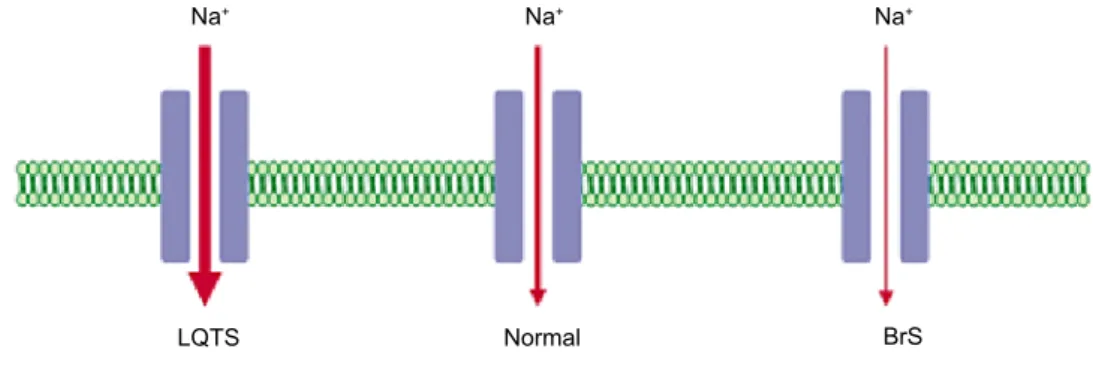
30,31BrS
In BrS the mutations in the Na
+channel gene
(SC-N5A
) cause reduced functionality and a decrease in
the I
Nacurrent.
32-35Some mutations lead to
com-pletely non-functional channels, while others alter
their biophysical properties.
35Frame-shift, nonsense,
and splice mutations lead to completely
non-tional channels, reducing by half the available
func-tional channels (haploinsufficiency).
13,36,37Many
mis-sense mutations have also been described that
ap-peared to reduce the permeability and conductivity of
the Na
+channel, or to lead to channels with altered
biophysical properties.
38The result in each case is a
decrease in the I
Nacurrent (Figure 1).
Clinical significance of the mutation type
LQTS
Mutations that, because of topology
(carboxy-termi-nal end), are associated with a milder (‘forme fruste’)
Table 1. The main genes of long-QT syndrome and Brugada syndrome.
Type Gene Chromosome Protein Function Frequency Reference
LQT1 KCNQ1 11p15.5 Kv7.1 α IKs↓ ~30-35% 77
LQT2 KCNH2 7q35-36 Kv11.1 α IKr↓ ~25-30% 2
LQT3 SCN5A 3p21-23 Nav1.5 α INa ↑ ~5-7% 3
LQT5 KCNE1 21q22.1-22.2 minΚ β IKs↓ ~1% 80
LQT6 KCNE2 21q22.1-22.2 MiRP1 β IKr↓ ~1% 79
BrS1 SCN5A 3p21-23 Nav1.5 α INa↓ ~18-30% 13
HJC
441
phenotype and clinical course have been described in
types LQT1, LQT2, and LQT5.
39-41A recent study
of LQT1 patients showed that patients who
car-ried transmembrane (which mainly affect the
chan-nel’s pore and voltage sensor), missense (which lead
to channels with altered biophysical properties), or
dominant-negative suppression mutations,
indepen-dently have a significantly increased risk of cardiac
events of all kinds, compared to those who have
car-boxy-terminal, non-missense or haploinsufficiency
mutations.
42BrS
A recent study of patients with mutations in the
SC-N5A
gene showed that the level of loss-of-function
caused by each mutation partially reflects the clinical
phenotype.
43Mutations that cause complete
loss-of-function (nonsense) have been found to be associated
with a significantly higher incidence of syncope and
more severe conduction disturbances (prolongation
of QRS on challenge and PR on challenge and rest)
compared to mutations that cause reduced function
(many of them missense). Research into this matter
is currently ongoing so that the genetic data can serve
as a new risk stratification index in the future.
Genetic overlap with other arrhythmogenic syndromes
Discrete mutations of the genes of cardiac ion
chan-nels have been described that, by affecting in various
ways the cardiac repolarisation currents I
Ks, I
Kr, I
K1and the depolarisation I
Nacurrent, form the
patho-logical substrate of discrete syndromes. Thus,
muta-tions in these genes are also implicated, apart from
LQTS and BrS, in short QT syndrome (SQTS), in
forms of familial atrial fibrillation, in systemic
con-duction disease, in idiopathic ventricular fibrillation,
and in congenital sick sinus syndrome (Figure 2).
44-45The recognition that mutations that cause
loss-of-function of the K
+channels lead to the clinical
phenotype of LQTS, while others, which cause
gain-of-function, lead to the clinical phenotype of SQTS,
starts to bring some kind of sense to the matter.
21,44-46Accordingly, it is understandable how mutations that
cause loss-of-function of the Na
+channel lead to the
clinical phenotype of BrS, while others, which cause
gain-of-function, lead to the clinical phenotype of
LQTS.
21,33,35This relatively simple interpretation has
validity, but becomes more complicated in light of
other findings.
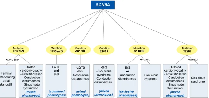
At the heart of the question of genetic
overlap-ping lies mainly the SCN5A gene. Discrete
muta-tions of the gene, most of which cause
loss-of-func-tion, form the common genetic denominator in
vari-ous syndromes, but in addition these same mutations
appear to lead in some cases to different phenotypes,
which, even within the same family, can be
manifest-ed in a mixmanifest-ed, combinmanifest-ed, or exclusive form (Figure
3).
55-64Furthermore, data from recent studies have
also implicated subclinical mutations of the gene in
ventricular fibrillation during the first hours of acute
myocardial infarction.
65,66In many cases, the
func-tional character of these mutations in cellular
expres-sion systems in vitro has been shown to affect a wide
assortment of the channel’s biophysical properties at
the same time, with simultaneous multilevel
loss-of-function, resulting in various clinical manifestations.
An example is the mutation 1795 insD, which leads to
the coexistence of LQTS and BrS, which is almost
in-comprehensible on the clinical level.
59Nevertheless, what these functional studies do
Na+ Na+ Na+
Normal
LQTS BrS
Figure 1. Gain- and loss-of-function of the Na+ channels in long-QT syndrome and Brugada syndrome, with an increase and decrease in
Familial atrial fibrillation (AF1) Familial atrial fibrillation (AF8) and SQTS (SQT2) Familial atrial fibrillation (AF3) Familial atrial fibrillation (AF1) and SQTS (SQT2) Familial atrial fibrillation (AF2) Conduction disturbances/ Lenegre-Lev disease Idiopathic ventricular fibrillation Sick sinus syndrome
Figure 2. Genetic overlap of the genes of the cardiac channels KCNQ1,44,47,48,77, KCNH2,2,45,49 KCNJ2,46,50,78 KCNE2,51,79, SCN5A.3,13,52-55
HJC
443
not explain is how various combinations of mixed and
exclusive phenotypes can appear among people who
are carriers of the same mutation of the SCN5A gene.
The same question also essentially applies to the
vary-ing degrees of penetrance and expressivity that the
diseases often exhibit among patients who are
car-riers of discrete mutations of the other genes of the
cardiac channels, which lead to classical forms of the
syndromes without significant genetic overlapping.
The factors of sex and age are often reported not to
contribute to these differences.
62Thus, the picture
that starts to take shape is one where the main
genet-ic substrate is the central axis of the clingenet-ical outcome,
but the way and the extent to which it is expressed
de-pend to a significant degree on other factors.
67,68By the term “modifying factors” we mean
main-ly those environmental and genetic factors whose
ef-fects shape the clinical outcome. The prevailing view
is that environmental factors are probably mainly
re-sponsible for the paroxysmal manifestations of familial
syndromes, while genetic factors are mainly implicated
in the interpersonal variations between patients with
a common main pathological substrate.
67The genetic
modifying factors mainly concern the single nucleotide
polymorphisms (SNPs), which are normally
responsi-ble for about 90% of our genetic diversity.
68Genetic modification via SNPs is applied either
via the interaction with the main pathological
sub-strate by the same gene, or by a different gene, while,
according to the case, this interaction may either
ex-acerbate the final pathological expression or mitigate
it.
68An indicative example of the exertion of
modify-ing action by the same gene is the SNP H558R of
SN-C5A
, where its presence in homozygous form (R558)
in combination with any of the mutations T512I,
M1766L, and R282H of SCN5A, significantly mitigates
their pathological expression (less loss of I
Na).
69-71Even
though the molecular mechanisms behind these
ef-fects are not known, such types of phenomenon
ap-pear also to contribute significantly to the degree of
penetrance and to the selective phenotypical
expres-sions of the diseases.
68The quest for a genetic variant with a modifying
action beyond that of the pathological reference gene
opens up a vast array of possibilities, and our
knowl-edge of this matter is still woefully scanty. To get
some kind of picture, we can turn to a recent study
that showed that certain SNPs of the genes of the α
2and β
1adrenergic receptors are associated with an
in-creased symptomatology and risk of cardiac events
among patients with LQTS who have the same
patho-logical mutation of LQT1.
72 Familial deteriorating atrial standstill Dilated cardiomyopathy - Atrial fibrillation - Conduction disturbances - Sinus node dysfunction (mixed phenotypes) LQTS and BrS (combined phenotypes) -LQTS -BrS -Conduction disturbances (mixed phenotypes) -BrS -Sick sinus syndrome -Conduction disturbances (mixed phenotypes) BrS or Conduction disturbances (exclusive phenotypes) Sick sinus syndrome - Dilated cardiomyopathy - Atrial fibrillation - Conduction disturbances - Sinus node dysfunction (mixed phenotypes) Sick sinus syndromeMutation Mutation Mutation Mutation Mutation Mutation
Figure 3. Genetic overlap of the SCN5A gene via the D1275N,56-58 1795insD,59-61 ΔK1500,62 E161K,63 G1408R,55,64 T220I55,58 mutations. Mixed phenotypes – one or more or all of the above. Combined phenotypes – all of the above. Exclusive phenotypes – one of the above. +Cx40 SNP – the phenotype is manifested only when there coexists a specific polymorphism of the connexin 40 gene. +P1298L/+R1623X – the phenotype is manifested only when these mutations also coexist (compound heterozygotes).
An understanding of the nature of genetic
mod-ifying factors is the next central goal in the
under-standing of inherited arrhythmogenic syndromes. To
achieve this goal it is likely that our gaze will need to
be less focused. The role of mutations of cardiac ion
channels appears to extend beyond the myocytes of
the ventricles and to those of the atria, to the
special-ised cells of the conduction system, the sinus node,
and the Purkinje fibres, where they exert their effect
through interactive and feedback processes with
doz-ens of other proteins that compose a complex, and in
places specialised, cellular environment. This theory
also has another dimension, that of a possible cellular
remodelling as a result of the cardiac ion channel
mu-tations’ additive pathological effect. Finally, the view
has been expressed that these mutations probably
al-so disturb the architecture of the intracellular
envi-ronment, leading locally to foci of fibrosis, apoptosis,
and cell death.
63,73,74This seems to concern mainly
the Na
+channels, which have complex natural roles
in the atria, the ventricles, and the conduction
sys-tem.
74Mutations of the SCN5A gene have recently been
implicated in cases of mixed phenotypes, with
con-comitant idiopathic dilated cardiomyopathy (Figure
3).
57,58In addition, in some cases of patients with a
clinical BrS phenotype, some of whom were carriers
of mutations of the SCN5A gene and had a “normal”
heart on non-invasive clinical examination, the
histol-ogy of samples from endomyocardial biopsy revealed
structural lesions.
75,76To what extent this genetic
sub-strate contributes to the development of structural
changes, or whether the two exist independently and
in combination are responsible for the clinical
pheno-type, remains to be elucidated.
73Clinical and basic research into inherited
car-diac diseases still has to overcome many challenges,
and it seems that this will require a combination of
approaches. The continual feedback of information
from bench to bedside, and vice versa, composes the
continuously cycling collaboration that, even today,
is leading to the provision of better health services
81and on which progress and understanding of these
diseases mainly depends. With each step we take
to-wards the future, this information steadily acquires
added value.
References
1. Schwartz PJ, Periti M, Malliani A. The long Q-T syndrome. Am Heart J. 1975; 89: 378-390.
2. Curran ME, Splawski I, Timothy KW, Vincent GM, Green ED, Keating MT. A molecular basis for cardiac arrhythmia: HERG mutations cause long QT syndrome. Cell. 1995; 80: 795-803.
3. Wang Q, Shen J, Splawski I, et al. SCN5A mutations asso-ciated with an inherited cardiac arrhythmia, long QT syn-drome. Cell. 1995; 80: 805-811.
4. Splawski I, Shen J, Timothy KW, et al. Spectrum of muta-tions in long-QT syndrome genes. KVLQT1, HERG, SC-N5A, KCNE1, and KCNE2. Circulation. 2000; 102: 1178-1185.
5. Chiang CE, Roden DM. The long QT syndromes: genetic basis and clinical implications. J Am Coll Cardiol. 2000; 36: 1-12.
6. Brugada P, Brugada J. Right bundle branch block, persistent ST segment elevation and sudden cardiac death: a distinct clinical and electrocardiographic syndrome. A multicenter re-port. J Am Coll Cardiol. 1992; 20: 1391-1396.
7. Brugada J, Brugada R, Brugada P. Right bundle-branch block and ST-segment elevation in leads V1 through V3: a marker for sudden death in patients without demonstrable structural heart disease. Circulation. 1998; 97: 457-460. 8. Brugada J, Brugada P, Brugada R. The syndrome of right
bundle branch block ST segment elevation in V1 to V3 and sudden death—the Brugada syndrome. Europace. 1999; 1: 156-166.
9. Brugada P, Brugada R, Brugada J. Sudden death in patients and relatives with the syndrome of right bundle branch block, ST segment elevation in the precordial leads V(1) to V(3) and sudden death. Eur Heart J. 2000; 21: 321-326.
10. Benito B, Brugada R, Brugada J, Brugada P. Brugada syn-drome. Prog Cardiovasc Dis. 2008; 51: 1-22.
11. Gussak I, Antzelevitch C, Bjerregaard P, Towbin JA, Chait-man BR. The Brugada syndrome: clinical, electrophysiologic and genetic aspects. J Am Coll Cardiol. 1999; 33: 5-15. 12. Grace AA. Brugada syndrome. Lancet. 1999; 354: 445-446. 13. Chen Q, Kirsch GE, Zhang D, et al. Genetic basis and
molec-ular mechanism for idiopathic ventricmolec-ular fibrillation. Nature. 1998; 392: 293-296.
14. Antzelevitch C, Brugada P, Brugada J, Brugada R. Brugada syndrome: from cell to bedside. Curr Probl Cardiol. 2005; 30: 9-54.
15. Napolitano C, Priori SG. Brugada syndrome. Orphanet J Ra-re Dis. 2006; 1: 35.
16. Schulze-Bahr E, Eckardt L, Breithardt G, et al. Hum Mutat. 2003; 21: 651-652.
17. Kotta CM, Anastasakis A, Gatzoulis K, Manolis AS, Stefa-nadis C. Novel sodium channel SCN5A mutations in Bruga-da syndrome patients from Greece. Int J Cardiol. 2010; 145: 45-48.
18. Ackerman MJ, Clapham DE. Ion channels—basic science and clinical disease. N Engl J Med. 1997; 336: 1575-1586. 19. Sansom MS. Ion channels: structure of a molecular brake.
Curr Biol. 1999; 9: R173-175.
20. Delisle BP, Anson BD, Rajamani S, January CT. Biology of cardiac arrhythmias: ion channel protein trafficking. Circ Res. 2004; 94: 1418-1428.
21. Roden DM, Lazzara R, Rosen M, Schwartz PJ, Towbin J, Vincent GM. Multiple mechanisms in the long-QT syn-drome. Current knowledge, gaps, and future directions. The SADS Foundation Task Force on LQTS. Circulation. 1996; 94: 1996-2012.
mecha-HJC
445
nisms of cardiac arrhythmias. Cell. 2001; 104: 569-580. 23. Tristani-Firouzi M, Chen J, Mitcheson JS, Sanguinetti MC.
Molecular biology of K(+) channels and their role in cardiac arrhythmias. Am J Med. 2001; 110: 50-59.
24. Sanguinetti MC, Curran ME, Spector PS, Keating MT. Spec-trum of HERG K+-channel dysfunction in an inherited car-diac arrhythmia. Proc Natl Acad Sci U S A. 1996; 93: 2208-2212.
25. Wollnik B, Schroeder BC, Kubisch C, Esperer HD, Wieacker P, Jentsch TJ. Pathophysiological mechanisms of dominant and recessive KVLQT1 K+ channel mutations found in in-herited cardiac arrhythmias. Hum Mol Genet. 1997; 6: 1943-1949.
26. Bianchi L, Shen Z, Dennis AT, et al. Cellular dysfunction of LQT5-minK mutants: abnormalities of IKs, IKr and traffick-ing in long QT syndrome. Hum Mol Genet. 1999; 8: 1499-1507.
27. Bianchi L, Priori SG, Napolitano C, et al. Mechanisms of I(Ks) suppression in LQT1 mutants. Am J Physiol Heart Circ Physiol. 2000; 279: H3003-3011.
28. Brunner M, Peng X, Liu GX, et al. Mechanisms of cardiac ar-rhythmias and sudden death in transgenic rabbits with long QT syndrome. J Clin Invest. 2008; 118: 2246-2259.
29. Wang DW, Yazawa K, George AL, Bennett PB. Character-ization of human cardiac Na+ channel mutations in the con-genital long QT syndrome. Proc Natl Acad Sci U S A. 1996; 93: 13200-13205.
30. Kass RS, Moss AJ. Long QT syndrome: novel insights into the mechanisms of cardiac arrhythmias. J Clin Invest. 2003; 112: 810-815.
31. Grant AO. Molecular biology of sodium channels and their role in cardiac arrhythmias. Am J Med. 2001; 110: 296-305. 32. Tan HL, Bezzina CR, Smits JP, Verkerk AO, Wilde AA.
Ge-netic control of sodium channel function. Cardiovasc Res. 2003; 57: 961-973.
33. Bezzina CR, Rook MB, Wilde AA. Cardiac sodium channel and inherited arrhythmia syndromes. Cardiovasc Res. 2001; 49: 257-271.
34. Balser JR. Sodium “channelopathies” and sudden death: must you be so sensitive? Circ Res. 1999; 85: 872-874. 35. Viswanathan PC, Balser JR. Inherited sodium
channelopa-thies: a continuum of channel dysfunction. Trends Cardio-vasc Med. 2004; 14: 28-35.
36. Baroudi G, Napolitano C, Priori SG, Del Bufalo A, Chahine M. Loss of function associated with novel mutations of the SCN5A gene in patients with Brugada syndrome. Can J Car-diol. 2004; 20: 425-430.
37. Keller DI, Barrane FZ, Gouas L, et al. A novel nonsense mu-tation in the SCN5A gene leads to Brugada syndrome and a silent gene mutation carrier state. Can J Cardiol. 2005; 21: 925-931.
38. Vatta M, Dumaine R, Antzelevitch C, et al. Novel mutations in domain I of SCN5A cause Brugada syndrome. Mol Genet Metab. 2002; 75: 317-324.
39. Berthet M, Denjoy I, Donger C, et al. C-terminal HERG mu-tations: the role of hypokalemia and a KCNQ1-associated mutation in cardiac event occurrence. Circulation. 1999; 99: 1464-1470.
40. Donger C, Denjoy I, Berthet M, et al. KVLQT1 C-terminal missense mutation causes a forme fruste long-QT syndrome. Circulation. 1997; 96: 2778-2781.
41. Ohno S, Zankov DP, Yoshida H, et al. N- and C-terminal KCNE1 mutations cause distinct phenotypes of long QT
syn-drome. Heart Rhythm. 2007; 4: 332-340.
42. Moss AJ, Shimizu W, Wilde AA, et al. Clinical aspects of type-1 long-QT syndrome by location, coding type, and bio-physical function of mutations involving the KCNQ1 gene. Circulation. 2007; 115: 2481-2489.
43. Meregalli PG, Tan HL, Probst V, et al. Type of SCN5A mu-tation determines clinical severity and degree of conduction slowing in loss-of-function sodium channelopathies. Heart Rhythm. 2009; 6: 341-348.
44. Bellocq C, van Ginneken AC, Bezzina CR, et al. Mutation in the KCNQ1 gene leading to the short QT-interval syndrome. Circulation. 2004; 109: 2394-2397.
45. Brugada R, Hong K, Dumaine R, et al. Sudden death associ-ated with short-QT syndrome linked to mutations in HERG. Circulation. 2004; 109: 30-35.
46. Priori SG, Pandit SV, Rivolta I, et al. A novel form of short QT syndrome (SQT3) is caused by a mutation in the KCNJ2 gene. Circ Res. 2005; 96: 800-807.
47. Chen YH, Xu SJ, Bendahhou S, et al. KCNQ1 gain-of-func-tion mutagain-of-func-tion in familial atrial fibrillagain-of-func-tion. Science. 2003; 299: 251-254.
48. Hong K, Piper DR, Diaz-Valdecantos A, et al. De novo KC-NQ1 mutation responsible for atrial fibrillation and short QT syndrome in utero. Cardiovasc Res. 2005; 68: 433-440. 49. Hong K, Bjerregaard P, Gussak I, Brugada R. Short QT
syn-drome and atrial fibrillation caused by mutation in KCNH2. J Cardiovasc Electrophysiol. 2005; 16: 394-396.
50. Xia M, Jin Q, Bendahhou S, et al. A Kir2.1 gain-of-function mutation underlies familial atrial fibrillation. Biochem Bio-phys Res Commun. 2005; 332: 1012-1019.
51. Yang Y, Xia M, Jin Q, et al. Identification of a KCNE2 gain-of-function mutation in patients with familial atrial fibrilla-tion. Am J Hum Genet. 2004; 75: 899-905.
52. Schott JJ, Alshinawi C, Kyndt F, et al. Cardiac conduction defects associate with mutations in SCN5A. Nat Genet. 1999; 23: 20-21.
53. Probst V, Kyndt F, Potet F, et al. Haploinsufficiency in com-bination with aging causes SCN5A-linked hereditary Lenègre disease. J Am Coll Cardiol. 2003; 41: 643-652.
54. Akai J, Makita N, Sakurada H, et al. A novel SCN5A muta-tion associated with idiopathic ventricular fibrillamuta-tion without typical ECG findings of Brugada syndrome. FEBS Lett. 2000; 479: 29-34.
55. Benson DW, Wang DW, Dyment M, et al. Congenital sick sinus syndrome caused by recessive mutations in the cardi-ac sodium channel gene (SCN5A). J Clin Invest. 2003; 112: 1019-1028.
56. Groenewegen WA, Firouzi M, Bezzina CR, et al. A cardiac sodium channel mutation cosegregates with a rare connexin 40 genotype in familial atrial standstill. Circ Res. 2003; 92: 14-22.
57. McNair WP, Ku L, Taylor MR, et al. SCN5A mutation asso-ciated with dilated cardiomyopathy, conduction disorder, and arrhythmia. Circulation. 2004; 110: 2163-2167.
58. Olson TM, Michels VV, Ballew JD, et al. Sodium channel mutations and susceptibility to heart failure and atrial fibril-lation. JAMA. 2005; 293: 447-454.
59. Bezzina C, Veldkamp MW, van Den Berg MP, et al. A single Na(+) channel mutation causing both long-QT and Brugada syndromes. Circ Res. 1999; 85: 1206-1213.
60. Veldkamp MW, Viswanathan PC, Bezzina C, Baartscheer A, Wilde AA, Balser JR. Two distinct congenital arrhythmias evoked by a multidysfunctional Na(+) channel. Circ Res.
2000; 86: E91-97.
61. Clancy CE, Rudy Y. Na(+) channel mutation that causes both Brugada and long-QT syndrome phenotypes: a simula-tion study of mechanism. Circulasimula-tion. 2002; 105: 1208-1213. 62. Grant AO, Carboni MP, Neplioueva V, et al. Long QT
syn-drome, Brugada synsyn-drome, and conduction system disease are linked to a single sodium channel mutation. J Clin Invest. 2002; 110: 1201-1209.
63. Smits JP, Koopmann TT, Wilders R, et al. A mutation in the human cardiac sodium channel (E161K) contributes to sick sinus syndrome, conduction disease and Brugada syndrome in two families. J Mol Cell Cardiol. 2005; 38: 969-981. 64. Kyndt F, Probst V, Potet F, et al. Novel SCN5A mutation
leading either to isolated cardiac conduction defect or Bru-gada syndrome in a large French family. Circulation. 2001; 104: 3081-3086.
65. Hu D, Viskin S, Oliva A, et al. Genetic predisposition and cellular basis for ischemia-induced ST-segment changes and arrhythmias. J Electrocardiol. 2007; 40: S26-29.
66. Hu D, Viskin S, Oliva A, et al. Novel mutation in the SCN5A gene associated with arrhythmic storm development during acute myocardial infarction. Heart Rhythm. 2007; 4: 1072-1080. 67. Priori SG. Inherited arrhythmogenic diseases: the
complex-ity beyond monogenic disorders. Circ Res. 2004; 94: 140-145. 68. Scicluna BP, Wilde AA, Wilde AW, Bezzina CR. The pri-mary arrhythmia syndromes: same mutation, different mani-festations. Are we starting to understand why? J Cardiovasc Electrophysiol. 2008; 19: 445-452.
69. Viswanathan PC, Benson DW, Balser JR. A common SC-N5A polymorphism modulates the biophysical effects of an SCN5A mutation. J Clin Invest. 2003; 111: 341-346.
70. Ye B, Valdivia CR, Ackerman MJ, Makielski JC. A common hu-man SCN5A polymorphism modifies expression of an arrhyth-mia causing mutation. Physiol Genomics. 2003; 12: 187-193. 71. Poelzing S, Forleo C, Samodell M, et al. SCN5A
polymor-phism restores trafficking of a Brugada syndrome mutation on a separate gene. Circulation. 2006; 114: 368-376.
72. Schwartz PJ, Vanoli E, Crotti L, et al. Neural control of heart rate is an arrhythmia risk modifier in long QT syndrome. J Am Coll Cardiol. 2008; 51: 920-929.
73. Saffitz JE. Structural heart disease, SCN5A gene mutations, and Brugada syndrome: a complex ménage à trois. Circula-tion. 2005; 112: 3672-3674.
74. Lehnart SE, Ackerman MJ, Benson DW, et al. Inherited ar-rhythmias: a National Heart, Lung, and Blood Institute and Office of Rare Diseases workshop consensus report about the diagnosis, phenotyping, molecular mechanisms, and ther-apeutic approaches for primary cardiomyopathies of gene mutations affecting ion channel function. Circulation. 2007; 116: 2325-2345.
75. Frustaci A, Priori SG, Pieroni M, et al. Cardiac histological substrate in patients with clinical phenotype of Brugada syn-drome. Circulation. 2005; 112: 3680-3687.
76. Coronel R, Casini S, Koopmann TT, et al. Right ventricular fibrosis and conduction delay in a patient with clinical signs of Brugada syndrome: a combined electrophysiological, ge-netic, histopathologic, and computational study. Circulation. 2005; 112: 2769-2777.
77. Wang Q, Curran ME, Splawski I, et al. Positional cloning of a novel potassium channel gene: KVLQT1 mutations cause cardiac arrhythmias. Nat Genet. 1996; 12: 17-23.
78. Plaster NM, Tawil R, Tristani-Firouzi M, et al. Mutations in Kir2.1 cause the developmental and episodic electrical phe-notypes of Andersen’s syndrome. Cell. 2001; 105: 511-519. 79. Abbott GW, Sesti F, Splawski I, et al. MiRP1 forms IKr
po-tassium channels with HERG and is associated with cardiac arrhythmia. Cell. 1999; 97: 175-187.
80. Splawski I, Tristani-Firouzi M, Lehmann MH, Sanguinetti MC, Keating MT. Mutations in the hminK gene cause long QT syndrome and suppress IKs function. Nat Genet. 1997; 17: 338-340.
81. Fowler SJ, Cerrone M, Napolitano C, Priori SG. Genetic testing for inherited cardiac arrhythmias. Hellenic J Cardiol. 2010; 51: 92-103.