THE
RETARDATION
OF
ERYTHROCYTE
SEDIMENTATION
IN
CONGESTIVE HEART FAILURE
BY
L. M. SANGHVI AND B. M. BOHRA
Fromthe Departmentof Cardiology, Sawai Man SinghHospital and Medical College, Jaipur,India
Received August7,1961
While it is generally believed that congestive heart failure retards erythrocyte sedimentation (E.S.R.) the data of McGinnis et al. (1953) indicated that the E.S.R. tends to be elevated in both
acuteand chronic congestive failure. From astudy of E.S.R. in 190 patients with cardiac failure ofvaried aetiology, Sanghvi (1960) concluded that heart failure has a retarding influence on the E.S.R. and tends to bring nearerto thenormal the E.S.R. in diseases known to be associatedwith abnor-mallyelevated values; he also found that elevation of the E.S.R. values after compensation is dueto
removal of this retarding influence. The cause of retardation of the E.S.R. during failure or of elevation after compensation is not yet known, and publications on the subject are few. McGinnis et al. (1953) suggested that absorptionof products of tissue necrosis as a result of passive congestion may play a part in the increase of E.S.R. in the period immediately after compensation. Parry (1961) in a study of factors which may influence the E.S.R. in heart failure found no correlation between the changes in the E.S.R. and in the plasma fibrinogen and serum protein fractions. The purpose of this paper is to report a study, during heart failure and after
establish-ment ofcompensation, of E.S.R. and various factors known to influence the E.S.R. in 35 patients.
MATERIAL AND METHODS
Thirty-five patients admitted to the S.M.S. Hospital for treatment of congestive heart failure duetocoronary,rheumatic, and emphysema heart disease, andhypertensiveheart diseasewith renal involvement, were selected for this study (Table I). The E.S.R. was determined by the method of Westergrenonadmission andatintervalsof 7to 10daysuntil after the relief of heartfailure; 1 *6 c.c. ofvenousblood was drawn in asyringe containing 0 4c.c. of 3-8 percentsodium citrate solution.
Sedimentation tubes were kept inanincubatorat370 C. and readings werenoted atthe endof one hour. Forthe purpose of thisstudy,areadingof 15mm. or moreatthe end ofonehour was con-sidered abnormally elevated andpathological, and anincreaseof 5 mm. ormorefrominitial value aftercompensationwasconsidered asignificantelevation. The group ofcasesinwhich the E.S.R. valuesrosesignificantly after compensation has been designated as SR+ group, and in which the values didnotchange significantly ordecreased has been designatedas SR- group.
Venouspressurewasdeterminedbydirectmethod, andarmtolungandarmtotonguecirculation
times by ether and magnesium sulphate methods, respectively. Other laboratory data were
ob-tainedin eachpatient,onadmission and after compensation, and in20 normalhealthyindividuals
to serve as normalcontrols. Thedata included determination of totalserumproteinsby the copper sulphate specific gravity method ofMoore andVan Slyke (1930), the normal control range being 6-0 to 8 1 gm. per cent; plasma fibrinogen by Quick's (1942) method, normal range being 190 to
380 mg. per cent; total and esterified serumcholesterol bythe lumetron photoelectriccolorimeter, 180
TABLE I
INCIDENCEOFNORMALAND ABNORMAL E.S.R. ONADMISSION ANDOF SIGNIFICANT ELEVATIONAFrERCOMPENSATION
Normal E.S.R. onadmission Abnormal E.S.R.on
admission
No.with elevation
Heartdisease Total No.with after compensation
cases elevation No.with further
Total Abnormal Total elevation after
cases Total elevation cases compensation
Rheumatic .. .. 7 4 3 2 2 4 2
Emphysema .. .. 13 11 10 8 6 3 3
Hypertensive .. .. 5 3 0 0 0 5 3
Coronary .. .. 10 10 4 4 2 6 6
Total .. .. .. 35 28 17 14 10 18 14
normal range being 110to 230 mg. for totalcholesterol and 50 to 70 per cent of the total for esterified cholesterol; total serum bilirubin by the method of Haslewood and King (1937), the normal range being
0*4
to 1 2 mg. per cent; thymol turbidity and flocculation tests by McLagan's (1947) method, normal being up to 5 units and one plus, respectively; carbon dioxide combining power of plasmaby thevolumetric method of Van Slyke and Cullen (1917), normal range being 25 to 30 mEq./I.; and packed cell volume of blood by Wintrobe's (1956) method, normal range being 38 to 45 c.c. Serumprotein fractionsweredetermined by the paper electrophoretic method of Flynn and Mayo
(1951); their range and mean values in normalcontrols are given in TableII. After compensation
eitheradecreaseor anincrease of total serumprotein and protein-fractions by 10 per cent or more of theinitial value was considered a significant alteration. An increase of more than 25 mg. in the
fibrinogenlevel aftercompensation wasconsidered a significant elevation.
RESULTS
The incidence of normal and abnormally elevated E.S.R. values on admission and after
compen-sation and of significant rise of E.S.R. from initial values after compensation is given in Table I. Onadmission the E.S.R. values were normal in 17 and abnormal in 18 patients, and after compen-sation, they were normal in 7 and abnormal in 28. After compensation the values showed no
signi-ficantchange in 5, a decrease in 2 (SR- group), and a significant increase in 28 cases (SR+ group).
Of 17patientswith normal initial values, in 10 these became abnormal and in 4 there was elevation with values still remaining normal. Of 18 cases with abnormal initial values there was further elevation in 14. In 7 instances the E.S.R. remained normal initially as well as after compensation. The venous pressure varied between 8 and22 cm. of normal saline and was raised in 32 patients,
beingmore than 18 cm. in 15. Arm to lungcirculation time varied between 7 and 14 secondsand wasprolonged in 28 cases, being more than 10 seconds in 12. Arm to tonguecirculation timevaried between 14 and 25 seconds and was prolonged in 33 instances, being more than 20 seconds in 9.
Packedcell volume of blood varied between 38and 62 per cent with an average of
45*5
per centonadmission, and 38 and 58 per cent with an average of 45 per cent aftercompensation. Intwo
cases ofchronicpulmonary heart disease there was polycythTmia. Thecarbon dioxidecombining
power ofplasma varied between 23 and 32-5mEq/l.withanaverage of 27 on admission, and 24and
29 mEq./l. with an average of 27 2 after compensation. There was no significant difference
between thevalues on admission and aftercompensation.
Serumbilirubin values on admission varied between0 4 to 3*0mg. per cent, andwereincreased in20cases, being more than
2*5
mg. in 4 ofthem. In all thepatients the values returnedtonormal aftercompensation. These results showed no relation to normal orabnormal values of E.S.R. onadmission; of 20 cases with raised levels and 15 cases with normallevels, 10and7cases,respectively, 181
TABLE IL
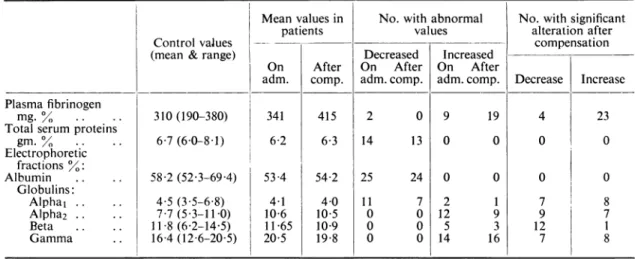
BLOOD PROTEINS: CONTROL VALUES, MEAN VALUES AND INCIDENCE OF ABNORMAL VALUES ON ADMISSION AND AFTER COMPENSATION, AND INCIDENCE OF SIGNIFICANT ALTERATION AFTERCOMPENSATION
Meanvalues in No.withabnormal No.with significant
patients values alteration after
ControlvaJues - compensation
(mean & range) Decreased Increased
On After On After On After
adm. comp. adm.comp. adm.comp. Decrease Increase Plasmafibrinogen
mg.% .. .. 310(190-380) 341 415 2 0 9 19 4 23
Totalserum proteins
gm.% .. .. 67(6-0-8 1) 6-2 6-3 14 13 0 0 0 0 Electrophoretic fractions0o: Albumin .. .. 58 2(52-3-69 4) 534 54-2 25 24 0 0 0 0 Globulins: Alpha1 .. .. 4-5 (3-5-6-8) 4 1 4-0 11 7 2 1 7 8 Alpha2 . .. 77 (53-11-0) 10 6 10 5 0 0 12 9 9 7 Beta .. .. 11*8(6-2-14-5) 11 65 10.9 0 0 5 3 12 I Gamma . 164(12 6-205) 20:5 198 0 0 14 16 7 8
hadnormal E.S.R. Bilirubin concentration was notfoundto show anyconstant relation to venous pressureorcirculation times. Totalserum cholesterol valuesweredecreased in 6 patients, in all of
whom they returned to normal after compensation. Esterified cholesterol values were decreased
in 26 cases on admission and in2after compensation. Thymol turbidity and flocculation tests of liver function were abnormal in 13 patients on admission and in 5 after compensation; in 11 of
these 13 casesthe serumbilirubinwasabnormally elevated. The incidence of abnormality of these
tests on admission in SR+ and SR- groups is given in Table I1I.
Meanvalues of totalserumproteinsandelectrophoretic protein fractions and incidence of
abnor-mal values on admission and after compensation are given in Table II. On admission the total
serum proteins varied between 5-0 and 8-1 gm. percent with anaverage of6-2 gm., and were
de-creased and less than 6 gm. in 14 instances. The serum albumin fraction varied between 48 and 605percentwithanaverageof 534percent,andwasless than 3-5gm.percentin 25cases.
Alpha,
globulinsweredecreased in 11 patients. Alpha2 globulinswereincreasedin 12cases,betaglobulins in5,andgammaglobulinsin 14. Totalglobulinswereincreased andmorethan 3- gm. percentin
7cases. Oneor moreglobulin fractions wereabnormalinatotal of27patients. Theincidence of
alterationsin proteins after compensation is giveninTableII. Therewas no significant difference in the incidence of abnormal values atthe time of admission and aftercompensation. After
com-pensation total proteins increased in 15 and decreased in 6 cases, and albumin increasedin 27 and
TABLE III
INCIDENCEOFABNORMAL LIVERFUNCTION TESTSONADMISSION
INSR+ AND SR- GROUPS
No.withabnormality Liverfunctiontest
SR+ group SR- group Total Thymol turbidity.. .... 8 5 13 Thymol flocculation .. .. 5 3 8 Serum bilirubin .. .. .. 15 5 20 Serumcholesterol Total .. .. .. .. 4 2 6 Esters .. .. .. .. 21 5 26 182
RETARDATION OF E.S.R. IN HEART FAILURE
decreased in 5. In no instance, however, did these values show significant alteration and total protein andalbumin remaineddecreased in13and24 cases,respectively. Thevaluesof theglobulin
fractionsincreasedsignificantly insome casesanddecreasedinothers, but again, therewas no signi-ficant difference in the incidence ofsignificant alteration of the values, except in the case of beta globulin whichdecreased in 12 cases but increased in only one. Total globulins remained elevated in all the 7 cases and gamma globulins in 13. Changes in the values of serum protein fractions showed no relationship to changes in E.S.R. values.
Plasma fibrinogen values onadmission varied between 160 and 705 mg. per cent with an average of 341 mg.; they were normal in 24 patients, decreased in 2, and abnormally elevated in 9. After compensation the values varied between 199 and 615 mg. with an average of 415 mg.; they were normal in 16 and abnormally elevated in 19 cases (Table IV). After compensation the values
TABLE IV
FIBRINOGENLEVELSANDTHEIR RELATIONTONORMALORABNORMAL E.S.R.ONADMISSIONAND
AFTERCOMPENSATION
Onadmission After compensation
Plasmafibrinogenmg.% No.. with No. with
Total abnormal Total abnormal
cases E.S.R. cases E.S.R.
<190 .. .. .. .. 2 1 0 0
190-380 .. .. .. .. 24 8 16 9
>380 .. .. .. .. 9 9 19 19
Total .. .. .. .. 35 18 35 28
increasedsignificantlyfrominitial values in 23 cases, all of them in SR+ group. They decreased in 4cases, 2 inthe SR- group in which the E.S.R. decreased, and 2 in SR+ group. They showed no
significant changein 8 cases, 3 casesin SR+ and 5 in SR- group. All patients with abnormally elevated values of fibrinogen, on admission as well as after compensation, showed abnormally elevated E.S.R. values. However, 8 cases on admission and 9 after compensation with abnormal
E.S.R.,showed normalfibrinogenvalues. Therelation betweenactualincreaseof E.S.R. valuesand
fibrinogenvalues from initial values which occurred aftercompensationisgiveninTable V. In all TABLE V
RELATION BETWEEN ACTUAL RISE OF E.S.R.AND OF CHANGE IN FIBRINOGEN VALUES AFTER COMPENSATION IN 28 CASES OF SR+ GROUP
Total No.withelevation of No. with
cases fibrinogen: mg.%/ decrease of
Rise of E.S.R.mm./hr. O fibrinogen
<25 25-100 100 <10 .. .. .. 7 2 4 0 1 10-15 .. .. .. 10 1 3 5 1 16-20 .. .. .. 7 0 3 4 0 >20 .. .. .. 4 0 0 4 0 Total .. .. .. 28 3 10 13 2
the4patients with arise of E.S.R. ofmorethan 20 mm.fibrinogenvaluesincreased by more than
140mg., and of7 caseswithrise of E.S.R. of less than 10mm., in 6 the increase offibrinogen was
less than 60 mg. and in 1 the fibrinogendecreased.
DISCUSSION
In the present series the E.S.R. values increased significantly after compensation in 28
(80%)
patients including 10 in whom the values became abnormallyelevated from initial normalvalues. Theheart diseases inpatientsinourseriesareknowntobe often associated withanabnormal E.S.R., and occurrence ofnormal E.S.R. initially with subsequent elevation to abnormal values strongly
suggestedthat the E.S.R.wasretardedbytheoccurrenceof heart failure and that elevation after com-pensation wasdue to removal of factors which retarded the E.S.R. duringfailure.
No correlation could be detected between elevation of E.S.R. values after compensation and changesinthe packedcell volume of blood and carbon dioxide combining power ofplasma.
Ab-normal thymol turbidityand flocculation tests in 37 per centof cases, abnormally elevated serum
bilirubin in 57 per cent, and decreased cholesterolesters in 74 percent ofcases onadmission, and
theirreturn tonormal inmostof them aftercompensation indicated impairment of liverfunctionas
a result ofheart failure (Altshule, 1938; Bernstein et al., 1942; Felder et al., 1950; Evans et al.,
1952). These changesalso showed no relation to changes in E.S.R. values.
Ithas been establishedthat inpatientswithcongestive heart failure total serum proteins are often
decreased,
mostly at the expense ofserum albumin, and total globulins are sometimes increased. The electrophoreticpattern ofserum proteinfractions in congestive heart failure has been studied(Sundermannand Sundermann, 1957;Hammond and Ross, 1960; Parry, 1961). Sundermann and Sundermann (1957) reported decreased total proteins and albumin, and increased alpha2 globulin and stated that the lattertwo changes represent anonspecific pattern and a common response in many infectious and in metabolic and neoplastic diseases. Hammond and Ross (1960) reported decreased total proteins and albumin, and increased
alpha,,
gamma and total globulins. Theseinvestigatorsdidnotcorrelate thesechanges to the changes in the E.S.R. Parry (1961) reported that
araised gammaglobulinwasthecommonest globulinfraction change in heart failure and the only
constantchangeseffectedby compensationin 8 caseswith adequate data were a rise of
alpha,
globulin fromalowto normal values in 5 patients and a persistently raised gamma globulin in 7. In the present series significantabnormalities of serum protein at the time of admission included decreased totalproteins, albumin,andalpha,
globulin,andincreasedalpha2,beta, and gamma globulins. Theseabnormalities, however, persisted in most ofthe cases after compensation. The only significant
alteration after compensation was decrease of beta globulin from initial values in 12 patients. Other fractions showed eitherslightdecreaseorincrease butnosignificant difference in the incidence of the two. The increase in albumin and totalproteins aftercompensationwas notsignificant in any instance despite a loss of7 to12kg. ofbodyweight in 9 cases, and of 5 kg. or more in 25, and showednodefinite relationto theactualweight loss. Inalmost all the patients in whom they were
abnormally decreased,
theycontinuedtoremain below the normal level. These findings confirm the observations of Herrmann(1946)
who also noted small differences in total proteins, albumin, andglobulin
in cardiac cedematous patients when they became cedema free. Ourfindings are not in agreementwith those of Hammond andRoss(1960)whostudiedserumproteins in7patients before and after treatment of heart failure and notedincrease in concentration of albumin in those whoquickly
lost their cedema. The data in the present seriesstrongly suggestthat thechanges in pro-teinsarenotthe result of heartfailure but representanonspecific pattern probably due to the diseasecausing
heartfailure,
assuggested
by Sundermann andSundermann(1957).In the present series abnormally high fibrinogenvalues were as a rule associated with a raised E.S.R., and normal values of E.S.R. with normalfibrinogen values. There were, however, several
patientswhohadaraised E.S.R. but normalfibrinogenvalues and thustherewas noparallel
relation-ship between thetwo. It is believed thattheabnormal elevation offibrinogenvalues in our cases
represents anonspecificresponse oftheorganism(Ham and Curtis, 1938) similar to the nonspecific
changesin serum proteins inour patients. The mostsignificant findingin this studywas a
signi-ficant increase offibrinogen values from initial values after establishment ofcompensation in 23 patients,in all of whom therewasalsoasignificantelevationoftheE.S.R.;in 13ofthem the increase
was more than 100 mg. In 2 casesin which the E.S.R. decreased fibrinogen also decreased signi-ficantly. It was also interesting to findthat of7cases with increase ofE.S.R. of less than 10mm.,
fibrinogen increased by less than 60 mg. in 6 and decreased in one of them; moreover, in all the 4
cases with increase of E.S.R. ofmorethan 20 mm. the fibrinogen increase was more than 140mg.
Thus a striking, though not a parallel, correlation was obtained between the changes in the E.S.R. and thefibrinogen values which occurred after compensation.
It has been clearly established that the E.S.R. is not dependent upon any one factor, but is the result of a complex interplay among various influences, the most important being plasma proteins, chiefly fibrinogen and globulins. It is also established that fibrinogen exerts a strong accelerating effect on the E.S.R. and that there may be a linear correlation between the E.S.R. and the plasma
fibrinogenconcentration, although there may be agreat increase in theE.S.R. without a significant
increase in the fibrinogen (Gilligan and Ernstene, 1934; Ham and Curtis, 1938; Nichols, 1942;
Meyersetal., 1953). Morrison (1946) suggested that qualitative changes in the fibrinogen fractions
were the basis of rapid erythrocyte sedimentation. McFarlane and O'brien (1946) stated that the
most active factors increasing the E.S.R. were fibrinogen and euglobulin while those lowering the E.S.R. werealbumin and nucleoprotein. Malmroos and Blix (1946) stated that although fibrinogen
was animportant factor, a rise of alpha2, betaorgammaglobulins might be associated with a raised E.S.R. Linko et al. (1955) found that in patients with myocardial infarction increased E.S.R. values were primarily determined by increase in fibrinogen and alpha2 globulin and decrease in albumin and
alpha,
globulin and that beta and gamma globulins did not contribute to changes in the E.S.R. Inpatients with heart failure, McGinnis et al. (1953) considered it unlikely that alteration inplasma fibrinogen wasacontributory factor in elevation of the E.S.R. after compensation. Parry
(1961)foundnorelationship between changes in the E.S.R. after compensation and the concentration ofplasma fibrinogenorserumglobulins. Inthe presentstudy itwasclear that changes in the E.S.R. aftercompensationcouldnotbecorrelatedtochangesintotalproteins, albumin, or globulin fractions. The data, however, strongly favour the assumption that increase of fibrinogen values is the most important factor in the rise of E.S.R. after compensation. The finding that fibrinogen values becameabnormally elevated from initial normalvalues in 10 cases and increased by more than 100 mg. in 13 is very significant. It warrants the assumption that during congestive heart failure the
fibrinogen level decreases andthat, withestablishment of compensation, it returns to the original level; it also suggests that the deficiency in the fibrinogen is responsible for the retardation of the E.S.R. during congestivefailure.
The question arises as to what is the mechanism of decrease of fibrinogen level during heart
failure. There are two possibilities. First, the increase in the circulating blood volume which
occursin congestive failure may lead to haemodilution, and consequently, decrease in the concen-tration offibrinogen. This doesnot seem to be the likelycause because, in that case, along with elevation of the E.S.R. and the fibrinogen, the protein fractions should have also increased after
compensation. This was,however,notfound and thechanges in protein fractions were inconstant.
Secondly,chronicpassive congestionof theliver may leadtodiminishedproduction of the fibrinogen
by the liver. Itis generally agreedthat congestive heart failure is usually associated with
impair-mentofliver function which hasatendencytoparallelismwith the severity of heart failure(Altschule,
1938; Bernstein et al., 1942; Felder et al., 1950; Evans et al., 1952). Parry (1961) noted some
relationship between the E.S.R. changes and the severity of heart failure which they stated might be fortuitous. In ourseries it isnoted that the E.S.R. valueswere20mm.orless in all18patients in whom either thevenouspressurewasincreasedtomorethan 18 cm. or inwhom the arm tolungand
the arm to tongue circulation times were prolonged to more than 10and 20 seconds, respectively. This pointed to some relationship between the low E.S.R. values and the severity ofheart failure althoughitwas not aparallel relationship. Liverfunction,asjudged byincreasedserum bilirubin,
decreased cholesterol, and abnormal thymol turbidity and flocculation tests, was abnormal inthe
majorityofourpatients. Itis, therefore,mostlikelythatimpairmentofliver functionas aresultof
chronicpassivecongestiondecreases
fibrinogen
production bythe liver. SuchchangesinfibrinogenSANGHVI AND BOHRA
can be definitely established if determinations are obtained before the onset of failure and after decompensation.
SUMMARY
With a view to determining the cause of retardation of E.S.R. in congestive heart failure, the
E.S.R. and some of the factors known to influence it have been studied in 35 patients during failure andafter establishment of compensation. These included liver function tests, total serum proteins
andelectrophoretic fraction of serum proteins, and plasma fibrinogen.
Onadmissionimpaired liver function was found in a majority of the patients. Decreased values
were found for total serum proteins, serum albumin, and
alpha,
globulins, while increased valueswereobtained in the case of alpha2, beta, and gamma globulins. After compensation these abnor-malities ofprotein concentration persisted in almost all of the cases. Alterations in their values showed no relation to changes in the E.S.R. after compensation.
Plasmafibrinogenvalues increased significantly in 23 of 28 patients in whom there was significant elevation of E.S.R. after compensation; in 13 cases the values increased by more than 100 mg. per cent. The data strongly suggest that during heart failure the fibrinogen level is decreased
owing to impairment of liver function with consequent retardation of E.S.R.; they also indicate that the increase of fibrinogen concentration is the most important factor in the elevation of the E.S.R. after compensation.
Dr. L. R. Sarin, Superintendent, S. M. S. Hospital, kindly permitted the publication of this report. REFERENCES
Altshule, M. D. (1938). Medicine, 17, 75.
Bernstein, M., Le Winn, E. B., and Simkins, S. (1942). J. Lab.clin. Med., 28, 1.
Evans, J. M.,Zimmerman, H. J., Wilmer, J. G., Thomas, L. J., and Ethridge, C. B. (1952). Amer. J. Med., 13, 704.
Felder, L., Mund, A., and Parker, J. G.(1950). Circulation, 2, 286.
Flynn, F. V.,and Mayo, P. de. (1951). Lancet, 2, 235.
Gilligan, D. R.,andEmstene, A. C. (1934). Amer. J. med. Sci., 187, 552.
Ham, T.H.,and Curtis, F. C.(1938). Medicine, 17, 413 and 447. Hammond, J. D. S., and Ross, R. S. (1960). Clin. Sci., 19, 119.
Haslewood, G. A. D., and King, E. J. (1937). Biochem. J., 31, 920. Cited by King E. J. (1951). Microanalysis in MedicalBiochemistry. J. & A. Churchill Ltd., London, p. 37.
Herrmann, G. R. (1946). Ann. intern. Med., 24, 893.
Linko,E., Waris, E., and Alikoski, H. A. (1955). Acta med. scand., 153, 389. Malmroos, H., and Blix, G. (1946). Acta med. scand. supp., 170, 280. McFarlane, R. G., and O'Brien, J. R. (1946). Practitioner, 157, 1.
McGinnis, A. E., Lansche, W. E., Glaser, R. J., and Loeb, L. H. (1953). Amer. J. med. Sci., 225, 599.
McLagan,N. F.(1947). Brit. med. J., 2, 197.
Meyers,A. J., Trevorrow, V., Washburn, A. H., and Mugrage, E. R. (1953). Blood, 3, 893.
Moore, S. S.,and Van Slyke, D. D. (1930). J. clin. Invest., 8, 337. Morrison, I. R. (1946). Amer. J. med. Sci., 211, 325.
Nichols,R. E.(1942). J. Lab.clin. Med., 27, 1317. Parry,E.H.0. (1961). Acta med. scand., 169, 79.
Quick, A. J. (1942). The Hemorrhagic Diseases and the Physiology of Hemostasis. C. C. Thomas, Springfield,
Illinois, Cited by Stefanini, M. and Dameshek, W. (1955). The Hemorrhagic Disorders. Grune & Stratton, NewYork, p. 308.
Sanghvi,L. M.(1960). Postgrad. med. J., 36, 620.
Sundermann, F. W., Jr., and Sundermann, F. W. (1957). Amer. J. clin.Path., 27, 125. VanSlyke,D. D., and Cullen, G. E. (1917). J. biol. Chem., 30, 289.
Wintrobe, M. M. (1956). Clinical Hematology. Lea and Febiger, Philadelphia, p. 367. 186