The Cerebellum and the Timing of Coordinated
Eye and Hand Tracking
R. C. Miall and G. Z. Reckess
University Laboratory of Physiology, Parks Road, Oxford OX1 3PT, United Kingdom Email: chris.miall@physiol.ox.ac.uk
Published online August 13, 2001
The cerebellum is known to have important functions in motor control, coordination, motor learning, and timing. It may have other ‘‘higher’’ functions as well, up to and including cogni-tive processing independent of motor behavior. In this article, we will review some of the evidence from functional imaging, lesion studies, electrophysiological recordings, and anat-omy which support the theory that the cerebellum provides a ‘‘forward model’’ of the motor system. This forward model would be used for control of movement; it could also underlie a cerebellar role in coordination. In this role, the forward model would generate time-specific signals predicting the motion of each motor effector, essential for predictive control of, for example, eye and hand movements. Data are presented from human eye and hand tracking that support this. Tracking performance is better if eye and hand follow the same spatial trajectory, but better still if the eye leads the hand by about 75 to 100 ms. This suggests that information from the ocular control system feeds into the manual control system to assist its tracking. 2001 Elsevier Science
A SENSORY ROLE FOR THE CEREBELLUM?
The cerebellum is an important neural structure, both because its dysfunction leads to pronounced disturbances in movement, posture, and balance and because it is a relatively massive structure in higher vertebrates. In humans, it represents about 10% of the volume of the brain, and even more striking, it has been estimated to hold more than half of all the neurones in the central nervous system. Such massive pro-cessing power must have a vital purpose, but this has proved difficult to pin down. The question ‘‘what does the cerebellum do?’’ has an implicit assumption, accepted by many but not all, that it performs a singular operation as it transforms its inputs into outputs. We accept this assumption, largely because of the uniformity of the cerebellar cortex, and would argue that the key to understanding the cerebellum is to find a single process that can account for the very diverse functions attributed to the cerebellum.
We will suggest that one of the fundamental functions of the cerebellum is to act as a ‘‘sensory predictor,’’ responsible for generating predictions about the sensory consequences of motor acts. Predictions about the sensory outcome of movement can be used to control motor outputs (Miall, Weir, Wolpert, & Stein, 1993). Sensory
R.C.M. is supported by a Wellcome Senior Research Fellowship. We thank David Rosenbaum for a thoughtful review of the paper.
Address correspondence and reprint requests to, R. C. Miall, University Laboratory of Physiology, Parks Roads, Oxford OX1 3PT, UK. E-mail: chris.miall@physiol.ox.ac.uk.
212
0278-2626/01 $35.00
2001 Elsevier Science All rights reserved.
predictions are also important for other functions more removed from on-line motor control (Miall & Wolpert, 1996), for example, in motor planning or mental imagery, or even for sensory discriminations, and there is a growing body of evidence which suggests that the cerebellum is involved in each of these different functions. We suggest that in each instance, the cerebellum is concerned with processing or generat-ing sensory predictions within a motor context. Of course, predictions must be time-specific, and it is no coincidence that many theories of cerebellar function propose that it is related to temporal processing (Braitenberg, 1967, 1983; Jueptner, Flerich, Weiller, Mualler, & Diener, 1996; Keele & Ivry, 1990).
Comparative anatomical studies have shown that the cerebellar cortex is a phyloge-netically ancient structure found in all vertebrates, although it is particularly large in primates. The cerebellar cortex has a clear orthogonal arrangement. Its length is very much greater than its width, forming in the human a sheet of about 2 m in anterior–posterior dimension, by 15 cm laterally. Its length is a plausible measure of its parallel processing power as it defines the number of transverse ‘‘beams’’ of parallel fibers which project onto the output cells, the Purkinje cells. Sultan and Braitenberg (1993) showed that the cerebellar surface area across 15 species is very strongly correlated with body mass raised to the power of about 2/3. The slope of 0.6 suggests a correlation with body surface area, rather than with body mass (slope 1.0), number of muscle fibers (slope 0.75),1or any other obvious attribute of the motor
system. This is important because the skin is a vital sensor affecting and affected by motor control. Any movement must distort the skin, and the skin of course is a crucial sensory interface between our actions and the external environment. Thus all motor acts have important consequences for sensory input to the skin, and perhaps the cere-bellum has a particular role in processing this reafferent information. The lateral dimensions of the cerebellum measured by Sultan and Braitenberg (1993) did not follow the same rule, however, and the width of the cerebellum in humans is dramati-cally increased, roughly in proportion to the massive expansion of the cerebral hemi-spheres. Perhaps the complexity of the process undertaken by each beam of Purkinje cells increases especially for humans, and this may indicate that more complex pre-dictions are generated by the primate cerebellum, or prepre-dictions further into the fu-ture.
In fish, relatively ancient vertebrates, the Purkinje cells—the very prominent cells that make up only output from the cerebellar cortex—are closely connected with the vibration-sensitive cells in the lateral lines on the fish’s flanks. In the mormyrid electric fishes, the cerebellum has a role in generating signals that predict the re-afferent signal produced by the electric organ (Bell, Bodznick, Montgomery, & Bastian, 1997), a sensory structure actually based on signals produced by modi-fied muscle fibers and closely coupled to the lateral line sensory systems. Thus even in early vertebrates, cerebellar structures appear to have important sensory func-tions.
Gao, Parsons, Bower, and Xiong (1996) reported a functional imaging study in which the cerebellum was shown to be most active in a haptic discrimination task, more active when attempting to discriminate shapes by touch than when performing the same movements without discrimination. Its specific role in processing reaffer-ence in human movement has recently been demonstrated in an fMRI experiment by Blakemore, Wolpert, and Frith (1998). They demonstrated cerebellar activation when subjects self-stimulated the skin on one of their hands by rhythmic movement
1The cross-sectional area of muscle can be shown to be proportional to body mass w to the power 3/4. Limb length L is proportional to 2/3 muscle diameter d, and weight is proportional to Ld2hence w
is proportional to d8/3
. Cross sectional area d2
is therefore proportional to w3/4
of the other hand, over and above the activation observed with external stimulation of the skin of the passive hand, or simply movement of the one hand without tactile stimulation of the other.
Thus the cerebellum seems to be involved in processing cutaneous or myoelectric sensory information that results from motor actions. It also processes visual feedback signals and is most important in the control of visually guided movements (move-ments toward visual targets), particularly important in move(move-ments made under visual feedback (Miall, Weir, & Stein, 1987; Van Donkelaar & Lee, 1994). Cerebellar pa-tients often display severe intention tremor at the end of movements toward visual targets, and poor control of the speed and path of the hand or joystick when tracing a moving object. Paradoxically, patients with pronounced cerebellar motor problems can actually do better in these tracking tasks without visual feedback (Beppu, Suda, & Tanaka, 1984; Haggard et al., 1995; Liu, Ingram, Palace, & Miall, 1999), suggesting that their symptoms may not be due to errors in the generation of the appropriate motor commands, but to errors in the guidance of the movement using sensory feed-back. The reason is probably that visual feedback pathways are slow, with delays of between 100 and 200 ms. Thus, normal visual guidance of movement must be based on a feedforward strategy that does not depend on current visual feedback, rather than on a feedback strategy. But the control strategy must also be able deal with this slow feedback signal, in order to guide the hand or arm accurately. Delaying visual feedback in healthy subjects by 200–300 ms can lead to visually guided tracking that is strikingly similar to that seen after cerebellar dysfunction (Fig. 1). Thus if the healthy subject tracks with added delays, the inappropriately delayed feedback de-grades their performance; for the cerebellar patient, the intrinsic delays in the sen-sory–motor control loop causes similar problems. This highlights the role of the intact cerebellum in dealing with intrinsic visual feedback delays.
FIG. 1. Typical traces of a monkey performing a visual tracking task. The upper two records show the effect of adding a delay of 300 ms between the joystick and the displayed cursor on a normal monkey. The lower two traces show the effect of temporary disruption of the lateral cerebellum with a cooling probe, on a monkey performing without added feedback delays.
THE CEREBELLUM AS A FORWARD MODEL
An important theory that has been gaining attention in recent years is that control systems (e.g., in robotic or industrial applications) benefit from ‘‘forward models.’’ A forward model is a representation of the object being controlled, and is used to predict the outcome of actions, a prediction of the future state of the robot or industrial process after a motor command has been issued. We have described (Miall & Wolpert, 1996) how, in physiological terms, this state estimate could be a prediction of the posture or motion of the body after following a motor command, or by a further transformation, a prediction of the sensory reafference that the command will cause. In a visual guided motor task, it could be a representation of the visual consequences of a movement. We have been recording from Purkinje cells in the intermediate/ lateral cerebellar cortex in monkeys performing visual tracking tasks, and have found a significant proportion of cells that do indeed code for the direction of a cursor moved by a joystick, even when the cursor is mirror reversed from the actual move-ment of the hand (Liu & Miall, 1998). Such cells could be used to predict the motion of the cursor—the visual consequence of hand movement. Through their projections from the cerebellar cortex via the thalamus to the motor cortical areas (Van Donke-laar, Stein, Passingham, & Miall, 1999), these signals would help to guide the move-ment of the cursor to the target. Imamizu et al. (2000) have shown that the human cerebellum is active in learning the use of a computer mouse, and stores information about novel mouse–cursor relationships.
Thus the cerebellum may function as a forward model, predicting the sensory out-come of movements. Hence, the suggestion that one role of the cerebellum is to process reafferent information is also compatible with its role controlling movement. This provides a good link between the evidence that cerebellar damage leads to motor deficits, especially in guided movement, and evidence that the cerebellum is closely involved in processing sensory information from movement.
MOTOR CONTROL AND THE CEREBELLUM
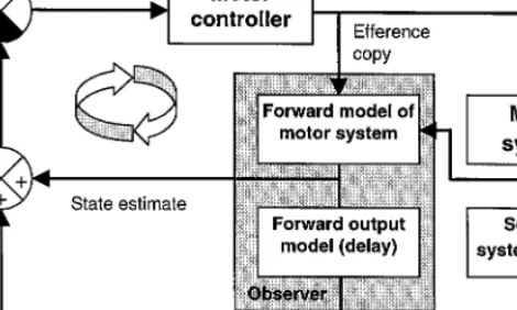
The preceding sections have stressed the role of the cerebellum in sensory estima-tion, or in the control of movement using sensory feedback. However, many authors have suggested a role for the cerebellum in the generation of feedforward motor commands (Kawato & Gomi, 1992), so it should be made clear that these are not exclusive operations: forward models can also be used for feedforward control. The forward model can be placed within an internal control loop, so that it provides an estimated negative feedback signal of the movement on which feedback error correc-tions are calculated. For linear systems, this control scheme can generate near optimal motor outputs. There may be difficulties in using the scheme for nonlinear feedfor-ward control, but we know of no situations in which physiological motor control is achieved without some feedback control. Thus a perfect controller is rarely, if ever, needed. We have proposed a particular form of this control strategy, known as a Smith Predictor (Smith, 1959), as a model for the cerebellum (Miall et al., 1993). This includes a forward model, servicing an internal feedback loop whose output can drive the arm toward the desired state (Fig. 2). As the forward model avoids the feedback delays in the real sensory–motor apparatus, this internal feedback loop can have a high open-loop gain, and function as a near-optimal feedforward controller (Schleck & Hanesian, 1978). The Smith Predictor also includes an explicit delay mechanism that delays a copy of the rapid sensory estimate to allow temporally
syn-FIG. 2. The Smith Predictor model: The outer loop of this figure indicates a feedback control loop in which motor commands from a motor controller (motor cortex) act on the motor system to alter its state, and cause reafferent sensory inputs. The forward model (in the cerebellum) receives the motor command (efference copy) and generates a state estimate that is used within an internal rapid feedback control loop, indicated by the circular arrow. In the visuo-motor system, the sensory and motor pathways introduce significant feedback delays, so the forward output model within the Smith Predictor is an internal model of these delays. Its output can be compared with the reafferent inputs caused by the motor action.
chronous comparison with the actual sensory consequences of the movement. This is important to allow any errors in the internal estimate to be detected and corrected. By ensuring synchrony between the delayed output of the forward model and the actual feedback, the Smith Predictor effectively isolates the feedback delays from the control loop.
CEREBELLAR CONTRIBUTION TO COORDINATION
A forward model used as a state estimator is also important for motor coordination. Temporal delays in the sensory and motor pathways suggest that for many human movements, coordination must depend on a predictive state estimate, rather than on feedback signals. For example, in eye–hand coordination tasks, there is near-synchro-nous tracking of the hand by the eye. This cannot be due to reactive following, as the eye can only follow unpredictable visual targets with a reaction time of about 100–150 ms. Even if the eye were using the more rapid signals provided by proprio-ceptors in the arm, this would still lead to a tracking lag larger than observed. Thus, it seems that the eye control system uses information derived from the motor com-mands being sent to the hand. Motor comcom-mands, which lead to contraction of muscles in the arm, cannot directly predict the position of the hand in 3-D space. They need to be translated into a representation of the final position of the hand in a visual coordinate system; this is the operation of a forward model.
There is strong evidence for the cerebellum’s role in eye–hand coordination. Mon-keys, like humans, can make eye movements to follow movement of a cursor con-trolled by the hand with very low latency. Vercher and Gauthier (1988) have shown that after inactivation of the cerebellum, the latency of the eye movements rose to a level seen when the monkey tracked an unpredictable visual target. They have also
recently demonstrated that sensory afferents from the arm are not required for the temporal linking of ocular and manual systems, while motor commands to voluntarily drive the arm movement are required (Vercher, Gauthier, 1996). This confirms that efferent copy is an important part of the coordination process: signals from the manual control system appear to be sent to the ocular control system, to provide predictive information about the required hand movement.
EYE AND HAND COORDINATION IN A VISUAL TRACKING TASK
Vercher and Gauthier (1988) could not test the temporal and spatial specificity of interactions between eye and hand in their experiments. In particular, it was not possi-ble to determine how precise was the timing of the predictive signals assumed to underlie the coordination process. The Smith Predictor model (Miall et al., 1993) suggests that the forward model should provide a prediction of the sensory feedback signals with minimal delay, to allow high gain in the internal control loop. In contrast, the delayed version of this prediction, required to cancel out the real reafference, must be precisely synchronized with these delayed signals.
We have recently shown that when the feedback delays are artificially increased, performance improves over a time course of several hours (Foulkes & Miall, 1999), suggesting that the visuo-motor control system can adapt to different delays. It has also been shown that the ticklish sensation caused by one hand stroking the other increases with the introduction of artificial delays between the two hands (by using a robotic interface, Blakemore, Frith & Wolpert, 1999). The authors suggest that the forward model in the cerebellum (Blakemore et al., 1998) used to cancel this expected reafference signal fails as the delays are increased, leaving some sensory input uncan-celled, and therefore more noticeable to the subject. Thus, there are behavioral data, theoretical reasons, and functional imaging results which suggest that the cerebellar forward model should be accurately timed, and we report below two behavioral ex-periments designed to begin to test this hypothesis.
EXPERIMENT 1—THE ROLE OF EYE MOVEMENT INFORMATION
The first experiment addressed the basic question of whether information about an unpredictable, external target can be derived from ocular tracking to provide dictive information about the desired trajectory of the hand. In the absence of a pre-dictable target trajectory, any performance benefits in hand movement must be di-rectly due to an internal predictor mechanism that incorporates eye movements into subsequent hand motor responses. We therefore designed a tracking task in which eye and hand tracking movements were performed simultaneously, but in which we could control the degree of coordination between the two. We predicted that improved performance would be seen if the eye and hand followed the same unpredictable trajectory, compared to when they followed separate trajectories or when only hand movement was required.
METHODS
Participants. Seven subjects (five male, two female), varying in age from 19–44 years, participated voluntarily. All subjects were right-handed and had normal or corrected-to-normal vision. Three of the participants were aware of the objectives of the experiment and were practiced at similar psychophysics tasks, but the remaining four subjects were completely naı¨ve at the time of participation.
Display. The computer VGA display showed a large yellow crosshair bisecting a blue screen back-ground. Superimposed on the crosshairs was a green square cursor, 16⫻16 pixels. A white circular target, 16 pixels in diameter, was positioned 50 pixels directly above the square, superimposed on the vertical crosshair (Fig. 3A).
Subjects used a small, lightweight joystick to control the cursor position. Sampling of the horizontal and vertical joystick positions was achieved with an A–D converter with 12-bit resolution, at intervals of 38 ms synchronized to the screen refresh rate (12.7 ms).
Task. The motion of the square, green cursor was controlled by a joystick, without any interposed dynamics or time lags. The subjects’ task was to keep the cursor superimposed on the stationary crosshairs in the center of the display. However, the cursor was continuously perturbed away from the crosshairs following a target waveform. The subjects had to compensate for this motion to return the cursor to the screen center (Poulton, 1974).
The paradigm consisted of four conditions. In the first ‘‘compensatory’’ condition, the hand was responsible for controlling a moving cursor while the eyes remained fixated on a stationary object. This was meant to serve as a ‘‘hands only’’ control condition, although even fixation of a stationary object can involve some small micro-saccadic eye movement. In the second (‘‘coordinated’’) condition, the hand and eye moved in the same direction and with the same velocity. In the third (‘‘independent’’) condition, the eye and hand moved in patterns unrelated to each other. The fourth condition was rest. The white circular target for ocular tracking was stationary during two of the four conditions, and in motion during the remaining trials. The two conditions during which the circle was motionless were the ‘‘rest’’ and the ‘‘compensated’’ conditions. The rest condition was the only explicitly signaled condition, with both the cursor and the target displayed as red, and the target stationary during this task. The rest condition was presented once every four trials. All other conditions were presented in a quasi-random sequence to prevent the influence of an order effect.
In Compensated tracking, the circular eye target also remained stationary. Thus, only the hand was moving in response to its continuous displacement from the center of the screen, and was therefore compensating for this movement without any cues from the ocular target. During the two remaining conditions, the white target for ocular tracking was also in motion, following either the same target motion as the square, or an unrelated trajectory. In Coordinated tracking, the ocular target and cursor followed movement patterns directly opposed to one another. Thus, in order to compensate for the cursor displacement from the central crosshair, the subject had to move his/her hand in the direction opposite, corresponding to the direction to the ocular target. Hence, eye and hand movements were spatially and temporally coordinated during accurate performance. During Independent tracking, the circular eye target was moving in a pattern unrelated to that of the square cursor. Ocular and manual responses were there-fore independent.
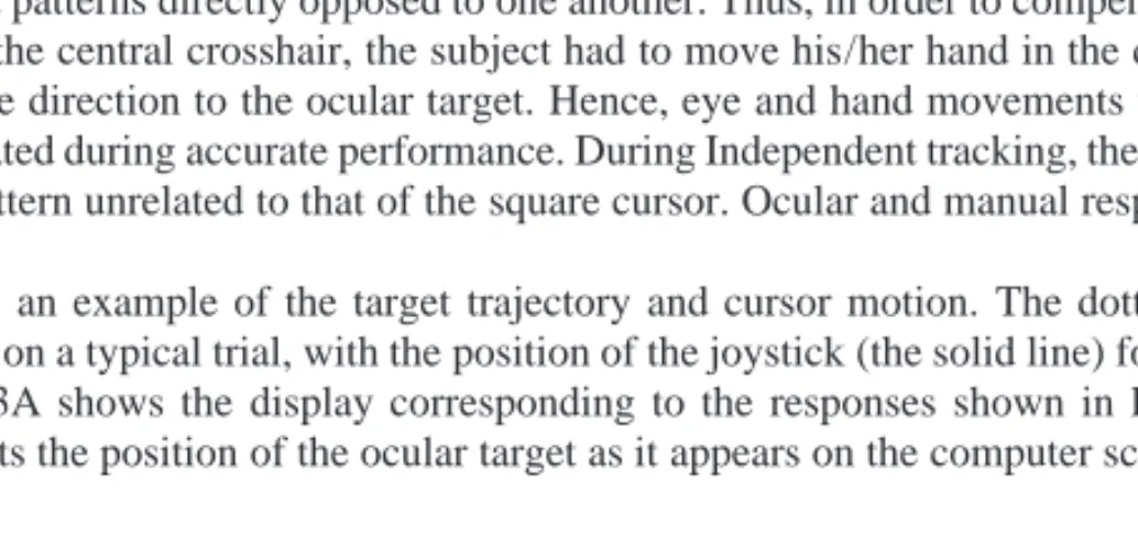
Figure 3B shows an example of the target trajectory and cursor motion. The dotted line represents the target trajectory on a typical trial, with the position of the joystick (the solid line) following relatively accurately. Figure 3A shows the display corresponding to the responses shown in Fig. 3B. Here, the dotted line represents the position of the ocular target as it appears on the computer screen, and the solid
FIG. 3. The screen display (A) and the corresponding movement of the joystick (B) during coordi-nated tracking. In Figure A, the small square represents a cursor controlled by the joystick, and its target is the crosshair; the circle is the ocular target, following the same trajectory as required for the joystick (B).
line indicates the cursor’s actual trajectory. Note that with the fairly accurate performance recorded in this trial, the cursor appears to hover around the crosshairs, because the motion of the joystick (Fig. 3B) is fairly similar to the required pattern of displacement.
There were 35 trials in each block, each trial lasting 15 s. Each block of trials therefore lasted 8.75 min. Performance was measured at three different target speeds: slow, medium, and fast, where the frequencies of the composite trajectory path were multiplied by a speed function (1.3, 1.65, and 2.0, respectively). These were tested in three separate blocks of trials, the order of which was systematically varied across subjects.
RESULTS
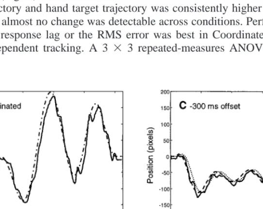
Figures 4A and 4B show typical tracking responses in Coordinated and Indepen-dent conditions, respectively. Although we could not record eye movements during this task, we have now recorded them with a single subject performing the tasks at the medium speed (Fig. 5). The joystick motion clearly followed the target quite accurately, with frequent small corrective movements. We calculated by crosscorrela-tion the mean lag between the mocrosscorrela-tion of the joystick and the target trajectory for hand movement, and the mean RMS error scores across seven subjects and for the three speeds (Fig. 6). The maximal correlation coefficient calculated between the joystick trajectory and hand target trajectory was consistently higher than r2⫽ .98,
and therefore almost no change was detectable across conditions. Performance mea-sured by the response lag or the RMS error was best in Coordinated (Fig. 6) and worst in Independent tracking. A 3 ⫻ 3 repeated-measures ANOVA revealed no
FIG. 4. Typical traces of joystick movement in Coordinated (A), Independent (B), and temporally offset conditions (C, D). The target for the eyes is indicated by the dotted line, that for the joystick is indicated by the dashed line, and the solid lines show the joystick movement. In (A) the eye and hand targets are identical.
FIG. 5. Typical eye position and joystick traces of from a single subject tracking in the Coordinated (A) and Compensated (B) conditions.
significant effect of speed for RMS errors (F(2) ⫽ 2.67, p ⫽ .11) or lag values (F(2) ⫽ 0.44, p ⫽.65). It did, however, confirm the existence of an overall main effect of condition (RMS errors: F(2)⫽20.10, pⱕ.0001; lag: F(2)⫽ 47.36, pⱕ .0001).
Because the data were normally distributed overall, post hoc paired t tests were calculated for lag and RMS error scores, comparing the differences between the three conditions for each speed. For the lag data from all three speeds, performance in Coordinated was significantly better than that in Independent, and performance in
FIG. 6. Manual tracking performance measures, Experiment 1. Three conditions were tested at three target speeds. Performance was best for the eye–hand Coordinated condition, and worst for Independent. The Compensated condition required hand movement alone.
Independent was significantly worse than in Compensated (Table 1). As expected, performance in Coordinated was consistently superior to performance in Independent. Both individual and grouped subject data showed that peak performance occurred in the Coordinated condition. This suggests that eye movement information that was consistent with the necessary hand movement response helped to predict the required velocity and direction of subsequent hand tracking, thereby decreasing lag time and error scores. Also, performance in Coordinated was better than in Compensated. Thus, moving eyes and hand together improved performance above hand movement alone, while performance was worst in Independent.
EXPERIMENT 2—OPTIMIZING THE PREDICTIVE POWER OF EYE MOVEMENT INFORMATION
In Experiment 1, the control of the hand was aided by eye target movements that occurred in spatial and temporal synchrony with the required joystick trajectory. This
TABLE 1
Results of Pairwise t-Test Comparisons of Tracking Lag and RMS Error between the Coordinated, Independent, and Compensated Conditions at Three Speeds
Condition Lag RMS error
Speed Comparison t p value t p value
Slow Coord–Comp ⫺3.32 .048* ⫺0.391 .709 ns Comp–Indep ⫺4.49 .012** ⫺2.681 .108 Coord–Indep ⫺7.59 .0003*** ⫺3.674 .030 Medium Coord–Comp ⫺4.35 .015* ⫺1.725 .405 ns Comp–Indep ⫺6.34 .003** ⫺4.035 .021* Coord–Indep ⫺8.20 .0003*** ⫺5.097 .006** Fast Coord–Comp ⫺4.78 .009** ⫺2.723 .105 Comp–Indep ⫺2.89 .084 ⫺5.402 .006** Coord–Indep ⫺6.90 .0003*** ⫺5.349 .006**
Note. n⫽7 subjects. p values have been Bonferoni-adjusted for the multiple comparisons within each speed condition.
was possible because eye tracking has a much smaller response lag than hand tracking. Although we cannot confirm that this was the case in Experiment 1 without repeating the task while recording eye movement response times and errors, we can assume this is true based on the large body of supporting evidence (Abrams, Meyer, & Kornblum, 1990; Koken & Erkelens, 1992; Vercher, Magenes, Prablanc, & Gauthier, 1994). We assume therefore that eye movements occur several tens of milliseconds before the hand response occurs, and can therefore provide the hand system with predictive clues (Fig. 5). But if there were an even greater time delay between ocular movement and hand target motion, would the information be more effectively used by the manual system?
The second experiment therefore introduced a time offset between the motion of the ocular and manual targets. We hypothesized that there should be an ideal delay that allows the hand system sufficient processing time to optimally use the informa-tion received from the ocular motor system. Such a finding, with peak performance when the eye significantly leads the hand, would argue against the possibility that the eye and hand controllers share the same predictor mechanism, and would instead support the prospect of two separate control systems that interact for behavioral coor-dination.
METHODS
The same equipment, display, and subjects were used for as for the first experiment. However, because there were more conditions, each subject was tested in two blocks of 45 trials (15-s duration per trial), each block lasting 11.25 min.
This second paradigm consisted of 16 different test conditions broken down into two blocks of 8, and a rest condition. Each block contained an independent condition, similar to the one in Experiment 1. The other 7 conditions comprised coordinated tracking, as in Experiment 1, but with varying temporal offsets between motion of the eye target and the hand target. For simplicity, these temporal shifts will be referred to as ‘‘offset’’ values. Offsets were staggered at approximately 50-ms intervals (⫺342,⫺304, ⫺266,⫺190,⫺152,⫺114,⫺38, 0, 38, 114, 152, 190, 266, 304), adjusted to match the joystick sampling interval (38 ms) and screen refresh rate (12.7 ms). Negative offsets indicate conditions where the hand target movement preceded that of the eye target, with a time lapse of zero representing perfect spatial and temporal synchrony between both targets (as in Coordinated, Experiment 1). To avoid any uneven biases between the two blocks of trials, conditions were allocated such that block one contained the 1st, 3rd, 5th, etc., offset value (e.g., block 1⫽ ⫺342,⫺266,⫺152 ms, etc.). Presentation of different offsets was pseudo-randomized, but this order of presentation was kept constant across subjects.2
RESULTS
Figure 4C is an example of the target and response trajectories during typical per-formance when the hand target leads the ocular target by 304 ms. Only the horizontal components of the hand target (dashed line) are shown, following the same path as the eye target (dotted line), but with a temporal offset. The solid line shows the horizontal component of manual response (the joystick position), and because of the typical 200-ms latency of manual tracking, it is nearly synchronous with the eye target trajectory. Figure 4D is identical except that in this example the eye target (dotted) precedes the hand target (dashed) by 304 ms, followed closely by the joystick position trace (solid). Here the joystick trajectory lags behind the eye target by about 300 ms.
There were large intersubject differences in performance of this task, although all
2Pilot data revealed a significant order effect, with performance best immediately after the rest
condi-tion. This forces a quasi-random presentation procedure so that every offset condition was tested at each stage after a rest.
FIG. 7. Manual tracking performance measures, Experiment 2. Eye–hand tracking with 14 time offsets between the targets for eye and hand movement and two independent conditions were tested at three target speeds, Performance was best for the condition with an offset of about⫹75 to⫹100 ms (eye target trajectory leading hand target), and worst for the independent condition.
followed the same general pattern. Response lag and RMS error values were therefore standardized by dividing each by the independent condition value for that subject. All analyses were performed for both the raw and the normalized data, and largely agreed. Figure 7 shows average, normalized lag and RMS error values for all subjects. Note the U-shaped curve, reaching a minimum near the zero-offset position, and the outlying data for the two independent conditions measured.
As in Experiment 1, a 3 ⫻ 14 repeated-measures ANOVA revealed that there was no significant effect of speed on overall performance (Table 2). Data from the independent condition were excluded from this analysis. However, there was a sig-nificant main effect of offset condition across the 14 tested offsets (F(13) ⬎ 3.2,
p⬍ .001; Table 2).
To estimate the offset value with maximal performance, a third-order polynomial curve was fit to each subject’s performance curve for both normalized and raw values, and the minimum position of the resulting smooth curve was recorded. Table 3 lists these results for each of the three speeds, where the ‘‘best offset’’ represents the estimated time delay between eye target and hand cursor movement that corresponds
TABLE 2
Main Effect of Speed (Fast, Medium, Slow) and of Condition (14 Values of Offset, Excluding the Independent Condition) on Overall Performance
Speed Condition
Data type F(df⫽2) p value F(df⫽13) p value
RMS Errors 1.390 .285 4.547 .0001***
Normalized RMS errors 1.608 .241 3.210 .001**
Lag 1.080 .370 6.288 .0001***
Normalized lag 1.186 .339 4.418 .0001***
TABLE 3
Average Location of the Minima of Best-Fit Curves for Performance across 14 Offset Conditions at Three Speeds
Data type Speed Best offset
RMS Errors Slow 117.5
Medium 120.44
Fast 62.58
Mean 61.00
Normalized RMS errors Slow 75.70 Medium 86.52 Fast 62.68 Mean 74.97 Lag Slow 98.71 Medium 143.57 Fast 109.43 Mean 117.24
Normalized lag Slow 69.00
Medium 119.14
Fast 106.12
Mean 98.09
to the best performance (e.g., the lowest point on the best-fit curve applied to the graphs in Fig. 7). The overall minimum values for normalized RMS error and lag data were 75 and 98 ms, respectively.
CONCLUSION
These behavioral results confirm earlier reports that coordinated eye and hand tracking is better than tracking using the hand alone, and much better than with inde-pendent but simultaneous eye and hand motion (Abrams et al., 1990; Biguer, Prablanc, & Jeannerod 1984; Koken & Erkelens, 1992; Vercher et al., 1994). This extends earlier descriptions to the tracking of unpredictable targets, rather than just to tracking of self-moved targets. Furthermore, the second experiment allows us to detect the causal flow of information from the oculomotor system to the manual control system, and indicated the time course of this transfer. The peak performance was achieved when the hand followed the same trajectory as the eyes, but with a latency of about 75–100 ms. This implies that internal knowledge of the motion of the eyes can feed through to the hand with the same latency. If the eye leads by a greater time offset, manual tracking performance is degraded, presumably because the hand then moves too early, compared to its own target. If the eyes lead by a lesser amount, or lag behind the hand, performance is again degraded, presumably because the predictive information is not available early enough to be used by the manual tracking system.
Of course, these behavioral data cannot yet tell us anything about the role of the cerebellum in this oculo-manual coordination task. For this, we are studying subjects in the same two tasks reported here, using a functional imaging magnetic scanner. However, it seems likely that the cerebellum is the site of this coordination process: it is known to be highly activated in these tasks, it has neural properties consistent with a prediction of the outcome of movement, and several lines of evidence suggest that it functions as a neural ‘‘forward model,’’ a necessary component of the process of oculomanual coordination.
The finding that peak performance for manual tracking occurs when the ocular target led by 75–100 ms, rather than zero ms, also suggests that the improvement in performance is indeed dependent on active coordination between eye and hand, rather than the effect of simultaneous movement of both systems. The quite steep trough in the performance curves (Fig. 7) suggests that the predictive signals are accurately timed: offset from the optimal time difference led to a rapid decline in performance, especially for the faster target speeds.
These results argue, although so far only indirectly, for accurate estimates of the outcome of eye movement commands, temporally specific, and—we suggest—based on a forward model. It is now necessary to confirm that these estimates are generated by the cerebellum.
REFERENCES
Abrams, R. A., Meyer, D. E., & Kornblum, S. (1990). Eye–hand coordination: Oculomotor control in rapid aimed limb movements. J. Exp. Psychol., 16, 248–267.
Bell, C., Bodznick, D., Montgomery, J., & Bastian, J. (1997). The generation and subtraction of sensory expectations within cerebellum-like structures. Brain Behav. Evol., 50(Suppl.), 17–31.
Beppu, H., Suda, M., & Tanaka, R. (1984). Analysis of cerebellar motor disorders by visually guided elbow tracking movement. Brain, 107, 787–809.
Biguer, B., Prablanc, C., Jeannerod, M. (1984). The contribution of coordinated eye and head movements in hand pointing accuracy. Exp. Brain Res., 55, 462–469.
Blakemore, S. J., Frith, C. D., & Wolpert, D. M. (1999). Spatio-temporal prediction modulates the perception of self-produced stimuli. J. Cogn. Neurosci, 11, 551–559.
Blakemore, S. J., Wolpert, D. M., & Frith, C. D. (1998). Central cancellation of self-produced tickle sensation. Nat. Neurosci., 1, 635–640.
Braitenberg, V. (1967). Is the cerebellar cortex a biological clock in the millisecond range? Prog. Brain
Res. 25, 334–346.
Braitenberg, V. (1983). The cerebellum revisited. J. Theor. Neurobiol, 2, 237–241.
Foulkes, A. J., & Miall, R. C. (1999). Adaptation to visual feedback delays in a human manual tracking task. Exp. Brain Res., 131, 101–110.
Gao, J.-H., Parsons, L. M., Bower, J. M., Xiong, J., Li, J., & Fox, P. T. (1996). Cerebellum implicated in sensory acquisition and discrimination rather than motor control. Science, 272, 545–547. Haggard, P. N., Miall, R. C., Wade, D. T., Fowler, S., Richardson, A., Anslow, P., & Stein, J. F. (1995).
Damage to cerebellocortical pathways following closed head injury: An MRI and behavioural study.
J. Neurol. Neurosurg. Psychiatry, 58, 433–438.
Imamizu, H., Miyauchi, S., Tamada, T., Sasaki, Y., Takino, R., Putz, B., Yoshioka, T., & Kawato, M. (2000). Human cerebellar activity reflecting an acquired internal model of a new tool. Nature, 403, 192–195.
Jueptner, M., Flerich, L., Weiller, C., Mueller, S. P., & Diener, H. C. (1996). The human cerebellum and temporal information processing—results from a PET experiment. Neuroreport, 7, 2761–2765. Kawato, M., & Gomi, H. (1992). A computational model of four regions of the cerebellum based on
feedback-error-learning. Biol. Cybern., 68, 95–103.
Keele, S. W., & Ivry, R. (1990). Does the cerebellum provide a common computation for diverse tasks? A timing hypothesis. Annals N. Y. Acad. Sci, 608, 179–211.
Koken, P. W., & Erkelens, C. J. (1992). Influences of hand movements on eye movements in tracking tasks in man. Exp. Brain Res., 88, 657–664.
Liu, X., Ingram, H. A., Palace, J. A., & Miall, R. C. (1999). Dissociation of ‘on-line’ and ‘off-line’ visuomotor control of the arm by focal lesions in the cerebellum and brainstem. Neurosci. Lett.,
264, 121–124.
Liu, X., & Miall, R. C. (1998). Neurons specifically related to visual representation of arm movements in the lateral cerebellar cortex. Soc. Neurosci. Abstr., 24, 555.4.
Miall, R. C., Weir, D. J., & Stein, J. F. (1987). Visuo-motor tracking during reversible inactivation of the cerebellum. Exp. Brain Res., 65, 455–464.
Miall, R. C., Weir, D. J., Wolpert, D. M. & Stein, J. F. (1993). Is the cerebellum a Smith Predictor? J.
Motor Behav., 25, 203–216.
Miall, R. C., & Wolpert, D. M. (1996). Forward models for physiological motor control. Neural
Net-works, 9, 1265–1279.
Poulton, E. C. (1974). Tracking skill and manual control. Academic Press: London.
Schleck, J. R. & Hanesian, D. (1978). An evaluation of the Smith Linear Predictor technique for control-ling deadtime dominated processes. ISA Transactions, 17, 39–46.
Smith, O. J. M. (1959). A controller to overcome dead time. ISA Journal, 6, 28–33.
Sultan, F., & Braitenberg, V. (1993). Shapes and sizes of different mammalian cerebella. A study in quantitative comparative neuroanatomy. J. Hirnforsch, 34, 79–92.
Van Donkelaar, P., & Lee, R. G. (1994). Interactions between the eye and hand motor systems: Disrup-tions due to cerebellar dysfunction. J. Neurophysiol., 72, 1674–1685.
Van Donkelaar, P., Stein, J. F., Passingham, R. E., & Miall, R. C. (1999). Neuronal activity in the primate motor thalamus during visually triggered and internally generated limb movements. J.
Neu-rophysiol., 82, 934–945.
Vercher, J. L., & Gauthier, G. M. (1988). Cerebellar involvement in the coordination control of the oculo-manual tracking system: Effects of cerebellar dentate nucleus lesion. Exp. Brain Res., 73, 155–166.
Vercher, J. L., Gauthier, G. M., Guedon, O., Blouin, J., Cole, J., & Lamarre, Y. (1996). Self-moved target eye tracking in control and deafferented subjects: Roles of arm motor command and proprio-ception in arm–eye coordination. J. Neurophysiol., 76, 1133–1144.
Vercher, J. L., Magenes, G., Prablanc, C., & Gauthier, G. M. (1994). Eye–head–hand coordination in pointing at visual targets: spatial and temporal analysis. Exp. Brain Res., 99, 507–523.